

A case of IgG-type heavy chain amyloidosis with membranous nephropathy-like changes with long-term survival
- PMID: 40021557
- PMCID: PMC12126444
- DOI: 10.1007/s13730-025-00982-7
A case of IgG-type heavy chain amyloidosis with membranous nephropathy-like changes with long-term survival
Abstract
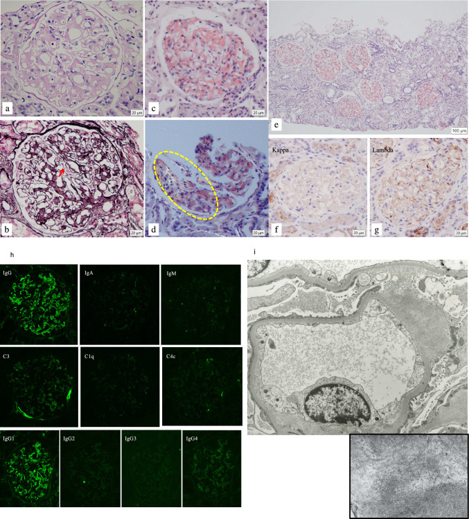
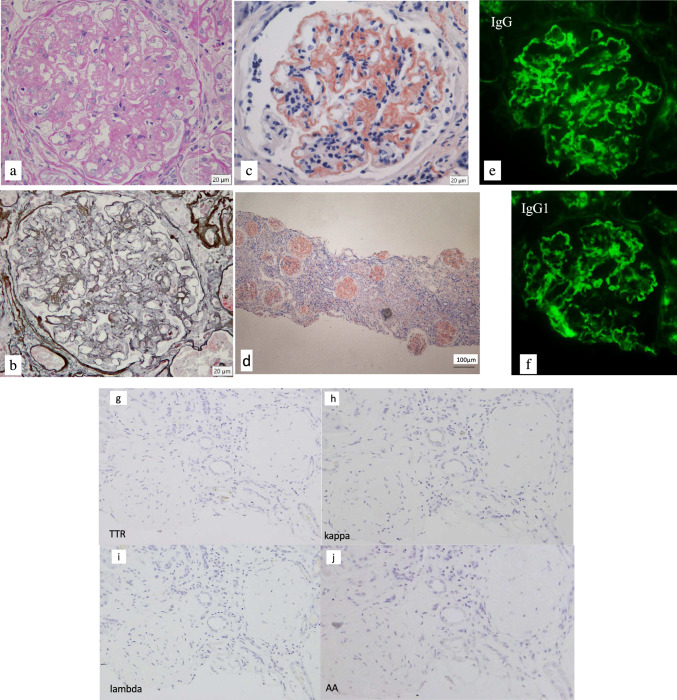
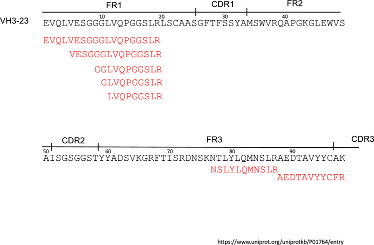
We performed a kidney biopsy on a 68-year-old man with 2.6 g/day proteinuria. Immunofluorescence (IF) study showed a positive finding of IgG (IgG1) along the glomerular capillary wall, suggesting membranous nephropathy. However, electron microscopy showed no subepithelial electron dense deposits and amorphous deposits of 8-12 nm fibrillar structure, and positive DFS/Congo red staining confirmed the diagnosis of amyloidosis. Amyloid deposits were localized only in the glomeruli. No overt extrarenal amyloid lesions were identified. Serum and urine were positive for monoclonal IgG-lambda, but IF was negative for kappa and lambda, ruling out AL-amyloidosis. At 75 years of age, he underwent a second kidney biopsy, and the IgG-positive findings at IF became more pronounced, with progressive glomerular sclerosing lesions. A proteomic analysis was performed focusing on the positive IgG finding in IF. The amyloid was proven to be composed of fragments of a heavy chain variable region sequence. AH-amyloidosis was the final diagnosis. He was treated according to the treatment for AL-amyloidosis, but hemodialysis was started at the age of 81, and died at the age of 86. We report a valuable case of AH-amyloidosis with an observed long-term prognosis.
Keywords: AH amyloidosis; Laser microdissection; Liquid chromatography‐tandem mass spectrometry (LMD/MS); Proteomic analysis.
© 2025. The Author(s), under exclusive licence to Japanese Society of Nephrology.
Conflict of interest statement
Declarations. Conflict of interest: The authors declare no competing financial interests. The authors also declare that they have no conflicts of interest. Ethical approval: The authors declare no competing financial interests. The authors also declare that they have no conflicts of interest.
Figures

References
-
- Yazaki M, Fushimi T, Tokuda T, Kametani F, Yamamoto K, Matsuda M, Shimojo H, Hoshii Y, Higuchi K, Ikeda S. A patient with severe renal amyloidosis associated with an immunoglobulin gamma-heavy chain fragment. Am J Kidney Dis. 2004;43(5):e23–8. - PubMed
-
- Nasr SH, Said SM, Valeri AM, Sethi S, Fidler ME, Cornell LD, Gertz MA, Dispenzieri A, Buadi FK, Vrana JA, Theis JD, Dogan A, Leung N. The diagnosis and characteristics of renal heavy-chain and heavy/light-chain amyloidosis and their comparison with renal light-chain amyloidosis. Kidney Int. 2013;83(3):463–70. 10.1038/ki.2012.414. - PubMed
-
- Nakagawa M, Yazaki M, Kametani F, Katoh N, Yoshinaga T, Higuchi K, Sekijima Y. Development of diagnostic antibodies against immunoglobulin heavy chain variable region for heavy chain amyloidosis (AH amyloidosis). Pathol Int. 2021;71(4):245–54. - PubMed
-
- Kono K, Sawa N, Wake A, Shintani-Domoto Y, Fujii T, Takazawa Y, Ubara Y, Ohashi K. Digital whole-slide imaging of changes in amyloid after peripheral blood stem cell transplantation in patients with amyloid light-chain amyloidosis. Pathol Int. 2024;74(9):508–19. - PubMed
Publication types
MeSH terms
Substances
LinkOut - more resources
Full Text Sources
Medical
Miscellaneous
