. 2025 Feb 28;16(1):2078.
doi: 10.1038/s41467-025-57290-y.

Nuclear retention of unspliced HIV-1 RNA as a reversible post-transcriptional block in latency

Affiliations

Nuclear retention of unspliced HIV-1 RNA as a reversible post-transcriptional block in latency

Agnieszka Dorman et al. Nat Commun. .

Abstract

HIV-1 latency is mainly characterized at transcriptional level, and little is known about post-transcriptional mechanisms and their contribution to reactivation. The viral protein Rev controls the nucleocytoplasmic export of unspliced and singly-spliced RNA that is central to proviral replication-competence and is therefore a prerequisite for efficient viral reactivation during the "shock-and-kill" cure therapy. Here we show that during infection and reactivation, unspliced HIV-1 RNA is a subject to complex and dynamic regulation by the Rev cofactor MATR3 and the MTR4 cofactor of the nuclear exosome. MATR3 and MTR4 coexist in the same ribonucleoprotein complex functioning to either maintain or degrade the RNA, respectively, with Rev orchestrating this regulatory switch. Moreover, we provide evidence of nuclear retention of unspliced HIV-1 RNA in ex vivo cultures from 22 ART-treated people with HIV, highlighting a reversible post-transcriptional block to viral RNA nucleocytoplasmic export that is relevant to the design of curative interventions.


Conflict of interest statement

Competing interests: A.O.P. received a research grant from Gilead Sciences Research Program. C.V.L. received a research grant from ViiV Healthcare. The funders had no role in study design, data collection and analysis, decision to publish, or preparation of the manuscript. The remaining authors declare no competing interests.

Figures
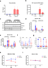
Fig. 1
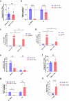
Fig. 1. MATR3 stabalizes Rev-dependent HIV-1 RNA during reactivation.
MATR3 depletion in J-Lat 9.2 cells was obtained with shMATR3 and compared to shLuc lentiviral transduction and three days after puromycin selection, cells were treated with TNFα [10 ng/ml] and after 24 h subjected to a flow cytometry to quantify the percentages of GFP+ cells, b RNA isolation from the supernatant followed by RT-qPCR for genomic HIV-1 RNA (primers for gag RNA), ce biochemical fractionation protocol to obtain nuclear and cytoplasmic extracts, fg immuno-RNA FISH followed by confocal microscopy analysis or h, i RNA stability test with actinomycin D (ActD). c Immunoblotting using anti-MART3, anti-histone H3, and anti-PSF as chromatin and nucleoplasm markers, respectively, and anti-GAPDH as a cytoplasmic marker. d, e Samples were subjected to RNA isolation followed by RT-qPCR for RRE-containing (d) and MS (e) HIV-1 transcripts. Values were normalized using 18S RNA primers and were presented as relative fold changes to the values measured in the untreated shLuc nuclear and cytoplasmic samples which were arbitrarily set at a value of 1. The number (f) and volume (g) of nuclear, cytoplasmic, and total gagHIV-1 spots were quantified from z-stacks obtained from 10 images/biological repetition, n = 3. Results are presented as box and whiskers with 5–95% confidence interval. Median value is shown as a bar, dots are points outside whiskers representing outliers, and the mean value is shown as “+”. Statistics were performed using a two-tailed unpaired Student’s t test. Statistical comparisons are indicated if p ≤ 0.01 (**), p ≤ 0.001 (***), and p ≤ 0.0001 (****), “ns” indicate no significant. hi Cells were collected at 0, 30, and 60 min and subjected to RNA isolation following RT-qPCR targeting RRE-containing (h) and MS (i) HIV-1 RNA. Values were normalized using 18S RNA primers and further normalized to DMSO control at each time point. All results from a, b, d, e, h, i are shown as mean values ± SEM, n = 3 (a, b, d, e) and n = 4 (h, i) biological replicates in duplicates. Statistics were performed using  a two-tailed paired Student’s t test (a, b, d, e, h, i). Statistical comparisons are indicated if p ≤ 0.5 (*), p ≤ 0.01 (**), p ≤ 0.001 (***), and “ns” indicate not significant. Source data are provided as a Source Data file.
Fig. 2
Fig. 2. MATR3/MTR4/Rev ribonucleoprotein complex regulates the fate of Rev-dependent HIV-1 RNA.
a 293 T cells were transfected with MS2-tagged pHIV-INTRO, pTat-GFP, and pMS2-FLAG and after 24 h whole cell lysates were subjected to anti-FLAG immunoprecipitation. b, c 293 T cells were transfected with pFLAG-MATR3, pHIV-INTRO, pTat, and pRev-GFP plasmids and after 24 h whole cell lysates were subjected to anti-FLAG (b), anti-MTR4/anti-IgG (c) immunoprecipitation followed by ±benzonase treatment or (d) anti-FLAG immunoprecipitation followed by elution with FLAG peptides for subsequent IP using either anti-IgG or anti-GFP antibodies. GAPDH is a loading control in input samples and indicates the specificity of IP. ECFP expressed from pHIV-INTRO is a transfection efficiency control in input samples and indicates the specificity of IPs. IgH (in b) refers to the immunoglobulin heavy chain from anti-flag mouse antibodies that are attached to beads. This was detected with a secondary anti-mouse antibody during the anti-GFP immunoblotting. e J-Lat 9.2 cells were stimulated with TNFα [10 ng/ml] and subjected to RNA FISH and immunostaining using antibodies against MTR4 and MATR3 for subsequent confocal microscopy analysis. MATR3 is shown in green, MTR4 in red, gagHIV-1 RNA in purple, and DAPI-stained nucleus in blue. Bright, large gagHIV-1 RNA spot corresponds to viral transcription site as described previously,,. White arrows indicate triple colocalization sites. Scale bar = 10 μm. f Colocalization between MATR3-HIV-1 RNA, MTR4-HIV-1 RNA, MATR3-MTR4, and MATR3-MTR4-HIV-1 RNA was quantified by counting the number of colocalizing spots and their volumes from z-stacks obtained from 10 images/biological repetition, n = 3. Results are presented as box and whiskers with 5–95% confidence interval. Median value is shown as a bar, dots are points outside whiskers representing outliers, mean value is shown as “+”. Statistics were performed using a two-tailed unpaired Student’s t test. Statistical comparisons are indicated if p ≤ 0.01 (**), p ≤ 0.001 (***), and p ≤ 0.0001 (****), “ns” indicate no significance. gi MTR4 depletion in 293 T cells was obtained using siRNA transfection. After 24 h, cells were transfected with plasmids pHIV-INTRO, pTat-GFP, and pRev-GFP and after 24 h collected for (g) total RNA isolation, (h) nuclear-cytoplasmic fractionation protocol followed by RT-qPCR for RRE-containing, TAR, and MS HIV-1 RNAs normalized using gapdh primers and presented as relative fold changes to the values measured in non-targeting control (siCTRL) condition, which was arbitrarily set at a value of 1. i total RNA isolation followed by RNA immunoprecipitation (IP) protocol using anti-MATR3 and anti-IgG isotype antibodies. RNA was purified from IPs and subjected to RT-qPCR targeting RRE-containing HIV-1 RNA. Values were normalized to input and shown as fold enrichment over IgG control. All results are shown as mean values ± SEM from n = 5 (g), n = 3 (h), n = 3 (I) biological replicates in duplicates. Statistics were performed using a two-tailed paired Student’s t test, p ≤ 0.5 (*), p ≤ 0.01 (**), p ≤ 0.001 (***). Source data are provided as a Source Data file.
Fig. 3
Fig. 3. Rev determines the MATR3 and MTR4 binding to viral RNA.
Primary CD4+ T cells were isolated from blood of healthy donors and infected with WT pNL4.3 HIV particles or Rev-mutated pNL4.3 HIV particles and collected after 3 days and used for (a) DNA extraction to quantify total HIV DNA by qPCR, for (b) total RNA extraction used for RT-qPCR quantification of RRE-containing RNA and Tat HIV-1 RNA, for (c) nuclear-cytoplasmic fractionation protocol followed by quantitative real-time RT-PCR for Tat and (d) RRE HIV-1 RNA, for (e) RNA immunoprecipitation using anti-MATR3, anti-MTR4 and anti-IgG isotype antibodies. RNA was purified from IPs and subjected to RT-qPCR targeting RRE-containing HIV-1 RNA. Values were normalized to input and shown as fold enrichment of IgG control. fh Jurkat T cells were infected with WT pNL4.3 HIV particles or Rev-mutated pNL4.3 HIV particles and collected after 3 days and used for (f) DNA extraction to quantify total HIV DNA by qPCR, for (g) total RNA extraction used for RT-qPCR quantification of RRE-containing and Tat HIV-1 RNA and (h) RNA immunoprecipitation using anti-MTR4 and anti-IgG isotype antibodies. RNA was purified from IPs and subjected to RT-qPCR targeting RRE-containing HIV-1 RNA. All results are shown as mean values ± SEM from n = 5 (a), n = 7 (b), n = 4 (c, d, f, g), n = 6 (e), n = 3 (h) biological replicates in duplicates. Statistics were performed using a two-sided Wilcoxon signed-rank test for HIV integration (a), a two-way ANOVA test (bg), and a two-tailed unpaired Student’s t test (h). Significant p values are indicated by the asterisks above the graphs p ≤ 0.05 [*], p ≤ 0.01 [**], ≤0.001 [***], ≤0.0001 [****]. Source data are provided as a Source Data file.
Fig. 4
Fig. 4. Nuclear retention of US HIV-1 RNA in PBMCs from 22 ART-treated PWH.
a PBMCs from healthy donors were left untreated or were treated with PHA [1.66 µg/ml] and after 24 h collected for immunoblotting with the use of anti-MATR3, anti-MTR4, anti-αtubulin and anti-GAPDH antibodies. αtubulin and GAPDH are loading controls. be Freshly isolated PBMCs from 16 ART-treated PWH were subjected to nucleocytoplasmic fractionation for subsequent nucleic acid isolation and RT-qPCR quantification of (b) US-short HIV-1 RNA, (c) gapdh mRNA, and (d) US-long HIV-1 RNA. Percentages of each of the measured RNAs in the cytoplasmic fraction are shown in (e). fk Ex vivo cultures of CD8+-depleted PBMCs from 6 ART-treated PWH were mock-treated, treated with SAHA [0.5 µM], disulfiram [0.5 µM], romidepsin [17.5 nM] or PHA [1.66 µg/ml] as a positive control in the presence of ARV [280 nM ritonavir, 180 nM azidothymidine, 200 nM raltegravir, 100 nM efavirenz]. Three days post-treatment cells were subjected to nucleocytoplasmic fractionation for subsequent nucleic acid isolation and RT-qPCR quantification of (f, g) US-short HIV-1 RNA, (h, i) gapdh mRNA, and (j, k) US-long HIV-1 RNA. Percentages of each of the measured RNAs in the cytoplasmic fraction are shown in (g, i, k). bk Medians are indicated as horizontal lines, and for (e, g, i, k) interquartile ranges are indicated as well. The RNA copy numbers were normalized to the cell numbers measured by the beta-actin qPCR assay. Open symbols in (b, d, f, j) indicate undetectable measurements of US RNA that were assigned the values corresponding to 50% of the corresponding assay detection limits. The detection limits depended on the amounts of the normalizer (input cellular DNA) and, therefore, differed between samples. Open symbols in e, g, k indicate samples where US RNA in the cytoplasmic fraction was undetectable and censored to 50% of the detection limits; because US RNA in the nuclear fraction in these samples was detectable, these circles depict the upper limits of the percentages of US RNA in the cytoplasm. Open crossed circles in e, g, k indicate samples where US RNA was undetectable in the nuclear fraction and censored to 50% of the detection limits; because US RNA in the cytoplasmic fraction in these samples was detectable, these circles depict the lower limits of the percentages of US RNA in the cytoplasm. Percentages of US RNA in the cytoplasmic fraction are not shown if US RNA was undetectable in both nuclear and cytoplasmic fractions. bd RNA levels were compared between nuclear and cytoplasmic fractions using a two-sided Wilcoxon signed-rank test. **, 0.001 < p < 0.01. ns, not significant. l A schematic view of the post-transcriptional regulation of US HIV-1 RNA by MATR3, MTR4, and Rev. Limiting levels of MATR3 and Rev cause nuclear retention of US HIV-1 RNA that is subjected to MTR4 for further degradation (left panel). During reactivation, MATR3 competes with MTR4 to stabilize the RNA. Rev hijacks the pathway to export the RNA (right panel). Created in BioRender. Wadas, J. (2025) https://BioRender.com/w90s107. Source data are provided as a Source Data file.

References

    1. Deeks, S. G., Lewin, S. R. & Havlir, D. V. The end of AIDS: HIV infection as a chronic disease. Lancet382, 1525–1533 (2013). - PMC - PubMed
    1. Dufour, C., Gantner, P., Fromentin, R. & Chomont, N. The multifaceted nature of HIV latency. J. Clin. Invest.130, 3381–3390 (2020). - PMC - PubMed
    1. Pasternak, A. O. & Berkhout, B. HIV persistence: silence or resistance? Curr. Opin. Virol.59, 101301 (2023). - PubMed
    1. Crowe, S., Zhu, T. & Muller, W. A. The contribution of monocyte infection and trafficking to viral persistence, and maintenance of the viral reservoir in HIV infection. J. Leukoc. Biol.74, 635–641 (2003). - PubMed
    1. Lewin, S. R. et al. HIV-1 DNA and mRNA concentrations are similar in peripheral blood monocytes and alveolar macrophages in HIV-1-infected individuals. AIDS12, 719–727 (1998). - PubMed

MeSH terms

Substances

LinkOut - more resources