

A customized bed based stand alone array of optically pumped magnetometers for fetal magnetocardiography measurements
- PMID: 40021708
- PMCID: PMC11871292
- DOI: 10.1038/s41598-025-90846-y
A customized bed based stand alone array of optically pumped magnetometers for fetal magnetocardiography measurements
Abstract
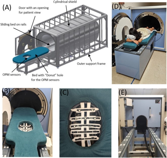
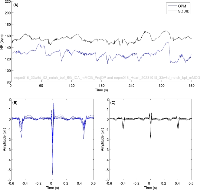
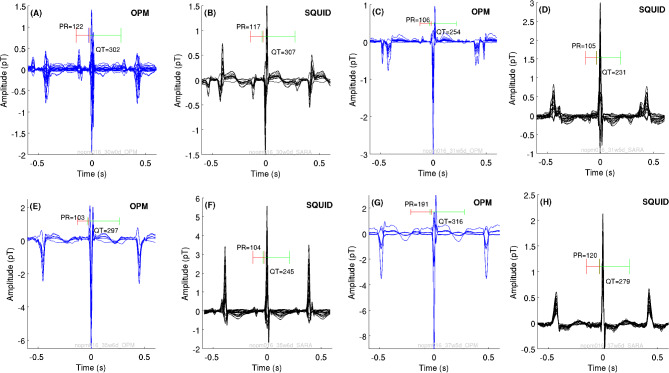
Fetal magnetocardiography (fMCG) is a non-invasive technique that measures the magnetic fields associated with fetal heart electrical activity outside of the maternal abdomen. fMCG has high temporal precision for measuring fetal heart rate and its variability which reflects fetal neurodevelopment. Free of cryogenics and low-cost sensors called microfabricated optically pumped magnetometers (OPMs) have emerged as an alternate to cryogenic SQUID (Superconducting Quantum Interference Device) systems to record fMCG. Previous research has demonstrated the ability of the OPMs to measure the fMCG at different maternal positions by taking the advantage of the conformal and geometric flexibility of the sensors. In this work, we designed and configured a bed-based stand-alone array of OPMs to obtain serial recordings of fMCG. 72 combined OPM-SQUID recordings were conducted at different gestational ages in 22 pregnant women. We were able to obtain fMCG with similar detectability as the gold standard SQUID from OPM sensors mounted on a novel belly-shape patient interface design with movable sensor holders. While additional translational research is needed, the outcome of this study can further facilitate the development of a non-cryogenic low-cost smaller footprint device to increase the use of OPMs for fetal research and clinical applications.
Keywords: Biomagnetism; Fetus; Magnetocardiography; Optically pumped magnetometers; Pregnancy.
© 2025. The Author(s).
Conflict of interest statement
Declarations. Competing interests: The authors declare no competing interests.
Figures


Similar articles
-
Recording and quantifying fetal magnetocardiography signals using a flexible array of optically-pumped magnetometers.Physiol Meas. 2021 Jan 1;41(12):125003. doi: 10.1088/1361-6579/abc353. Physiol Meas. 2021. PMID: 33086201 Free PMC article.
-
Fetal magnetocardiographic recordings with a prototype bed-based array system of optically-pumped magnetometers.Med Eng Phys. 2024 Jun;128:104175. doi: 10.1016/j.medengphy.2024.104175. Epub 2024 May 8. Med Eng Phys. 2024. PMID: 38789219 Free PMC article.
-
Adaptable Sensor Arrays for Fetal Magnetocardiographic Measurements Using Optically-Pumped Magnetometers: A Pilot Study.Annu Int Conf IEEE Eng Med Biol Soc. 2020 Jul;2020:1803-1806. doi: 10.1109/EMBC44109.2020.9175691. Annu Int Conf IEEE Eng Med Biol Soc. 2020. PMID: 33018349 Free PMC article.
-
[Fetal magnetocardiography: a promising way to diagnose fetal arrhytmia and to study fetal heart rate variability?].Ceska Gynekol. 2015 Jan;80(1):58-63. Ceska Gynekol. 2015. PMID: 25723081 Review. Czech.
-
Contribution of Fetal Magnetocardiography to Diagnosis, Risk Assessment, and Treatment of Fetal Arrhythmia.J Am Heart Assoc. 2022 Aug 2;11(15):e025224. doi: 10.1161/JAHA.121.025224. Epub 2022 Jul 29. J Am Heart Assoc. 2022. PMID: 35904205 Free PMC article. Review.
References
-
- Lowery, C. L. et al. Noninvasive antepartum recording of fetal ST segment with a newly developed 151-channel magnetic sensor system. Am. J. Obstet. Gynecol.188(6), 1491–1497 (2003). - PubMed
-
- Vrba, J. Multichannel SQUID biomagnetic systems 61–138 (Springer, 2000).
-
- Alem, O. et al. Fetal magnetocardiography measurements with an array of microfabricated optically pumped magnetometers. Phys. Med. Biol.60(12), 4797 (2015). - PubMed
-
- Eswaran, H., Escalona-Vargas, D., Bolin, E. H., Wilson, J. D. & Lowery, C. L. Fetal magnetocardiography using optically pumped magnetometers: a more adaptable and less expensive alternative?. Prenat. Diagn.37(2), 193–196 (2017). - PubMed
MeSH terms
Grants and funding
LinkOut - more resources
Full Text Sources
