. 2025 Feb 28;16(1):101.
doi: 10.1186/s13287-025-04243-3.

Thrombin-preconditioned mesenchymal stromal cell-derived extracellular vesicles attenuate experimental necrotizing enterocolitis

Affiliations

Thrombin-preconditioned mesenchymal stromal cell-derived extracellular vesicles attenuate experimental necrotizing enterocolitis

Sein Hwang et al. Stem Cell Res Ther. .

Abstract

Background: Necrotizing enterocolitis (NEC) is a critical gastrointestinal disease in preterm infants, for which no specific treatment is established. We previously demonstrated that thrombin-preconditioned mesenchymal stromal cell-derived extracellular vesicles (thMSC-EVs) enhance protection against other neonatal tissue injuries. Therefore, this study aimed to evaluate the therapeutic potential of thMSC-EVs in modified in vitro, in vivo, and organoid models of NEC.

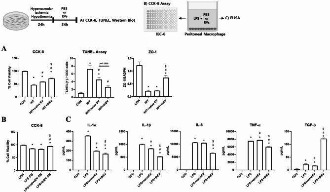
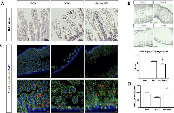
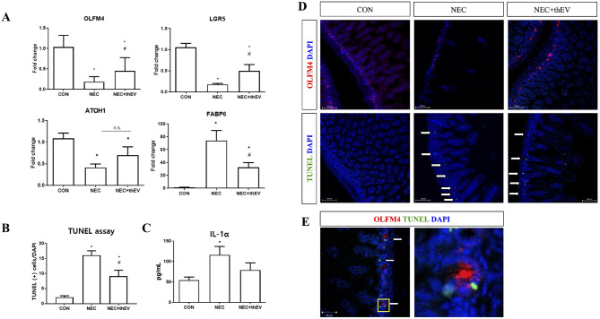
Methods: In vitro, the effects of thMSC-EVs and naïveMSC-EVs were compared in hyperosmotic, ischemic, and hypothermic (HIT)-stressed IEC-6 cells and LPS-treated peritoneal macrophages. In vivo, NEC was induced in P4 mouse pups by three cycles of formula feeding, oral LPS administration, hypoxia, and hypothermia, followed by overnight dam care. 2 × 10⁹ thMSC-EVs were intraperitoneally administered daily for three days, and the therapeutic effects were assessed macroscopically, histologically, and biochemically. NEC mouse-derived organoids were established to evaluate the thMSC-EVs' effect in mature enterocytes. LC-MS/MS was performed to analyze the EV proteomics.

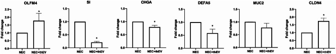
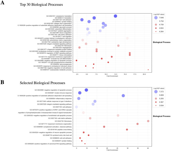
Results: In vitro, compared with naïveMSC-EVs, thMSC-EVs significantly improved cellular viability in HIT-induced IEC-6 cells and reduced pro-inflammatory (IL-1α, IL-1β, TNF-α) but increased anti-inflammatory (TGF-b) cytokine levels in LPS-treated peritoneal macrophages. In vivo, thMSC-EVs significantly attenuated clinical symptoms, reduced intestinal damage, and retained intestinal stem cell markers, showing more significant localization in NEC-induced intestines than in healthy intestines. In NEC mouse-derived organoids, thMSC-EVs significantly increased OLFM4 and claudin-4 expression and reduced stress-related markers such as sucrase-isomaltase, defensin, and chromogranin A. Proteomic analysis revealed that thMSC-EVs were greater enriched in anti-apoptotic, anti-inflammatory, cell adhesion, and Wnt signaling pathways than naïveMSC-EVs.

Conclusion: thMSC-EVs improved cellular viability, reduced apoptosis, attenuated inflammation, and upregulated key intestinal stem cell markers, collectively suggesting their tissue-protective effects and highlighting their potential as a treatment for NEC.

Keywords: Extracellular vesicles; Mesenchymal stromal cells; Necrotizing enterocolitis.


Conflict of interest statement

Declarations. Ethics approval and consent to participate: Human WJ-MSCs from a single donor, which were collected with informed consent for the use of the study, were provided by the Samsung Medical Center Good Manufacturing Practice Facility (IRB approval number: 2016-07-102-043; Date of approval: 2016-09-20; Approval expiration date: 2025-09-15; Study name: Study on the Selection of Optimal Mesenchymal Stem Cells from Different Sources for the Treatment of Chronic/Intractable Diseases including Alzheimer’s Disease and Musculoskeletal Disorders). All animal studies were reviewed and approved by the Institutional Animal Care and Use Committee (Approval numbers: 20231115001 and 20230116001. Approval Date: 2023.12.22 and 2023.02.22) of the Samsung Biomedical Research Institute, an Association for Assessment and Accreditation of Laboratory Animal Care-accredited facility, following the National Institutes of Health Guidelines for Laboratory Animal Care. Animal studies followed the ARRIVE guidelines. Consent for publication: Not applicable. Conflict of interest: The funders had no role in the study design; collection, analyses, or interpretation of data; writing of the manuscript; or decision to publish the results. Yun Sil Chang, Se In Sung, Young Eun Kim, Sein Hwang, and Ara Koh declare potential conflicts of interest arising from an issued patent titled “A composition for the prevention and treatment of necrotizing enterocolitis containing extracellular vesicles derived from thrombin-treated mesenchymal stem cells” (10-2024-0051684) as co-inventors, not as patentees.

Figures

Fig. 1
Fig. 1
thMSC-EVs significantly improved cell viability, cell integrity, and inflammation in NEC in vitro models.(A) Bar graph representation of CCK-8 cell viability assay, TUNEL assay, and western blot protein expression levels of ZO-1 in IEC-6 cells. n = 16, 16, 16, and 16 for the CCK-8 assay; n = 10, 10, 10, and 10 for the TUNEL assay; and n = 3, 3, 3, and 3 for western blot analysis in the CON, HIT, HIT + naïveEV, and HIT + thEV groups, respectively. *, p < 0.01 vs. CON; #, p < 0.01 vs. HIT; $, p < 0.01 vs. HIT + naïveEV. (B) Bar graph representation of CCK-8 cell viability assay in LPS-induced peritoneal macrophage CM-treated IEC-6 cells. n = 16, 16, 16, and 16 in the CON, LPS CM, LPS + naïveEV CM, and LPS + thEV CM groups, respectively. (C) Bar graph showing inflammatory cytokine levels in LPS-induced peritoneal macrophages. n = 4, 4, 4, and 4 in the CON, LPS, LPS + naïveEV, and LPS + thEV groups, respectively. *, p < 0.01 vs. CON; #, p < 0.01 vs. LPS; $, p < 0.01 vs. LPS + naïveEV. Data are presented as mean ± SEM. One-way ANOVA and the post-hoc Tukey test were used in the analysis. CON, normal control; HIT, hyperosmolar stress, ischemia, and hypothermia-induced IEC-6 cells; LPS, lipopolysaccharide; naïve MSC-EVs, naïve MSC-derived EVs; thMSC-EVs, thrombin-preconditioned MSC-derived EVs
Fig. 2
Fig. 2
Intraperitoneal administration of thMSC-EVs improved the clinical symptoms in NEC-induced mouse pups.(A) Graphical representations of daily body weight measurements and percent survival of mouse pups. n = 23, 40, 43 in the CON, NEC, and NEC + thEV groups, respectively. (B) Bar graph representation of clinical sickness score assessment at P7. n = 8, 16, and 13 in the CON, NEC, and NEC + EV groups, respectively. (C) Bar graph representation of GI length measured at P7. n = 8, 16, and 13 in the CON, NEC, and NEC + thEV groups, respectively. Data are presented as mean ± SEM. *, p < 0.01 vs. CON; #, p < 0.01 vs. NEC. The log-rank test was used to compare the survival rate between the groups. One-way ANOVA and the post-hoc Tukey test were used in all other analyses. CON, normal control; NEC, necrotizing enterocolitis control group; NEC + thEV, thMSC-EVs treated NEC group
Fig. 3
Fig. 3
Intraperitoneal administration of thMSC-EVs attenuated the histological damage in NEC-induced mouse pups.(A) Representative microscopic images of H&E-stained histology slides. (B) Bar graph representation of histological damage score assessment. n = 11, 21, and 25 in the CON, NEC, and NEC + thEV groups, respectively. (C) Representative microscopic images of immunofluorescent staining. (D) Bar graph representation of quantification of MUC2 (+) cells. n = 6, 7, and 11 in the CON, NEC, and NEC + EV groups, respectively. Data are presented as mean ± SEM. *, p < 0.01 vs. CON; #, p < 0.01 vs. NEC. One-way ANOVA and the post-hoc Bonferroni test were used in the analysis. CON, normal control; NEC, necrotizing enterocolitis control group; NEC + thEV, thMSC-EVs treated NEC group
Fig. 4
Fig. 4
Intraperitoneal administration of thMSC-EVs increased goblet cells and ISC marker gene expression in NEC-induced mouse pups.(A) Bar graph representation of the expression levels of intestinal marker genes LGR5, OLFM4, ATOH1, and FABP6 analyzed via RT-qPCR. n = 16, 11, and 16 in the CON, NEC, and NEC + thEV groups, respectively. The Mann–Whitney test was used for the analysis. (B) Bar graph representation of the count of TUNEL (+) cells. n = 11, 21, and 25 in the CON, NEC, and NEC + thEV groups, respectively. One-way ANOVA and the post-hoc Bonferroni test were used in the analysis. (C) Bar graph representation of pro-inflammatory cytokine IL-1α levels measured using ELISA. n = 18, 23, and 19 in the CON, NEC, and NEC + EV groups, respectively. (D) Representative microscopic image of immunofluorescent-stained OLFM4 (+) cells and TUNEL (+) cells. (E) Representative microscopic image of merged OLFM4 and TUNEL (+) cells. One-way ANOVA and the post-hoc Bonferroni test were used in the analysis. White arrows point to TUNEL (+) cells. Data are presented as mean ± SEM. *, p < 0.01 vs. CON; #, p < 0.01 vs. NEC. CON, normal control; NEC, necrotizing enterocolitis control group; NEC + thEV, thMSC-EVs treated NEC group
Fig. 5
Fig. 5
thMSC-EVs’ effect in gene expression levels of differentiated NEC organoids.Bar graph representation of intestinal organoid marker gene expression levels analyzed using RT-qPCR. n = 4 and 4 in the NEC and NEC + thEV groups, respectively. Data are presented as mean ± SEM. *, p < 0.01 vs. NEC. The Mann–Whitney test was used in the analysis. CON, normal control; NEC, necrotizing enterocolitis control group; NEC + thEV, thMSC-EVs treated NEC group
Fig. 6
Fig. 6
Proteomic comparison of naïveMSC-EVs and thMSC-EVs.(A) Bubble plot representation of top 30 Biological Processes enriched in thMSC-EVs than in naïve MSC-EVs. (B) Bubble plot representation of selected Biological Processes. Proteins that had more than two-fold greater abundance value in thMSC-EVs than in naïveMSC-EVs with p < 0.05 were analyzed. Fisher’s exact statistic was used to analyze protein enrichment. The size of the bubble represents protein counts

References

    1. Alsaied A, Islam N, Thalib L. Global incidence of necrotizing Enterocolitis: a systematic review and Meta-analysis. BMC Pediatr. 2020;20(1):344. - PMC - PubMed
    1. Fitzgibbons SC et al. Mortality of necrotizing enterocolitis expressed by birth weight categories. J Pediatr Surg, 2009. 44(6): pp. 1072-5; discussion 1075-6. - PubMed
    1. Yee WH, et al. Incidence and timing of presentation of necrotizing Enterocolitis in preterm infants. Pediatrics. 2012;129(2):e298–304. - PubMed
    1. Mekonnen SM, et al. The prevalence of necrotizing Enterocolitis and associated factors among enteral fed preterm and low birth weight neonates admitted in selected public hospitals in addis Ababa, Ethiopia: A Cross-sectional study. Glob Pediatr Health. 2021;8:2333794X211019695. - PMC - PubMed
    1. Patel BK, Shah JS. Necrotizing enterocolitis in very low birth weight infants: a systemic review. ISRN Gastroenterol, 2012. 2012: p. 562594. - PMC - PubMed

LinkOut - more resources