. 2025 Feb 28;26(1):48.
doi: 10.1186/s40360-025-00883-6.

Prodelphinidin B-2,3,3"-O-gallate via chemical oxidation of epigallocatechin-3-gallate shows high efficacy inhibiting triple-negative breast cancer cells

Affiliations

Prodelphinidin B-2,3,3"-O-gallate via chemical oxidation of epigallocatechin-3-gallate shows high efficacy inhibiting triple-negative breast cancer cells

Jing Wang et al. BMC Pharmacol Toxicol. .

Abstract

Background: Triple-negative breast cancer is a clinically aggressive malignancy with poorer outcomes versus other subtypes of breast cancer. Numerous reports have discussed the use of epigallocatechin-3-gallate (EGCG) against various types of cancer. However, the effectiveness of EGCG is limited by its high oxidation and instability. The Notch pathway is critical in breast cancer development and prognosis, and its inhibition is a potential treatment strategy.

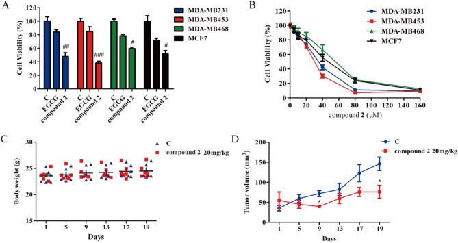
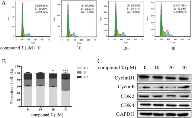
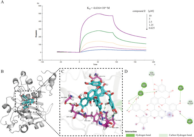
Results: In this study, we investigated the effects of prodelphinidin B-2,3,3''-O-gallate (named PB2,3,3''/OG or compound 2) via chemical oxidation of EGCG on cell viability and the Notch1 signaling pathway in breast cancer cells. We found that compound 2 showed significant cytotoxicity against triple-negative breast cancer cells, with the half maximal inhibitory concentration (IC50) values ranging 20-50 µM. In MDA-MB453 cells, compound 2 inhibited proliferation, clone formation, and the expression of proteins involved in the Notch1 signaling pathway. Furthermore, compound 2 induced cell cycle arrest and apoptosis. Consistent with the results of in-vitro experiments, treatment with compound 2 significantly reduced tumor growth. Mechanistically, compound 2 directly bound to Notch1 with high binding affinity (dissociation constant: KD=4.616 × 10- 6 M).

Conclusion: Our finding suggested that compound 2 may be a promising agent for the development of novel anti-cancer therapy options.

Keywords: Apoptosis; Cell viability; Notch1; Prodelphinidin B-2,3,3''-O-gallate; Triple-negative breast cancer.


Conflict of interest statement

Declarations. Ethical approval: All experiments on nude mice comply with Yunnan Agricultural University guidelines. The experiments carried out in this work have been approved by ethics committee. Competing interests: The authors declare no competing interests.

Figures

Fig. 1
Fig. 1
Chemical structures of EGCG (A) and PB2,3,3’’/OG (B)
Fig. 2
Fig. 2
Effects of EGCG and its oxide (compound 2) on the proliferation of breast cancer cells in vitro and in vivo. (A) Three TNBC cell lines (MDA-MB231/453/468) and an ERα-positive breast cancer cell line (MCF7) were treated with EGCG (40 µM) or compound 2 (40 µM) for 24 h. Cell proliferation was measured using the MTS assay. Results are expressed as the mean ± SD (n = 3). #p < 0.05, ##p < 0.01, ###p < 0.001 versus EGCG. (B) The cells were treated with compound 2 (5, 10, 20, 40, 80, and 160 µM) for 24 h. All values are expressed as the mean ± SD (n = 3). (C) Data on the body weight of mice in each group were analyzed. (D) Tumor volumes were measured with calipers and analyzed. *p < 0.05 versus control
Fig. 3
Fig. 3
Effects of compound 2 on the clone formation of MDA-MB453 cells. (A) Cells were treated with compound 2 (10, 20, 40 µM) for 24 h. After 14 days, colony formation was stained with 0.01% crystal violet solution and assessed. (B) The cell colonies were dissolved with 10% glacial acetic acid, and the optical densities of the samples were measured at 560 nm. The ratio of clone formation in each group is expressed as percentage. All values are expressed as the mean ± SD (n = 3). ***p < 0.001 versus control. (C) Cells were treated with compound 2 (10, 20, 40 µM). The protein expression of Ki67 was detected by western blotting. GAPDH was used as the loading control
Fig. 4
Fig. 4
Compound 2 induced S-phase cell cycle arrest in MDA-MB453 cells. (A) Cells were treated with compound 2 (10, 20, 40 µM) for 24 h. The cell cycle distribution was analyzed using the FlowJo software. (B) The quantification of the cell populations in each phase of the cell cycle is presented. All values are expressed as the mean ± SD (n = 3). *p < 0.05, ***p < 0.001 versus control. (C) Cells were treated with compound 2 (10, 20, 40 µM), and the expression levels of cell cycle-related proteins were determined through western blotting
Fig. 5
Fig. 5
Compound 2 induced apoptosis in MDA-MB453 cells. (A) Flow cytometry of MDA-MB453 cells after treatment with compound 2 (10, 20, 40 µM) for 24 h. (B) The ratio of apoptotic cells in each group is expressed as percentage. All values are expressed as the mean ± SD (n = 3). **p < 0.01, ***p < 0.001 versus control. (C) Cells were treated with compound 2 (10, 20, 40 µM), and the expression levels of cleaved-caspase3/9, caspase3/9, cleaved-PARP1, and PARP1 were detected by western blotting. GAPDH was used as the loading control
Fig. 6
Fig. 6
Effect of compound 2 on Notch1 processing in MDA-MB453 cells. Cells were treated with compound 2 (10, 20, 40 µM) for 24 h. The protein expression levels of Notch1 and cleaved-Notch1 were detected by western blotting
Fig. 7
Fig. 7
Binding mode of compound 2 and Notch1. (A) Compound 2 directly interacted with Notch1, with a KD value of 4.616 × 10− 6 M. (B, C) Molecular docking model of compound 2 bound to the Notch1. Ligand and key residues are shown as sticks, and interactions are shown as dashed lines. (D) Two-dimensional diagram of compound 2-Notch1 interactions. KD, dissociation constant

References

    1. Siegel RL, Miller KD, Wagle NS, et al. Cancer statistics, 2023. CA Cancer J Clin. 2023;73(1):17–48. - PubMed
    1. Hammond ME, Hayes DF, Dowsett M, et al. American society of clinical oncology/college of American pathologists guideline recommendations for immunohistochemical testing of Estrogen and progesterone receptors in breast cancer. J Clin Oncol. 2010;28(16):2784–95. - PMC - PubMed
    1. Kalimutho M, Parsons K, Mittal D, et al. Targeted therapies for Triple-Negative breast cancer: combating a stubborn disease. Trends Pharmacol Sci. 2015;36(12):822–46. - PubMed
    1. Reinert T, Barrios CH. Optimal management of hormone receptor positive metastatic breast cancer in 2016. Ther Adv Med Oncol. 2015;7(6):304–20. - PMC - PubMed
    1. Carey LA, Dees EC, Sawyer L, et al. The triple negative paradox: primary tumor chemosensitivity of breast Cancer subtypes. Clin Cancer Res. 2007;13(8):2329–34. - PubMed

MeSH terms

LinkOut - more resources