. 2025 Apr 2;33(4):1407-1419.
doi: 10.1016/j.ymthe.2025.02.036. Epub 2025 Feb 28.

A differentiated β-globin gene replacement strategy uses heterologous introns to restore physiological expression

Affiliations

A differentiated β-globin gene replacement strategy uses heterologous introns to restore physiological expression

Kirby A Wallace et al. Mol Ther. .

Abstract

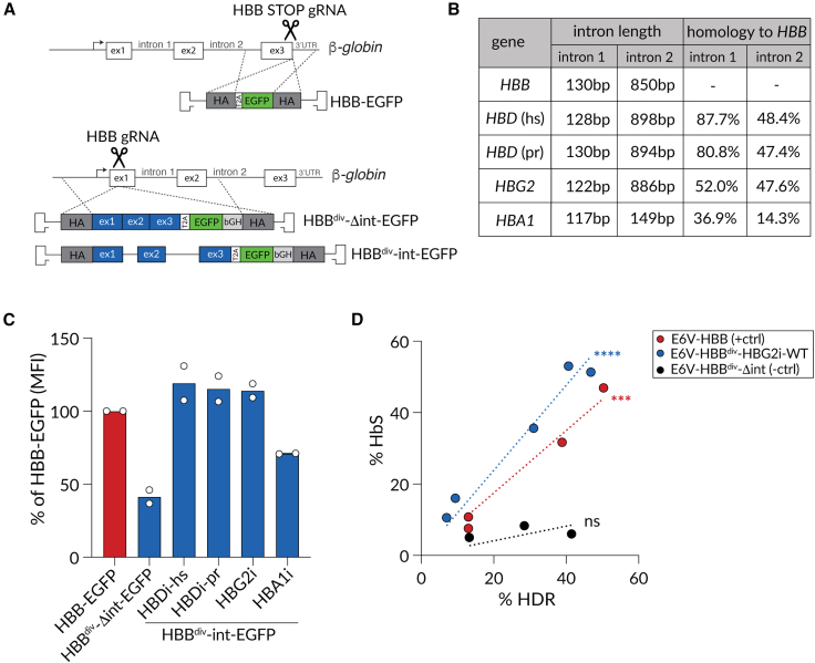
β-Hemoglobinopathies are common monogenic disorders. In sickle cell disease (SCD), a single mutation in the β-globin (HBB) gene results in dysfunctional hemoglobin protein, while in β-thalassemia, over 300 mutations distributed across the gene reduce β-globin levels and cause severe anemia. Genetic engineering replacing the whole HBB gene through homology-directed repair (HDR) is an ideal strategy to restore a benign genotype and rescue HBB expression for most genotypes. However, this is technically challenging because (1) the insert must not be homologous to the endogenous gene and (2) synonymous codon-optimized, intron-less sequences may not reconstitute adequate β-globin levels. Here, we developed an HBB gene replacement strategy using CRISPR-Cas9 that successfully addresses these challenges. We determined that a DNA donor containing a diverged HBB coding sequence and heterologous introns to avoid sequence homology provides proper physiological expression. We identified a DNA donor that uses truncated γ-globin introns, results in 34% HDR, and rescues β-globin expression in in vitro models of SCD and β-thalassemia in hematopoietic stem and progenitor cells (HSPCs). Furthermore, while HDR allele frequency dropped in vivo, it was maintained at ∼15%, demonstrating editing of long-term repopulating HSPCs. In summary, our HBB gene replacement strategy offers a differentiated approach by restoring naturally regulated adult hemoglobin expression.

Keywords: CRISPR-Cas9; HBB; HDR; gene editing; gene replacement; hematopoietic stem cells; hemoglobinopathies; sickle cell disease; β-thalassemia.


Conflict of interest statement

Declaration of interests All authors are former employees of Graphite Bio, Inc. and may own stock/options in the company, and all work was done while authors were employed at Graphite Bio, Inc.. A patent application encompassing aspects of this work has been filed, with C.B., A.R.C., D.P.D., and B.W. included as inventors and Graphite Bio, Inc. as the applicant (PCT/US2022/024477, pending). M.H.P. serves on the scientific advisory board of Allogene Tx and is an advisor to Versant Ventures; he has equity in CRISPR Tx and serves on the board of directors of Kamau Therapeutics; and he is affiliated with Stanford University. J.L.G. is an advisor to Kamau Therapeutics. K.A.W. and M.H.P. are current employees and shareholders of Kamau Therapeutics. B.W. is affiliated with and has equity in Evercrisp Biosciences. T.L.G. and L.C. are affiliated with Scribe Therapeutics. C.L.E. is affiliated with the University of California, San Francisco. J.A.P.-B. is affiliated with Genentech. J.R.P. is affiliated with Novartis. W.M.M. and A.R.C. are affiliated with Enceladus Biosciences. K.K. is affiliated with Bio-Rad Laboratories. S.K. is affiliated with and has equity in CRISPR Tx. C.D.L. is affiliated with OmniAb. G.M.C. is affiliated with Caribou Biosciences. J.E.V. is affiliated with Alector. B.J.S. is affiliated with Senti Biosciences. B.J.Q. is affiliated with the University of California, San Diego. K.A.H. is affiliated with Amber Biosciences. J.L.G. is affiliated with Biogen.

Figures

None
Graphical abstract
Figure 1
Figure 1
Heterologous globin introns rescue β-globin expression to physiological levels after gene replacement (A) Schematic of two different gene-editing strategies at the HBB locus. Insertion of a T2A-EGFP sequence at the STOP codon of endogenous HBB (top, HBB-EGFP), and an HBB gene replacement cassette with diverged codons (HBBdiv; see Figure S1B) containing either no introns (HBBdiv-Δint-EGFP) or introns from different heterologous globin genes followed by a T2A-EGFP-bGH sequence (HBBdiv-int-EGFP). (B) Table outlining the length and homology of globin introns tested relative to native HBB introns. (C) EGFP mean fluorescence intensity (MFI) of EGFP+ populations of in vitro differentiated erythroid progenitors edited with the AAV6 DNA donors outlined in (A). Shown is the MFI of EGFP+ cells normalized to the MFI for HBB-EGFP control for each experiment, n = 2. (D) HbS expression by HPLC and HDR allele frequency by ddPCR (E6V-HBBdiv-HBG2i-WT and E6V-HBBdiv-Δint) or amplicon NGS (E6V-HBB) of in vitro differentiated erythroid progenitors edited at the HBB locus to introduce the E6V SCD mutation (Figure S3A). The dotted line represents a linear regression for each construct. Statistical significance of experimentally determined slopes’ difference to zero was calculated using a simple linear regression. Each data point represents a biological HSPC replicate. hs, Homo sapiens; ns, not significant; pr, primate. ∗∗∗p < 0.001; ∗∗∗∗p < 0.0001; bar graphs represent the mean.
Figure 2
Figure 2
Truncating HBG2 intron 2 maintains protein expression and reduces off-target recombination (A) Schematic outlining the HBBdiv-HBG2i-WT and the truncated (HBBdiv-HBG2i-i2v2) AAV6 DNA donors and the assays used to compare their performance. (B) HDR frequencies in HSPCs edited with HBBdiv-HBG2i-WT or HBBdiv-HBG2i-i2v2 AAV6 donors determined by ddPCR (3′ assay; Figure S3B). Statistical significance was determined by unpaired two-tailed t test. n = 25 and 29 HSPC biological replicates, respectively. (C) HbS production by HPLC and HDR allele frequency by ddPCR (3′ assay) in HSPC-derived erythroid progenitors edited with E6V-HBBdiv-HBG2i-WT or E6V-HBBdiv-HBG2i-i2v2 AAV6 donors (n = 5 HSPC donors for each data point). Statistical significance was determined by first performing a non-linear regression for each construct. The analysis determined that the relationship between %HDR and %HbS was linear for each construct and is represented by the dotted lines. Next, the slopes were defined, and statistical significance was determined by an unpaired two-tailed t test comparing the slopes of the regression. (D) CFU frequency from in vitro cultured HSPCs edited with HBBdiv-HBG2i-WT or HBBdiv-HBG2i-i2v2 AAV6 donors. Shown is the percentage of colonies out of the total HSPCs plated. Statistical significance was determined by ordinary one-way ANOVA using Šídák’s multiple comparisons test. (E) Distribution of lineages formed by colonies shown in (D) normalized to the total number of colonies formed (D and E) n = 9 HSPC replicates for all sample types, and each data point represents a biological replicate. (F) Frequency and types of events identified by PacBio NoAmp sequencing of HSPCs edited with RNP-only, HBBdiv-HBG2i-WT, or HBBdiv-HBG2i-i2v2 AAV6 donors, respectively. Structural variant (SV) analysis workflow (Figure S5B) classifies indels <35 bp and unedited reads as no SV. n = 2–4 HSPC donors. (G) ddPCR outcomes quantifying recombination events between HBG and HBB genes in HSPCs edited with HBBdiv-HBG2i-WT or HBBdiv-HBG2i-i2v2 AAV6 donors, respectively (HBG2-HBB assay). Cells were cultured according to the method for culture and gene editing of CD34+ HSPCs. n = 16 biological replicates; statistical significance was determined by ordinary one-way ANOVA using Šídák’s multiple comparisons test. ns, not significant; ∗p < 0.05; ∗∗p < 0.01; ∗∗∗∗p < 0.0001; bar graphs represent the mean and the error bars represent standard deviation.
Figure 3
Figure 3
HBB gene replacement rescues disease phenotypes in vitro (A) Schematic of the gene editing workflow for SCD HSPC HBB gene replacement. (B) HDR allele frequencies measured by amplicon NGS (SNP donor) or ddPCR (HBBdiv-HBG2i-i2v2, 3′ assay). Each data point represents a biological replicate, and the shape (circle or triangle) indicates different SCD HSPC donors as follows: n = 6 for RNP-only and SNP donor or n = 4 for HBBdiv-HBG2i-i2v2 biological replicates in two SCD HSPC donors. (C) Hemoglobin tetramer composition of SCD and gene-corrected SCD erythroid progenitors (day 14 of differentiation) measured by HPLC. Shown is the area for HbS, HbA, and HbF as a fraction of total area. Biological replicates were measured as n = 8 each for untreated, RNP-only, and SNP donor or n = 6 for HBBdiv-HBG2i-i2v2 in two SCD HSPC donors. (D) Exon and intron transcript read coverage by RNA-seq of in vitro differentiated SCD erythroid progenitors (day 10). Shown is the fraction of HBB transcripts that contained sequences from each exon or intron, and within those, the fraction of transcripts that were gene edited (HBBEdit). For SNP donor only exon 1 contains notable sequence differences after gene editing. Shown are data for n = 6 biological replicates in two SCD HSPC donors for each sample type. (E) Schematic overview of gene-editing workflow to rescue HBB KO (β-thalassemia-like) in bulk HSPCs. (F) HDR frequencies of edited HSPCs by ddPCR (HBBdiv-HBG2i-i2v2, 3′ assay) (G) RP-HPLC ratios of β-globin to α-globin chains in gene-edited HSPC-derived erythroid progenitors. (F and G) Each data point represents a biological replicate, n = 8 for all groups in four healthy HSPC donors. Different shapes represent different HBB KO sgRNAs (circle = scramble, square = KO-HBB sgRNA 1, triangle = KO-HBB sgRNA 2). KO, knockout; RP-HPLC, reverse-phase HPLC. Bar graphs represent the mean and the error bars represent standard deviation.
Figure 4
Figure 4
HBBdiv-HBG2i-i2v2 alleles are retained after HSPC engraftment into immunodeficient mice (A) Schematic of the transplantation experiment workflow. (B) At 16 weeks post-transplantation, mouse bone marrow was analyzed for human cell chimerism (%hCD45+ as percentage of live) by flow cytometry. Gating strategy in Figure S9A. (C) Multilineage engraftment of human B (CD19+) and myeloid (CD33+) cells. Other cell types are further analyzed in Figures S9B and S9C. Circles = HSPC donor 1; triangles = HSPC donor 2. (D) HDR allele frequencies by ddPCR (3′ assay) in bulk hCD45+ cells from bone marrow 16 weeks after transplant. (E) Comparison of editing outcomes in HSPCs pre- and post-engraftment. HDR was determined by ddPCR (3′ assay), and NHEJ was obtained by amplicon NGS. For post-engraftment samples the average of all mice within the respective group is shown. Donor 1, circles; donor 2, triangles. (F) Number of unique indel (NHEJ) events observed in samples pre-engraftment and in engrafted cells in each mouse represented in >0.001% of reads. (B–D and F) Statistical significance was calculated using a two-way ANOVA and Šídák’s multiple comparisons test; ∗∗∗∗p < 0.0001; error bars represent standard deviation. Each data point represents data from one mouse, unless otherwise noted. ns, not significant; bar graphs represent the mean and error bars represent standard deviation.

References

    1. Tisdale J.F., Thein S.L., Eaton W.A. Treating sickle cell anemia. Science. 2020;367:1198–1199. - PMC - PubMed
    1. Kato G.J., Piel F.B., Reid C.D., Gaston M.H., Ohene-Frempong K., Krishnamurti L., Smith W.R., Panepinto J.A., Weatherall D.J., Costa F.F., Vichinsky E.P. Sickle cell disease. Nat. Rev. Dis. Primers. 2018;4 - PubMed
    1. Thein S.L. The molecular basis of β-thalassemia. Cold Spring Harb. Perspect. Med. 2013;3 - PMC - PubMed
    1. Thein S.L. Pathophysiology of beta thalassemia--a guide to molecular therapies. Am. Soc. Hematol. Educ. Program. 2005:31–37. - PubMed
    1. Charache S., Terrin M.L., Moore R.D., Dover G.J., Barton F.B., Eckert S.V., McMahon R.P., Bonds D.R. Effect of hydroxyurea on the frequency of painful crises in sickle cell anemia. N. Engl. J. Med. 1995;332:1317–1322. - PubMed

LinkOut - more resources