. 2025 Mar-Apr;33(2):e70009.
doi: 10.1111/wrr.70009.

Buprenorphine blunts inflammatory response and wound progression after skin exposure to nitrogen mustard

Affiliations

Buprenorphine blunts inflammatory response and wound progression after skin exposure to nitrogen mustard

Jingbo Pang et al. Wound Repair Regen. 2025 Mar-Apr.

Abstract

Opioid analgesics are often used to alleviate pain in rodent models of skin wound injury and repair. However, previous studies have demonstrated that opioids can alter the inflammatory response to injury and subsequent wound healing. The purpose of this study was to determine the impact of different formulations of buprenorphine on mouse behaviour, inflammatory response and wound progression following skin exposure to nitrogen mustard (NM). Administration of either short-acting or long-acting formulations of buprenorphine in conjunction with skin NM exposure resulted in body weight loss, reduced activity and behavioural changes. Both short-acting and long-acting formulations also dampened aspects of the inflammatory response to NM exposure, including reduced levels of the chemokines CCL3 and CXCL2 and reduced accumulation of pro-inflammatory macrophages. The diminished inflammatory response was associated with reduced skin injury assessed both externally and histologically. These results have important implications for the use of opioid analgesics in studies involving vesicant exposure as well as the potential for the use of opioids as a countermeasure after NM exposure.

Keywords: inflammation; mouse model; opioid analgesic; vesicant injury.


Conflict of interest statement

The authors have no conflicts of interest to declare.

Figures

FIGURE 1
FIGURE 1
Extended release buprenorphine formulations negatively impact mouse body weight and clinical score following nitrogen mustard (NM) exposure. (A) Schematic of experimental procedures. (B) Impact of extended release buprenorphine formulations (ER and XR) on body weight without and with NM exposure. CTL: Not treated with NM controls. (C) Impact of ER and XR on clinical score. Clinical score based on observation of eyes, posture and activity. α: Significant main effect of treatment, β: Significant main effect of time, * ER + NM significantly different from NM alone. ^XR + NM significantly different from NM alone. p < 0.05. N = 4–8 per group.
FIGURE 2
FIGURE 2
Extended release buprenorphine negatively impacts mouse behaviour following nitrogen mustard (NM) exposure. (A) Impact of extended release buprenorphine formulations (ER and XR) on time elapsed before interacting with new nesting material. Mice are given a time of 600 s if no response. (B) Impact of ER and XR on time elapsed for complete incorporation of new nesting material into the nest. Mice are given a time of 600 s if incorporation is not complete. (C) Impact of ER and XR on yes/no response to nesting material stimuli without and with NM exposure. Response is monitored for 10 min; mice are given a score of 1 for response, 0 for no response. CTL: Not treated with NM controls. α: Significant main effect of treatment, β: Significant main effect of time, *ER + NM significantly different from NM alone. ^XR + NM significantly different from NM alone. p < 0.05. N = 4–8 per group.
FIGURE 3
FIGURE 3
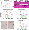
Extended release buprenorphine inhibits wound progression following nitrogen mustard (NM) exposure. (A) Impact of extended release buprenorphine formulations (ER and XR) on wound progression assessed in digital images of skin surface after NM exposure. (B) Representative images of haematoxylin and eosin stained cryosections of skin on Day 4 after NM exposure showing the impact of ER and XR on wound progression. (C) Impact of ER and XR on structural disruption assessed in haematoxylin and eosin stained cryosections. (D) Impact of ER and XR on inflammatory infiltrate assessed in haematoxylin and eosin stained cryosections. α: Significant main effect of treatment, β: Significant main effect of time. (E) Representative images of immunohistochemical staining for CD11b in cryosections of skin on Day 4 after NM exposure without or with ER or XR treatment. (F) Impact of ER and XR on CD11b staining on Day 4 after NM exposure. * ER + NM significantly different from NM alone. #significant difference between groups. p < 0.05. N = 4–8 per group.
FIGURE 4
FIGURE 4
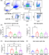
Extended release buprenorphine reduces skin inflammatory cell accumulation following nitrogen mustard (NM) exposure. (A) Gating strategy for identifying different inflammatory cell populations in skin wounds. (B) Impact of extended release buprenorphine formulations (ER and XR) on CD11b+ myeloid cell (Live CD11b+) accumulation on Day 4 after NM exposure. CTL: Not treated with NM controls. (C) Impact of ER and XR on Ly6G+ neutrophil (Live CD11b + Ly6G + Ly6C+) accumulation on Day 4 after NM exposure. (D) Impact of ER and XR on pro‐inflammatory Ly6C+ monocyte/macrophage (Live CD11b + Ly6G‐CD64hiLy6Chi, Mo/Mϕ) accumulation on Day 4 after NM exposure. (E) Impact of ER and XR on mature Ly6C‐ macrophage (Live CD11b + Ly6G‐CD64hiLy6Clo/−, Mϕ) accumulation on Day 4 after NM exposure, #significant difference between groups. p < 0.05. N = 4–8 per group. FSC‐A, forward scatter‐area; FSC‐H, forward scatter‐height; FSC‐W, forward scatter‐width.
FIGURE 5
FIGURE 5
Extended release buprenorphine reduces levels of selected chemokines in skin following nitrogen mustard (NM) exposure. (A) Impact of extended release buprenorphine formulations (ER and XR) on skin CCL3 levels on Day 4 after NM exposure. CTL: Not treated with NM controls. (B) Impact of ER and XR on skin CXCL2 levels on Day 4 after NM exposure. (C) Impact of ER and XR on skin TNF levels on Day 4 after NM exposure. (D) Impact of ER and XR on skin IL‐6 levels on Day 4 after NM exposure, #significant difference between groups. p < 0.05. N = 4–8 per group.
FIGURE 6
FIGURE 6
Short‐acting buprenorphine blunts early inflammatory response in skin following nitrogen mustard (NM) exposure. (A) Impact of extended release (ER and XR) and short‐acting (BU) buprenorphine formulations on body weight without and with NM exposure. CTL: Not treated with NM controls. (B) Impact of ER, XR and BU on yes/no response to nesting material stimuli without and with NM exposure. Response monitored for 10 min; mouse given a score of 1 for response, 0 for no response. (C) Impact of ER, XR and BU on wound progression assessed in digital images of skin surface after NM exposure. (D–G) Impact of ER, XR and BU on CD11b + myeloid cell (Live CD11b + Ly6G‐Ly6C+) accumulation (D), Ly6G+ neutrophil (Live CD11b + Ly6G + Ly6C+) accumulation (E), pro‐inflammatory Ly6C+ monocyte/macrophage (Live CD11b + Ly6G‐CD64hiLy6Chi, Mo/Mϕ) accumulation (F) and mature Ly6C‐ macrophage (Live CD11b + Ly6G‐CD64hiLy6Clo/−, Mϕ) accumulation on Day 2 after NM exposure. (H, I) Impact of ER, XR and BU on skin CCL3 (H) and CXCL2 (I) levels on Day 2 after NM exposure. α: Significant main effect of treatment, β: Significant main effect of time, * ER + NM significantly different from NM alone. ^XR + NM significantly different from NM alone, #significant difference between groups. p < 0.05. N = 4–8 per group.
FIGURE 7
FIGURE 7
Skin nitrogen mustard (NM) exposure enhances the persistence of circulating buprenorphine levels. Buprenorphine levels were measured in the peripheral blood of mice administered extended release (ER and XR) formulations without and with NM exposure. CTL: Not treated with NM controls. Note that buprenorphine levels are higher in the ER and XR groups with NM exposure than in the groups without NM exposure, indicating a #significant difference between groups. p < 0.05. N = 4–8 per group.

References

    1. Koh TJ, DiPietro LA. Inflammation and wound healing: the role of the macrophage. Expert Rev Mol Med. 2011;13:e23. - PMC - PubMed
    1. Singer AJ, Clark RA. Cutaneous wound healing. N Engl J Med. 1999;341(10):738‐746. - PubMed
    1. Pena OA, Martin P. Cellular and molecular mechanisms of skin wound healing. Nat Rev Mol Cell Biol. 2024;25(8):599‐616. - PubMed
    1. Mirza R, DiPietro LA, Koh TJ. Selective and specific macrophage ablation is detrimental to wound healing in mice. Am J Pathol. 2009;175(6):2454‐2462. - PMC - PubMed
    1. Goren I, Allmann N, Yogev N, et al. A transgenic mouse model of inducible macrophage depletion: effects of diphtheria toxin‐driven lysozyme M‐specific cell lineage ablation on wound inflammatory, angiogenic, and contractive processes. Am J Pathol. 2009;175(1):132‐147. - PMC - PubMed

Publication types

LinkOut - more resources