. 2025 Apr 3;188(7):1807-1827.e34.
doi: 10.1016/j.cell.2025.01.046. Epub 2025 Feb 28.

HIF regulates multiple translated endogenous retroviruses: Implications for cancer immunotherapy

Affiliations

HIF regulates multiple translated endogenous retroviruses: Implications for cancer immunotherapy

Qinqin Jiang et al. Cell. .

Abstract

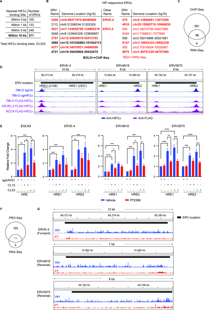
Clear cell renal cell carcinoma (ccRCC), despite having a low mutational burden, is considered immunogenic because it occasionally undergoes spontaneous regressions and often responds to immunotherapies. The signature lesion in ccRCC is inactivation of the VHL tumor suppressor gene and consequent upregulation of the HIF transcription factor. An earlier case report described a ccRCC patient who was cured by an allogeneic stem cell transplant and later found to have donor-derived T cells that recognized a ccRCC-specific peptide encoded by a HIF-responsive endogenous retrovirus (ERV), ERVE-4. We report that ERVE-4 is one of many ERVs that are induced by HIF, translated into HLA-bound peptides in ccRCCs, and capable of generating antigen-specific T cell responses. Moreover, ERV expression can be induced in non-ccRCC tumors with clinical-grade HIF stabilizers. These findings have implications for leveraging ERVs for cancer immunotherapy.

Keywords: ERV; HIF; cancer vaccine; ccRCC; immunopeptidomic; immunotherapy; kidney cancer; neoantigen.


Conflict of interest statement

Declaration of interests D.A.B. reports share options in Elephas; advisory board, consulting, or personal fees from DLA Piper, AbbVie, Compugen, Link Cell Therapies, Scholar Rock, NeoMorph, Nimbus, Exelixis, AVEO, Eisai, and Elephas; and research support from AstraZeneca, outside of the submitted work. N.N., T.P., M.C.-R., C.G., and G.M. are employees of and own equity in T-Scan Therapeutics, and G.M. is a board member of T-Scan Therapeutics. S.K. is currently an employee of Genentech. W.J.L. reports advisory board and consulting fees from One Lambda Inc. and CareDx Inc. and royalties from Thermo Fisher Scientific Inc. S.A.S. is a consultant for Jivanu Therapeutics and has equity in Agenus Inc., Agios Pharmaceuticals, Breakbio Corp., Bristol-Myers Squibb, and Lumos Pharma. E.M.V.A. provides advisory/consulting to Enara Bio, Manifold Bio, Monte Rosa, and the Novartis Institute for Biomedical Research; receives research support from Novartis, BMS, Sanofi, and NextPoint; and owns equity in Tango Therapeutics, Enara Bio, and Monte Rosa. T.K.C. reports involvement in NeoVax clinical trials and Gateway for Cancer Research; advisory board, consulting, or personal fees from Merck and BMS; institutional patents (outside the submitted work) filed on molecular alterations and immunotherapy response/toxicity and ctDNA; and equity in Tempest, Osel, Precede Bio, CureResponse, InnDura Therapeutics, Primium, Abalytics, and Faron Pharma. S.A.C. is a member of the scientific advisory boards of Kymera, PTM BioLabs, Seer, and PrognomIQ. C.J.W. holds equity at BioNTech, Inc. W.G.K. has financial interests in Lilly Pharmaceuticals, Fibrogen, Nextech Invest, and Casdin Capital and receives belzutifan royalties.

Figures

Figure 1.
Figure 1.. Regulation of ERV3.2 and ERV4700 by pVHL and HIF2
(A, C, E, G) ERV RNA levels in 786-O cells (A, C, E) or the indicated ccRCC cell lines (G) that were stably infected to express pVHL (A, E, G) or treated for 72 hours with 2 μM PT2399 (PT) (C) where indicated in red. The cells first underwent CRISPR-based gene editing with a HIF2α sgRNA (sgHIF2α) or a control sgRNA (sgCtrl) (A) or were stably infected to express the HIF2α dPA (P405A, P531A) variant (E) where indicated. (N=3) (B, D, F, H) Immunoblots of cells as in (A, C, E, G). (I and M) ERV4700 RNA levels (I) (N=3) and anti-HIF2α ChIP-seq of the ERV4700 locus (M) in 786-O cells treated with 0.5 μM of the GSK3685032 (DNMT1i) or vehicle (Veh) for 9 days. In (I) the cells were first edited with a sgHIF2α or a sgCtrl guide. In (M) NDRG1 locus was included as a control. See also Figure S2G. (J) Immunoblots of cells as in (I). (K and L) ERV4700 RNA levels (K) (N=3) and immunoblot analysis (L) of 786-O(VHL) or 786-O(EV) cells treated with 0.5 μM GSK3685032 (DNMT1i), 100 μM FG-4592,or both for 9 days. (N, P) ERV RNA levels of WM266–4 melanoma cells (N) and BT-549 TNBC cells (P) treated with 50 μM FG-4592 or Veh for 9 days. The cells were first edited with a HIF1α sgRNA or a sgCtr guide. (N=6) For all RT-qPCR panels, ERV RNA levels were normalized to ACTB RNA levels and then to the first condition listed. (O and Q) Immunoblots of cells of (N and P) on day 3.
Figure 2.
Figure 2.. Identification of HIF2-responsive ERVs by RNA-seq
(A-D) Venn Diagram (A) and RT-qPCR validation (B-D) of HIF2-responsive ERVs in 786-O(VHL) or 786-O(EV) cells, treated with 2 μM PT or Veh for 72 hours, or edited with a sgHIF2α or a sgCtrl guide. NDRG1 mRNA levels served as controls. ERV RNA levels were normalized to ACTB RNA levels and then to the EV, Veh or sgCtrl cells. (E) Correlation matrix (Pearson’s R) between the expression of 15 ERVs and the expression of a validated HIF2 signature, the HIF2-responsive genes EGLN3 and NDRG1, and a 42-gene housekeeping gene signature, based on RNA-seq data of 250 advanced ccRCCs from the CheckMate 025 trial. (F) Examples of four ERVs from (E). Shown are Pearson’s correlations between ERV expression levels and expression of a HIF2 pathway signature (upper) and a housekeeping gene set signature (lower).
Figure 3.
Figure 3.. Identification of ERVs that are Directly Regulated by HIF2
(A) Number of ERVs with neighboring HIF2α binding sites. (B) HIF2-responsive ERVs identified by RNA-seq from Figure 2A. ERVs in bold are located within 10 kb of a HIF2α binding site based on anti-HIF2α ChIP-seq. ERVs in red are direct HIF2α targets based on PRO-seq. (C) Venn Diagram showing overlap of 16 HIF2-responsive ERVs (B) and ERVs located within 10 kb of a HIF2α binding site. (D) Representative Anti-HIF2α ChIP-seq tracks (blue) in 786-O cells that underwent CRISPR-based gene editing with a HIF2α sgRNA or a control sgRNA (sgCtrl) and representative Anti-FLAG ChIP-seq tracks (purple) in 786-O, OS-RC-2 and RCC4 cells in which a FLAG-HA epitope tag coding sequence was inserted at the 5’ end of the endogenous HIF2α open reading frame by CRISPR-HDR (FLAG-HIF2α). See also Figure S2A. (E) Anti-FLAG ChIP-qPCR assays using two different clones (CL15 and CL53) of 786-O FLAG-HIF2α cells compared to parental cells that were infected with a sgAAVS1 control guide. Cells were treated with 2 μM PT or Veh for 72 hours prior to ChIP-qPCR with primers specific for the putative HREs for the indicated ERVs. EglN3 served as a control. Data were normalized to the untreated sgAAVS1 cells. (F) Venn Diagram showing overlap of 16 HIF2-responsive ERVs (B) and ERVs scored as HIF2α direct targets by PRO-seq. (G) Representative nascent RNA tracks in OS-RC-2 cells that were treated with 2 μM PT2399 or vehicle for 2 hours.
Figure 4.
Figure 4.. Identification of HLA-bound ERV-derived Peptides
(A) Venn Diagram showing overlap of 16 HIF2-responsive ERVs with 26 pVHL-responsive translated ERVs based on Polysome-seq of 786-O(VHL) or 786-O(EV) cells. (B) Filtering approach applied to LC-MS/MS peptide identification for ERV-derived peptides. SA(spectral angle); LC(liquid chromatography); dRT(delta retention time). (C) HLA-bound peptides derived from the human proteome or from the 57 ERVs that scored in two of the three pairwise 786-O cell comparisons (high HIF2 versus low HIF2) and/or scored as pVHL-responsive by Polysome-seq. The volcano plot depicts the relative amount of each peptide as determined by anti-HLA immunoprecipitation of 786-O(VHL) or 786-O(EV) cells treated with 100 ng/ml IFNγ for 72 h, followed by TMT labeling and MS. Note the enrichment of pVHL-derived peptides in the 786-O(VHL) cells relative to the 786-O(EV) cells (right side of volcano plot) and enrichment of peptides from the HIF-responsive gene product NDRG1 in the 786-O(EV) cells (left side of the volcano plot). (D) ERV-derived peptides identified in (C). Note that some peptides could be encoded by more than one homologous ERV from among the 57 ERVs interrogated in (C). The ERVs scored as HIF2-responsive by RNA-seq and/or pVHL-responsive by Polysome-seq as indicated by the “+”. (C and D) Shown in red are two potentially pVHL-responsive ERV-derived peptides. (E and F) MS/MS spectra comparing HLA-bound peptides determined to be KLIAGLIFLK (E) and ATFLGSLTGK (F) compared to the corresponding synthetic peptides bearing a heavy lysine (Lys8).
Figure 5.
Figure 5.. Human HIF2-responsive ERVs are Largely Derived from Cancer Cells
(A and C) Volcano plots describing differential chromatin accessibility (A) and differential expression (C) derived from scATAC-seq data and scRNA-seq data from 16 ccRCCs for the 81 HIF2-responsive ERVs nominated by our cell line analyses. The cancer cell-specific CA9 and immune cell-specific PTPRC genes (highlighted in red), served as controls. The vertical dashed lines indicate |Log(2)FC| of 1 and p-adj of 0.05. (B) Coverage plot showing single-cell ATAC-seq tracks for the indicated genes (± 3 kb). (D) UMAP of scRNA-seq data by cell type, followed by feature plots for ERV5875 and ERV3797. (E) Correlation of ERV expression with ERV chromatin accessibility based on the data in (A) and (C). The vertical and horizontal dashed lines indicate |Log(2)FC| of 1 and p-adj of 0.05. (F-G) Violin plots of all ERV expression by scATAC-seq peaks defined as ‘All’, ‘Unchanged’, ‘Down’ or ‘Up’ (cancer vs immune) (F) or all ERV expression and chromatin accessibility by HIF2-responsiveness (G).
Figure 6.
Figure 6.. HLA-Bound and Immunogenic ERV-derived Peptides are Present in Human Kidney Cancer Samples
(A) Workflow for identifying ERV-derived peptides in ccRCCs. (B and C) 22 ERV-derived peptides identified from 11 ccRCC patients. * indicates peptides that were tumor-specific based on analysis of available paired normal kidney tissue. “HIF2-responsive ERV” column indicates potential source ERV(s) for each peptide from the 81 ERVs identified in 786-O and A-498 cells. “No. of Possible Coding ERVs” column indicates number of potential source ERVs for the peptides. Peptides that uniquely mapped to one HIF2-responsive ERV are shown in (B). (D and G) Schema for quantifying IFNγ secreting T cells derived from PBMCs (D) and splenocytes (G) by ELISpot assay. (E and F) ELISpot images (E) and quantification (spot count) (F) of IFNγ secreting PBMCs derived from ccRCC patient 110 incubated with the indicated peptides. An HIV peptide and a pool of viral peptides (CMV, EBV and Influenza) (CEF peptides) served as negative and positive controls respectively. (N=3) (H and I) Five HLA-A*11:01 transgenic mice were immunized with a pool of 11 peptides as in (G). Ten days post second vaccination, splenocytes were harvested and incubated with individual peptides ex vivo and monitored by ELISpot assays. A HIV peptide and CD3 served as negative and positive controls respectively. Representative ELISpot images (H) and spot counts (I) are shown. (N=5)
Figure 7.
Figure 7.. ERV-derived Peptides Recognized by T cells from Human ccRCC Patients
(A-C) ELISpot quantification of IFNγ secreting PBMCs derived from ccRCC Allo-SCT patients after stimulation with the indicated ERV-derived peptides. DMSO and an HIV peptide served as negative controls. CEF peptides and PHA served as positive controls. (N=3) Significance was determined by comparing the response to indicated peptides and to the HIV peptide for the corresponding patient. IST(immunosuppressive therapy); GVHD(graft-versus-host-disease); CsA(Cyclosporine A). (D) Schema for T-Scan antigen discovery screen, adapted from Ferretti et al. MACS(magnetic activated cell sorting); IFP(infrared fluorescent protein); FACS (fluorescence activated cell sorting); NGS(next generation sequencing). (E) Patient samples with ERV-reactive TILs. (F) Epitope mapping of ERV antigens. Reporter activity (% IFP) of HEK293T reporter cells that were either pulsed with the indicated peptides or transduced with the target protein fragment clone (for 1514 only; 63AA) and co-cultured with the relevant TCR. Sequences for scoring peptides are shown.

References

    1. Postow MA, Callahan MK, and Wolchok JD (2015). Immune Checkpoint Blockade in Cancer Therapy. J Clin Oncol 33, 1974–1982. 10.1200/JCO.2014.59.4358. - DOI - PMC - PubMed
    1. Baumeister SH, Freeman GJ, Dranoff G, and Sharpe AH (2016). Coinhibitory Pathways in Immunotherapy for Cancer. Annu Rev Immunol 34, 539–573. 10.1146/annurev-immunol-032414-112049. - DOI - PubMed
    1. Sharma P, Siddiqui BA, Anandhan S, Yadav SS, Subudhi SK, Gao J, Goswami S, and Allison JP (2021). The Next Decade of Immune Checkpoint Therapy. Cancer Discov 11, 838–857. 10.1158/2159-8290.CD-20-1680. - DOI - PubMed
    1. Rizvi NA, Hellmann MD, Snyder A, Kvistborg P, Makarov V, Havel JJ, Lee W, Yuan J, Wong P, Ho TS, et al. (2015). Cancer immunology. Mutational landscape determines sensitivity to PD-1 blockade in non-small cell lung cancer. Science 348, 124–128. 10.1126/science.aaa1348. - DOI - PMC - PubMed
    1. Yarchoan M, Hopkins A, and Jaffee EM (2017). Tumor Mutational Burden and Response Rate to PD-1 Inhibition. N Engl J Med 377, 2500–2501. 10.1056/NEJMc1713444. - DOI - PMC - PubMed

MeSH terms

Substances