. 2025 Apr;24(4):100935.
doi: 10.1016/j.mcpro.2025.100935. Epub 2025 Feb 28.

Embryo-Induced Changes in the Protein Profile of Bovine Oviductal Extracellular Vesicles

Affiliations

Embryo-Induced Changes in the Protein Profile of Bovine Oviductal Extracellular Vesicles

Rosane Mazzarella et al. Mol Cell Proteomics. 2025 Apr.

Abstract

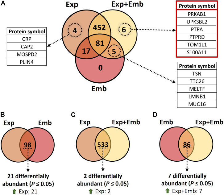
The study of early maternal-embryonic cross-talk remains one of the most challenging topics in reproductive biology. Understanding the physiological mechanisms involved in the interactions between the maternal reproductive tract and the developing embryo is essential for enhancing bovine reproductive efficiency. This complex communication starts within the oviduct, where the modulation of biological processes important for ensuring embryo quality is partially facilitated through extracellular vesicles (EVs). Utilizing a combination of in vivo and in vitro models this study had three main objectives: 1) to examine the protein cargo of EVs isolated from the oviductal fluid (OF) of cyclic and pregnant heifers to understand their role in maternal-embryonic communication in vivo; 2) to characterize the protein profile of EVs in conditioned medium (CM) resulting from the culture of oviductal explants alone (Exp) or in the presence of 8- to 16-cell stage embryos (Exp + Emb); and 3) to compare the protein cargo of EVs from Exp with EVs from cyclic heifers and EVs from Exp + Emb with EVs from pregnant heifers. Proteins were considered "identified" if detected in at least three out of five replicates and considered "exclusive" if detected in at least three out of five replicates within one group but absent in all samples of other groups. We identified 659 and 1476 proteins in the OF-EVs of cyclic and pregnant heifers, respectively. Among these, 644 proteins were identified in OF-EVs from both cyclic and pregnant heifers, and 40 proteins were exclusive to OF-EVs from the pregnant group. Within the 644 proteins identified in both groups, 31 were identified as differently abundant proteins (DAPs). In pregnant heifers, DAPs were mainly related to genome activation, DNA repair, embryonic cell differentiation, migration, and immune tolerance. In vitro, we identified 841 proteins in the CM-EVs from Exp alone, 613 from Exp + Emb, and 111 in the CM-EVs from Emb alone. In the qualitative analysis between the three in vitro groups, 81 proteins were identified in all groups, 452 were common to Exp and Exp + Emb, 17 were common to Exp and Emb, 5 were common to Exp + Emb and Emb, 4 were unique to Exp, 6 were unique to Exp + Emb, and none were unique to Emb. Proteins identified when there is an interaction between the oviduct and the embryo in vitro, corresponding to the Exp + Emb group, were associated with immune tolerance, structural activity, binding, and cytoskeletal regulation. In vivo and in vitro EVs exhibit distinct qualitative and quantitative protein contents, both when comparing EVs produced in the absence of an embryo (Cyclic and Exp) and those that have undergone embryo-oviduct interaction (Pregnant and Exp + Emb). The observed changes in the protein cargo of EVs due to maternal-embryonic communication in vivo and in vitro suggest that the interaction between the embryo and the maternal milieu initiates within the oviduct and is potentially facilitated by EVs and their protein contents.

Keywords: bovine; embryo-maternal interaction; exracellular vesicles; in vitro; in vivo; oviduct; pregnancy.


Conflict of interest statement

Conflicts of interest The authors declare that they have no conflicts of interest with the contents of this article.

Figures

None
Graphical abstract
Fig. 1
Fig. 1
Experimental Model and Group Comparisons. Heifers were synchronized, artificially inseminated (Pregnant) or not (Cyclic), and slaughtered 3.5 days after insemination. Oviducts ipsilateral to the corpus luteum were flushed to obtain the oviductal fluid (OF), and the presence of an embryo confirmed pregnancy. For the in vitro model, six 0.25 mm2 oviductal explants were obtained from each cyclic heifer and cultured individually in 750 μl protein-free synthetic oviduct fluid (SOF): three were cultured alone, and three were co-cultured with 10 in vitro-produced 8- to 16-cell stage bovine embryos. Also, a group of 100 in vitro-produced 8- to 16-cell stage bovine embryos were cultured alone in 500 μl of SOF. After 6 h, the conditioned media (CM) was collected for extracellular vesicles (EV) isolation. The protein content of EVs from the following four comparisons was analyzed (1): OF-EVs from non-pregnant (Cyclic) heifers compared with pregnant heifers (Pregnant) (2); CM-EVs from oviductal explants cultured alone (Exp) versus those co-cultured with 8- to 16-cell stage embryos (Exp + Emb) versus the EVs from the CM of 8- to 16-cell stage embryos cultured alone (Emb) (3); OF-EVs from Cyclic heifers compared with EVs from the CM of oviductal explants cultured alone (Exp); and (4) OF-EVs from Pregnant heifers compared with EVs from the CM of oviductal explants co-cultured in vitro with 8- to 16-cell stage embryos (Exp + Emb). Created in BioRender (https://BioRender.com/i44h277).
Fig. 2
Fig. 2
Characterization of Oviductal Fluid Extracellular Vesicles (OF-EVs). Nanoparticle tracking analysis showed no difference in particle size (A) and concentration (B) between the groups Cyclic and Pregnant. C, flow cytometry identified the CD63, CD81, and CD44 markers in both groups. D, transmission electron microscopy image from OF-EVs showing cup-shaped particles with characteristic sizes resembling EVs. Error bars represent the standard error of the mean (SEM). White arrows indicate EVs.
Fig. 3
Fig. 3
Characterization of Conditioned Media (CM) Extracellular Vesicles (EVs). Nanoparticle tracking analysis showing no difference in particle size (A) among CM-EVs from oviductal explants cultured alone (Exp), oviductal explants co-cultured with 8- to 16-cell stage embryos (Exp + Emb) and embryos cultured alone (Emb) (B). C, flow cytometry showing the identification of the CD63, CD81, and CD44 markers in all groups. D, transmission electron microscopy image showing cup-shaped particles with characteristic sizes resembling EVs in the CM by Exp (D), Exp + Emb (E), and Emb (F). Error bars represent SEM. White arrows indicate EVs. Different letters indicate significative differences (p ≤ 0.05).
Fig. 4
Fig. 4
Protein Profile of Oviductal Fluid Extracellular Vesicles (OF-EVs) from Cyclic and Pregnant heifers.A, the table indicates the number of proteins identified in each group, and the Venn diagram represents the 644 proteins common to both and the 40 proteins exclusively detected in Pregnant heifers. B, principal Component Analysis of differentially abundant proteins. C, three proteins were less abundant, and 28 were more abundant in OF-EVs from Pregnant compared to Cyclic heifers. Proteins were considered ‘identified’ if detected in at least three out of five replicates and considered ‘exclusive’ if detected in at least three out of five replicates within one group but absent in all samples of other groups. Error bars represent SEM. p ≤ 0.05 was considered as significant.
Fig. 5
Fig. 5
Functional enrichment of the proteins exclusive to oviductal fluid extracellular vesicles of pregnant heifers.A, protein class, (B) gene ontology, and (C) pathways identified using the PANTHER 18.0 Classification System (https://pantherdb.org/). Darker bars indicate the number of genes associated with each category name, while lighter bars represent the percentage of these genes relative to the total number of genes in that category.
Fig. 6
Fig. 6
Functional enrichment of the proteins overabundant abundant in oviductal fluid extracellular vesicles of pregnant compared with cyclic heifers.A, protein class, (B) gene ontology, and (C) pathways identified using the PANTHER 18.0 Classification System (https://pantherdb.org/). Darker bars indicate the number of genes associated with each category name, while lighter bars represent the percentage of these genes relative to the total number of genes in that category.
Fig. 7
Fig. 7
The protein profile of extracellular vesicles from conditioned media following the culture of oviductal explants in the absence (Exp) or presence (Exp + Emb) of 8- to 16-cell stage embryos, or from embryos cultured alone (Emb).A, Venn diagram represents the number of proteins associated with CM-EVs from Exp, Exp + Emb, and Emb. Red boxes indicate the list of the six proteins identified as only present when there is an interaction between the oviduct and the embryo in vitro. B, Venn diagrams representing the number of proteins in common among Exp versus Emb, (C) Exp versus Exp + Emb, and (D) Emb versus Exp + Emb, as well as the number of differentially abundant proteins amongst the common proteins in each comparison. Proteins were considered ‘identified’ if detected in at least three out of five replicates and considered “exclusive” if detected in at least three out of five replicates within one group but absent in all samples of other groups.
Fig. 8
Fig. 8
Number of proteins identified between the in vivo and the in vitro model.A, Venn diagram representing the number of proteins associated with oviductal fluid extracellular vesicles (OF-EVs) from Cyclic heifers versus conditioned media extracellular vesicles (CM-EVs) from oviductal explants cultured alone in vitro (Exp). B, Venn diagram representing the number of proteins associated with OF-EVs from Pregnant heifers versus CM-EVs from oviductal explants cultured with 8- to 16-cell embryos (Exp + Emb) in vitro, and three proteins (MDH2, HEBP1, and A0A4W2DFR9) unique to the OF-EVs of Pregnant heifers also identified in CM-EVs from Exp + Emb.
Fig. 9
Fig. 9
Functional membership analysis for proteins matching with “embryo development” term. Proteins identified in the oviductal fluid extracellular vesicles from Cyclic (green pie chart) and Pregnant Heifers (blue pie chart) and in the conditioned media extracellular vesicles from explants cultured alone (Exp; orange pie chart) and explants cocultured with embryos (Exp + Emb; yellow pie chart). The outer pie chart illustrates the count and proportion of proteins within the background dataset that are affiliated with the “embryo development” membership, whereas the inner pie chart presents the count and proportion of proteins in the specific input gene list associated with this membership. The p-value positioned above the pie charts indicates a statistically significant enrichment of the membership across all groups.

References

    1. Rizos D., Ward F., Duffy P., Boland M.P., Lonergan P. Consequences of bovine oocyte maturation, fertilization or early embryo development in vitro versus in vivo: implications for blastocyst yield and blastocyst quality. Mol. Reprod. Dev. 2002;61:234–248. - PubMed
    1. Lonergan P., Rizos D., Gutierrez-Adán A., Moreira P.M., Pintado B., de la Fuente J., et al. Temporal divergence in the pattern of messenger RNA expression in bovine embryos cultured from the zygote to blastocyst stage in vitro or in vivo. Biol. Reprod. 2003;69:1424–1431. - PubMed
    1. Khurana N.K., Niemann H. Energy metabolism in preimplantation bovine embryos derived in vitro or in Vivo1. Biol. Reprod. 2000;62:847–856. - PubMed
    1. Rizos D., Clemente M., Bermejo-Alvarez P., De La Fuente J., Lonergan P., Gutiérrez-Adán A. Consequences of in vitro culture conditions on embryo development and quality. Reprod. Domest. Anim. 2008;43:44–50. - PubMed
    1. Rizos D., Fair T., Papadopoulos S., Boland M.P., Lonergan P. Developmental, qualitative, and ultrastructural differences between ovine and bovine embryos produced in vivo or in vitro. Mol. Reprod. Dev. 2002;62:320–327. - PubMed

LinkOut - more resources