. 2025 Mar 3;13(1):29.
doi: 10.1038/s41413-025-00409-0.

CD47 is required for mesenchymal progenitor proliferation and fracture repair

Affiliations

CD47 is required for mesenchymal progenitor proliferation and fracture repair

Robert L Zondervan et al. Bone Res. .

Abstract

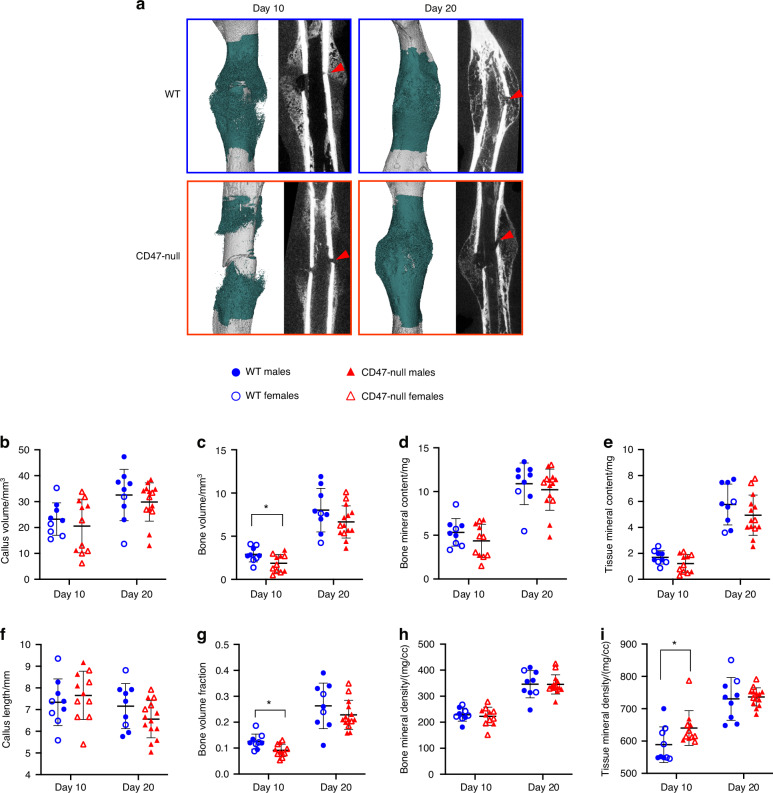
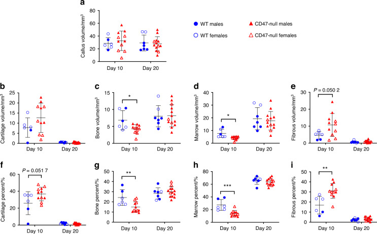
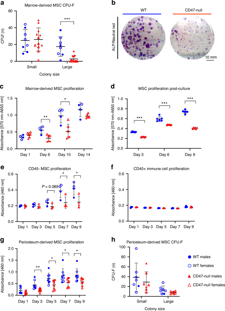
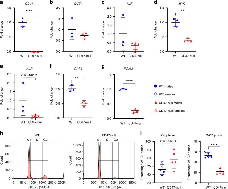
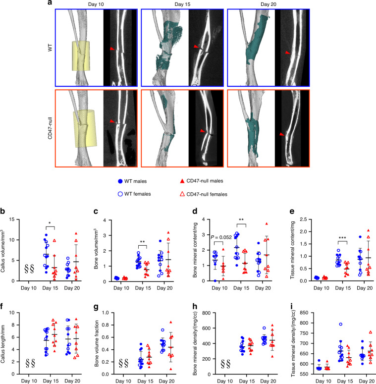
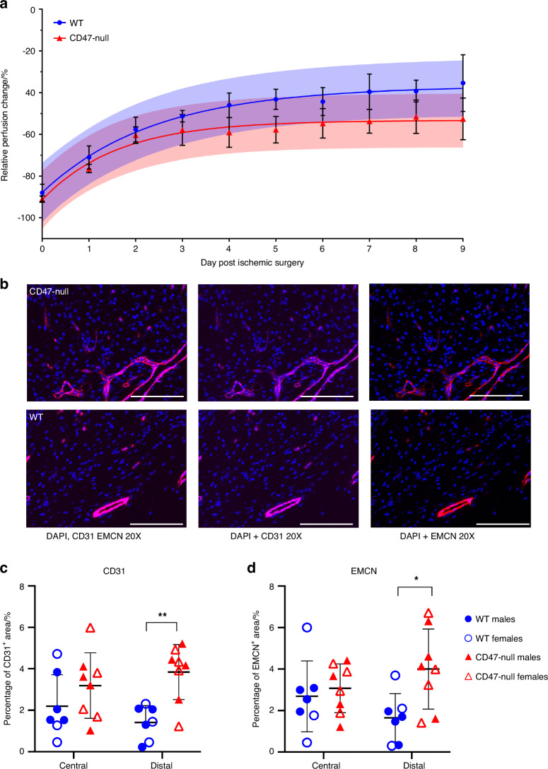
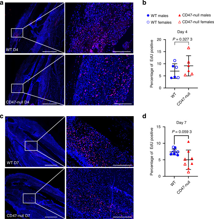
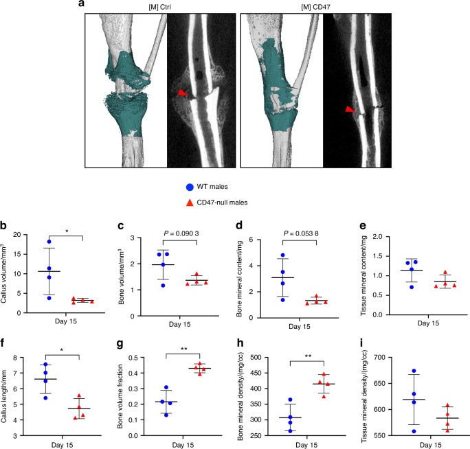
CD47 is a ubiquitous and pleiotropic cell-surface receptor. Disrupting CD47 enhances injury repair in various tissues but the role of CD47 has not been studied in bone injuries. In a murine closed-fracture model, CD47-null mice showed decreased callus bone formation as assessed by microcomputed tomography 10 days post-fracture and increased fibrous volume as determined by histology. To understand the cellular basis for this phenotype, mesenchymal progenitors (MSC) were harvested from bone marrow. CD47-null MSC showed decreased large fibroblast colony formation (CFU-F), significantly less proliferation, and fewer cells in S-phase, although osteoblast differentiation was unaffected. However, consistent with prior research, CD47-null endothelial cells showed increased proliferation relative to WT cells. Similarly, in a murine ischemic fracture model, CD47-null mice showed reduced fracture callus size due to a reduction in bone relative to WT 15 days-post fracture. Consistent with our in vitro results, in vivo EdU labeling showed decreased cell proliferation in the callus of CD47-null mice, while staining for CD31 and endomucin demonstrated increased endothelial cell density. Finally, WT mice with ischemic fracture that were administered a CD47 morpholino, which blocks CD47 protein production, showed a callus phenotype similar to that of ischemic fractures in CD47-null mice, suggesting the phenotype was not due to developmental changes in the knockout mice. Thus, inhibition of CD47 during bone healing reduces both non-ischemic and ischemic fracture healing, in part, by decreasing MSC proliferation. Furthermore, the increase in endothelial cell proliferation and early blood vessel density caused by CD47 disruption is not sufficient to overcome MSC dysfunction.


Conflict of interest statement

Competing interests: The authors declare no competing interests.

Figures

Fig. 1
Fig. 1
CD47-null femoral fractures show reduced bone formation, but increased tissue mineral density. µCT analysis of transverse femoral fracture in WT (n = 9) and CD47-null (n = 11-14) mice at 10- and 20-days post-fracture. Each data point represents an individual mouse. a Representative 3D reconstructions (white background) and sagittal-plane reconstruction (black background) of WT and CD47-null mice at days 10 and 20 post-fracture. Location of fracture (red arrowhead) is marked on the sagittal reconstructions. Representative 3D reconstructions include callus mineralization (teal). Quantitative assessments of various bone healing parameters determined by µCT (mean ± SD) at day 10 and 20 post-fracture. b Callus volume, c Bone volume, d bone mineral content, e Tissue mineral content, f Callus length, g Bone volume fraction, h. Bone mineral density, i Tissue mineral density. *P < 0.05, two-sided t test performed at each timepoint
Fig. 2
Fig. 2
Absence of CD47 during femoral fracture healing results in alterations in fracture tissue composition. Histomorphometry of transverse femoral fractures in WT (n = 6-7), and CD47-null (n = 11–13) mice at 10- and 20-days post-fracture. Each data point represents an individual mouse. Callus composition was quantified through histomorphometry (mean ± SD) at day 10 and 20 post-fracture. a Callus volume, b Cartilage volume, c Bone volume, d Marrow volume, e Fibrous volume, f Cartilage percent, g Bone percent, h Marrow percent, i Fibrous percent. *P < 0.05, **P < 0.01, ***P < 0.001, two-sided t test performed at each timepoint
Fig. 3
Fig. 3
CD47 is required for cell colony expansion and proliferation. Whole marrow and periosteum were harvested from the femur and tibia of WT and CD47-null mice. a CFU-F of WT (n = 8) and CD47-null (n = 12) marrow-derived cells. b Representative CFU-f plate from WT (left) and CD47-null (right) mice. c MTT assay of primary marrow-derived MSC from WT (n = 4) and CD47-null (n = 4) mice at 1, 6, 10 and 14 days in culture. d, MTT assay of marrow-derived MSC pooled from two to three mice (equivalent ratios of males and females), WT (n = 6 pools), and CD47-null (n = 6 pools) after the 1st passage at 3, 6 and 9 days in culture. e CCK8 assay of magnetically sorted CD45- MSC from WT (n = 4) and CD47-null (n = 4) after 1st passage at 1, 3, 5, 7 and 9 days. f CCK8 assay of magnetically sorted CD45+ immune cells from WT (n = 4) and CD47-null (n = 4) after 1st passage at 1, 3, 5, 7 and 9 days. g CCK8 assay of periosteal MSC from WT (n = 8) and CD47-null (n = 8) after the 1st passage at 1, 3, 5, 7 and 9 days in culture. h CFU-F of periosteal MSC from WT (n = 6) and CD47-null (n = 6) mice. Unless otherwise stated, each data point represents an individual mouse. Mean ± SD, *P < 0.05, **P < 0.01, ***P < 0.001 two-sided t tests
Fig. 4
Fig. 4
Loss of CD47 decreases MSC pluripotency, proliferation, and apoptosis genes and decreased CD47-null MSC are in S/G2 phase. Gene expression analysis of MSC harvested from the femur and tibia of WT (n = 3) and CD47-null (n = 4) after the 1st passage. Each data point represents the pooling of two to three mice. a Cd47 gene expression. Pluripotent stem cell genes, b Oct4, c Klf4, d c-Myc. Pre-osteoblast gene e alkaline phosphatase (ALP), apoptosis associated gene, f caspase 3 (CAP3), and proliferation marker g forkhead box M1 (FOXM1). Graphs indicate WT and CD47-null gene expression normalized to the housekeeping gene, GAPDH, relative to WT (n = 3) mice. Cell cycle analysis of marrow-derived MSC harvested from the femur and tibia of WT (n = 5) and CD47-null (n = 5) following first passage. h Representative histograms of WT (left) and CD47-null (right) with cell cycle analysis overlays. i Percent of cells in G1 phase or S/G2. Mean ± SD, *P < 0.05, **P < 0.01, ***P < 0.001, two-sided t test
Fig. 5
Fig. 5
CD47-null mice show reduced ischemic fracture callus formation. µCT analysis of ischemic tibia fracture callus of WT (n = 7–11) and CD47-null (n = 6–9) mice at day 10, 15 and 20 post-fracture. Each data point represents an individual mouse. a Representative 3D reconstructions (white background) and sagittal-plane reconstruction (black background) of WT (top row) and CD47-null (bottom row) mice at day 10, 15 and 20 post-fracture. Location of fracture (red arrowhead) is marked on the sagittal reconstructions. Day 10 3D reconstruction includes representative cylindrical ROIs (transparent yellow cylinder) used to calculate callus morphology. Day 15 and 20 representative 3D reconstruction include highlighted callus mineralization (teal). Callus morphology (mean ± SD) at days 10, 15 and 20 post-fracture. b Callus volume, c Bone volume, d bone mineral content, e Tissue mineral content, f Callus length, g Bone volume fraction, h Bone mineral density, i Tissue mineral density. *P < 0.05, **P < 0.01, ***P < 0.001, two-sided t tests performed at each timepoint; § no data
Fig. 6
Fig. 6
Genetic knockout of CD47 limits recovery of whole limb perfusion, but shows local increases in endothelial cells after induced ischemia. Relative limb perfusion of WT and CD47-null mice at days 0–9 post-ischemic surgery using laser Doppler flowmetry. a Perfusion data was fit to a one-phase non-linear curve for WT (fit is the solid blue line with 95% CI in transparent blue; R2 = 0.760 9) and CD47-null (fit is the solid red line with 95% CI in transparent red; R2 = 0.622 3). The rate of recovery was faster, but maximum recovery was significantly lower in CD47-null compared to WT mice (P < 0.000 1, extra sum-of-squares F test). b Representative immunofluorescence staining of CD31 and EMCN in CD47-null (n = 8) and WT (n = 7) ischemic fractures at 20× (Scale bar = 100 µm). c CD31 quantification in WT and CD47-null ischemic fracture calluses at day 4 post-fracture at peripheral and central regions of the fracture callus. d EMCN quantification in WT and CD47-null ischemic fracture calluses at day 4 post-fracture at peripheral and central regions. Each data point represents an individual mouse. Mean ± SD, *P < 0.05, **P < 0.01, two-sided t test
Fig. 7
Fig. 7
Genetic knockout of CD47 reduces cell proliferation in the early fracture callus. EdU incorporation at 4 and 7 days post-ischemic tibia fracture in WT (n = 6-9) and CD47-null (n = 6–8) mice. Each data point represents an individual mouse. a Representative images of WT and CD47-null fracture calluses at day 4 post-fracture. b Percentage of EdU positive cells in WT and CD47-null fracture callus at 4 days post-fracture. c Representative images of WT and CD47-null fracture callus at day 7 post-fracture. d Percentage of EdU positive cells in WT and CD47-null fracture calluses at 7 days post-fracture. Two-sided t-test. 10× stitched images, scale bar = 1 000 µm. 10X zoomed images, scale bar = 200 µm
Fig. 8
Fig. 8
Disruption of CD47 using a morpholino inhibits early ischemic fracture callus formation. µCT analysis of ischemic tibia fracture callus of morpholino-control ([M]Ctrl) (n = 4) and morpholino-CD47 ([M]CD47) (n = 4) at day 15 post-fracture. Each data point represents an individual mouse. a 3D reconstructions (white background with teal mineralized callus highlight) and sagittal-plane reconstruction (black background) of [M]Ctrl (left) and [M]CD47 (right) mice at day 15 post-fracture. Location of fracture (red arrowhead) is marked on the sagittal reconstructions. Callus morphology (mean ± SD) at day 15 post-fracture. b Callus volume, c Bone volume, d bone mineral content, e Tissue mineral content, f Callus length, g Bone volume fraction, h Bone mineral density, i Tissue mineral density. *P < 0.05, **P < 0.01, two-sided t test

Update of

References

    1. Bahney, C. S. et al. Cellular biology of fracture healing. J. Orthop. Res.37, 35–50 (2019). - PMC - PubMed
    1. Zhang, N., Hu, L., Cao, Z., Liu, X. & Pan, J. Periosteal skeletal stem cells and their response to bone injury. Front. Cell Dev. Biol.10, 812094 (2022). - PMC - PubMed
    1. Muire, P. J., Mangum, L. H. & Wenke, J. C. Time course of immune response and immunomodulation during normal and delayed healing of musculoskeletal wounds. Front. Immunol.11, 1056 (2020). - PMC - PubMed
    1. Chen, M. et al. Skeleton-vasculature chain reaction: a novel insight into the mystery of homeostasis. Bone Res.9, 21 (2021). - PMC - PubMed
    1. Lu, C., Miclau, T., Hu, D. & Marcucio, R. S. Ischemia leads to delayed union during fracture healing: a mouse model. J. Orthop. Res.25, 51–61 (2007). - PMC - PubMed

LinkOut - more resources