. 2025 Mar 2;16(1):2110.
doi: 10.1038/s41467-025-57241-7.

Molecular glues of the regulatory ChREBP/14-3-3 complex protect beta cells from glucolipotoxicity

Affiliations

Molecular glues of the regulatory ChREBP/14-3-3 complex protect beta cells from glucolipotoxicity

Liora S Katz et al. Nat Commun. .

Abstract

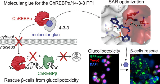
The Carbohydrate Response Element Binding Protein (ChREBP) is a glucose-responsive transcription factor (TF) with two major splice isoforms (α and β). In chronic hyperglycemia and glucolipotoxicity, ChREBPα-mediated ChREBPβ expression surges, leading to insulin-secreting β-cell dedifferentiation and death. 14-3-3 binding to ChREBPα results in cytoplasmic retention and suppression of transcriptional activity. Thus, small molecule-mediated stabilization of this protein-protein interaction (PPI) may be of therapeutic value. Here, we show that structure-based optimizations of a 'molecular glue' compound led to potent ChREBPα/14-3-3 PPI stabilizers with cellular activity. In primary human β-cells, the most active compound retained ChREBPα in the cytoplasm, and efficiently protected β-cells from glucolipotoxicity while maintaining β-cell identity. This study may thus not only provide the basis for the development of a unique class of compounds for the treatment of Type 2 Diabetes but also showcases an alternative 'molecular glue' approach for achieving small molecule control of notoriously difficult to target TFs.


Conflict of interest statement

Competing interests: US provisional patent application No. 63/523,289—Pennings M.A.M., Katz L.S. Ottmann C., Scott D.K., Visser E.J., Plitzko K.F., Kaiser M., Cossar P.J., Brunsveld L. Small-molecule stabilizers of the ChREBP–14-3-3 interaction with cellular activity. United States Patent and Trademark Office, June 2023. The authors declare the following competing financial interest(s): L.B. and C.O. are founders of Ambagon Therapeutics. L.B. is a member of Ambagon’s scientific advisory board, C.O. is employee of Ambagon. All other Authors declare no competing interests.

Figures

Fig. 1
Fig. 1. Protein-protein interaction between 14-3-3 and ChREBPα regulates β-cell fate.
a Under normoglycemic conditions, ChREBPα remains mostly cytoplasmic by binding to 14-3-3. Some elements were made using BioRender.com. ChREBPα is one of very few phosphorylation-independent 14-3-3 partner proteins and binds via a pocket containing a phosphate or sulfate ion, ketone, or AMP. b In acute hyperglycemia, ChREBPα dissociates from 14-3-3 and transiently translocates into the nucleus where it binds multiple carbohydrate response elements (ChoREs) and promotes adaptive β-cell expansion. Some elements were made using BioRender.com. c In prolonged hyperglycemia or hyperglycemia combined with hyperlipidemia (glucolipotoxicity), ChREBPα initiates and maintains a feed-forward surge in ChREBPβ expression, leading to β-cell demise. Some elements were made using BioRender.com. d A novel class of molecular glue drugs specifically stabilize ChREBPα/14-3-3 interaction, prevent a surge of ChREBPβ expression in glucolipotoxicity, and protect β-cell identity and survival. Some elements were made using BioRender.com.
Fig. 2
Fig. 2. SAR around analog 1 results in improved stabilizer 30.
a Crystal structure of compound 1 (blue sticks) in complex with 14-3-3σ (white surface) and ChREBPα (red sticks and surface). Final 2Fo-Fc electron density contoured at 1.0 σ. b Interactions of 1 (blue sticks) with 14-3-3σ (white) and ChREBPα (red) residues (relevant side chains are displayed in stick representation, polar contacts are shown as black dashed lines). c FA 2D protein titration of 14-3-3β in FITC-labeled ChREBPα peptide (10 nM) and varied but fixed concentrations of 1 (0–500 µM) (mean, n = 2), including the cooperativity factor (α) determined by fitting, using a thermodynamic equilibrium model and intrinsic affinity of 1 to 14-3-3 (KDII). Source data are provided as a Source Data file. d Structure and activity analogs of 1. The two best compounds are marked in cyan and yellow. EC50 in parenthesis with mean ± SD, n = 2 (IA = inactive). For FA titration graphs see Supplementary Figs. 4, 5. e, f Crystallographic overlay of 1 (blue sticks) with 30 (yellow sticks) in complex of 14-3-3σ (white cartoon) and ChREBPα (red cartoon). g Interactions of 30 (yellow) with 14-3-3 σ (white) and ChREBPα (red) (relevant side chains are displayed in stick representation, polar contacts are shown as black dashed lines).
Fig. 3
Fig. 3. Fluorination of compounds enhances stabilizing potency.
a Structures and bar graphs of pEC50 values derived from FA compound titrations, for Y = H (blue bars) and Y = F (yellow bars). (For graphs see Supplementary Figs. 7, 8, for EC50 values see Supplementary Table 3) (bars represent mean, n = 2). b, c Titration of 14-3-3β to FITC-labeled ChREBPα peptide (10 nM) against varying fixed concentrations of 30 or 43 (0–500 µM) (mean, n = 2), including the cooperativity factor (α, determined, by fitting, using a thermodynamic equilibrium model) and intrinsic affinity of the stabilizers to 14-3-3 (KDII). d Selectivity studies by titrating 43 to 14-3-3β and eight different 14-3-3 interaction FITC-labeled peptides (all 10 nM) (mean, n = 2). e Crystallographic overlay of 30 (yellow) and 43 (purple) in complex with 14-3-3σ (white cartoon) and ChREBPα (red cartoon). Helix 9 of 14-3-3σ is colored in the same color as the corresponding compound, showing a helical ‘clamping’ effect when 43 (purple) is present. f Surface representation of 43 (purple) in complex with 14-3-3σ (white) and ChREBPα (red), showing the distances (black dashes) of the 43 m-F substitution to the residues (sticks) of 14-3-3σ and ChREBPα. g Interactions of 43 (purple) with 14-3-3σ (white) and ChREBPα (red) (relevant side chains are displayed in stick representation, polar contacts are shown as black dashed lines). Source data are provided as a Source Data file.
Fig. 4
Fig. 4. Active compounds protect human β-cells from glucolipotoxicity.
a Overview of compounds included in cellular assays. Table shows results of cytotoxicity and β-cell rescue from glucolipotoxicity in the presence of the compounds (green indicates positive outcome and red cytotoxicity). b Schematic of adaptation to the SPARKL assay in human islets to specifically monitor β-cells. Some elements were made using BioRender.com. c Representative figures from (d) at 48 h with 43. The results are representative from 4 different human cadaveric donors. d Representative kinetics of β-cell death in glucolipotoxicity (20 mM glucose+500 μM palmitate), in the presence of 10 µM of the indicated compounds. e Quantification of β-cell death (assessed by Yoyo3 + % of GFP+ cells) at 24 h from (d). f Human islets were treated for 24 h as indicated, followed by quantification of glucose-stimulated insulin secretion (GSIS) in KREBS buffer (2.8 mM glucose, 1% BSA) over 30 min. The corresponding GSIS-stimulation index (SI) was obtained by determining the ratio of insulin release at high vs low glucose. Data are means +/- SEM; n = 4; p < 0.01**, p < 0.01; ***, p < 0.005; ****, p < 0.001 by 2-Way ANOVA. Source data are provided as a Source Data file.
Fig. 5
Fig. 5. 43 stabilizes ChREBPα/14-3-3 interaction and thus retains cytoplasmic ChREBPα localization in response to glucose and glucolipotoxicity.
a Proximity ligation assay demonstrating increased interaction between 14-3-3 and ChREBPα. INS-1 cells were cultured overnight (ON) at low (5.5 mM) glucose and exposed to high (20 mM) glucose for the indicated times, representative images from three independent experiments. b, d Representative figures showing the nuclear localization of ChREBPα after exposure to high glucose (b) or glucolipotoxic (d) conditions. c (n = 5), (e) (n = 3). Time course of nuclear localization of ChREBPα based on figures (b, d) respectively in addition to 1 mM AICAR. f, g CRISPR/Cas9 engineered INS-1 cells treated with the indicated compounds for 24 h; Low-5.5 mM glucose, High-20 mM glucose, glucolipotoxicity-20 mM glucose+500 μM palmitate. f Representative images at 24 h n = 3. g Quantification of % nuclear ChREBP at 24 h n = 3. Data are the means +/- SEM, n = 3-5, *p < 0.05, **p < 0.01, ***p < 0.005 by Two-Way ANOVA. Source data are provided as a Source Data file.
Fig. 6
Fig. 6. 43 preserves β-cell identity in glucolipotoxicity and prevents upregulation of ChREBPβ in high glucose and glucolipotoxicity.
a, b, f, g mRNA fold-enrichment over control low glucose in human islets, treated with 10 µM 43 for 24 h; Low-5.5 mM glucose, High-20 mM glucose, glucotox-20 mM glucose+500 μM palmitate. c, d, h. Immunostaining for Pdx1, c-term ChREBP, and insulin in dispersed islets treated for 48 h with the indicated treatments. e INS-1 cells expressing luciferase driven by the human TXNIP promoter were incubated for 24 h at the indicated glucose concentrations, in the presence or absence of 10 μM 43. Some elements were made using BioRender.com. a, b n = 5, d n = 6, e n = 3, f-9 n = 5, h n = 3. Data are the means +/- SEM, n = 3–7, *p < 0.05, **p < 0.01***, p < 0.005, ****, p < 0.001 by One-Way ANOVA. Source data are provided as a Source Data file.
Fig. 7
Fig. 7. The mechanism by which small molecule stabilizers of the ChREBPα/14-3-3 protein-protein interaction (PPI) protect insulin-secreting β-cells from glucolipotoxicity.
The optimized “molecular glue” compounds retain ChREBPα in the cytoplasm, preventing its transcriptional activity and subsequent β-cell dedifferentiation and death. This approach highlights a therapeutic strategy for maintaining β-cell identity and function in the context of Type 2 Diabetes. Some elements were made using BioRender.com.

Update of

References

    1. Ogurtsova, K. et al. IDF diabetes atlas: global estimates of undiagnosed diabetes in adults for 2021. Diabetes Res. Clin. Pr.183, 109118 (2022). - PubMed
    1. Collaborators, G. B. D. D. Global, regional, and national burden of diabetes from 1990 to 2021, with projections of prevalence to 2050: a systematic analysis for the Global Burden of Disease Study 2021. Lancet402, 203–234 (2023). - PMC - PubMed
    1. Tancredi, M. et al. Excess mortality among persons with type 2 diabetes. N. Engl. J. Med.373, 1720–1732 (2015). - PubMed
    1. Chatterjee, S., Khunti, K. & Davies, M. J. Type 2 diabetes. Lancet389, 2239–2251 (2017). - PubMed
    1. Schellenberg, E. S., Dryden, D. M., Vandermeer, B., Ha, C. & Korownyk, C. Lifestyle interventions for patients with and at risk for type 2 diabetes: a systematic review and meta-analysis. Ann. Intern. Med.159, 543–551 (2013). - PubMed

MeSH terms

Substances

LinkOut - more resources