. 2025 Mar 1;16(1):104.
doi: 10.1186/s13287-025-04242-4.

The role of oxidative stress-mediated fibro-adipogenic progenitor senescence in skeletal muscle regeneration and repair

Affiliations

The role of oxidative stress-mediated fibro-adipogenic progenitor senescence in skeletal muscle regeneration and repair

Yuqing Yao et al. Stem Cell Res Ther. .

Abstract

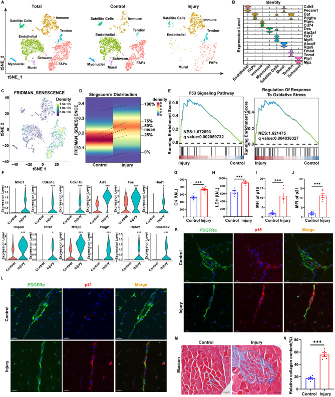
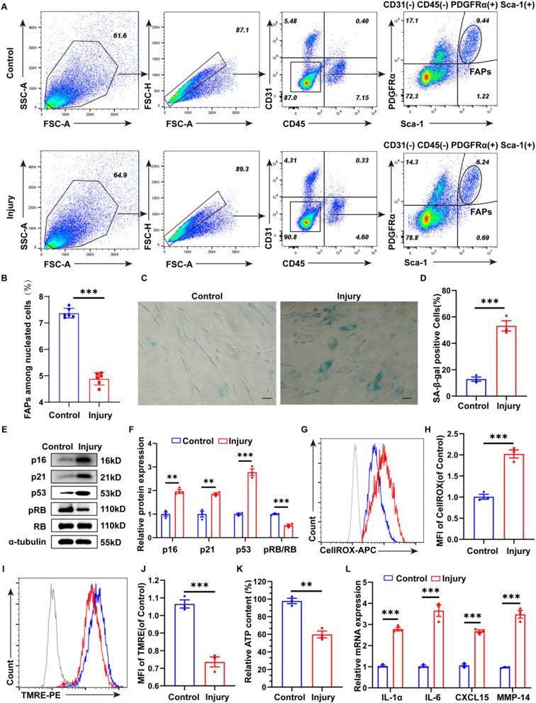
Background: Stem cells play a pivotal role in tissue regeneration and repair. Skeletal muscle comprises two main stem cells: muscle stem cells (MuSCs) and fibro-adipogenic progenitors (FAPs). FAPs are essential for maintaining the regenerative milieu of muscle tissue and modulating the activation of muscle satellite cells. However, during acute skeletal muscle injury, the alterations and mechanisms of action of FAPs remain unclear.

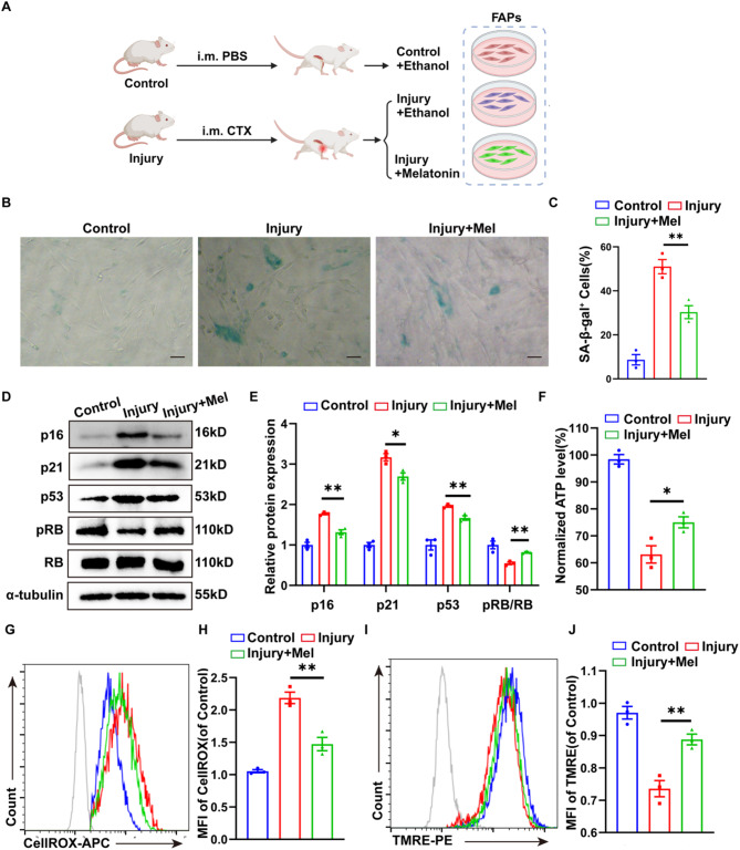
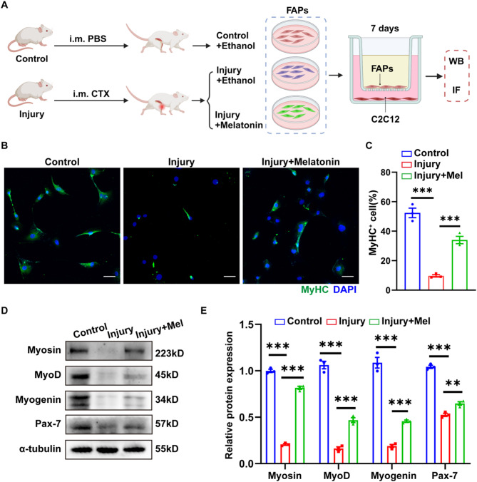
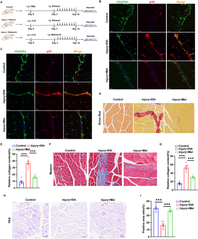
Methods: we employed the GEO database for bioinformatics analysis of skeletal muscle injury. A skeletal muscle injury model was established through cardiotoxin (CTX, 10µM, 50µL) injection into the tibialis anterior (TA) of C57BL/6 mice. Three days post-injury, we extracted the TA, isolated FAPs (CD31-CD45-PDGFRα+Sca-1+), and assessed the senescence phenotype through SA-β-Gal staining and Western blot. Additionally, we established a co-culture system to evaluate the capacity of FAPs to facilitate MuSCs differentiation. Finally, we alleviated the senescent of FAPs through in vitro (100 µM melatonin, 5 days) and in vivo (20 mg/kg/day melatonin, 15 days) administration experiments, confirming melatonin's pivotal role in the regeneration and repair processes of skeletal muscle.

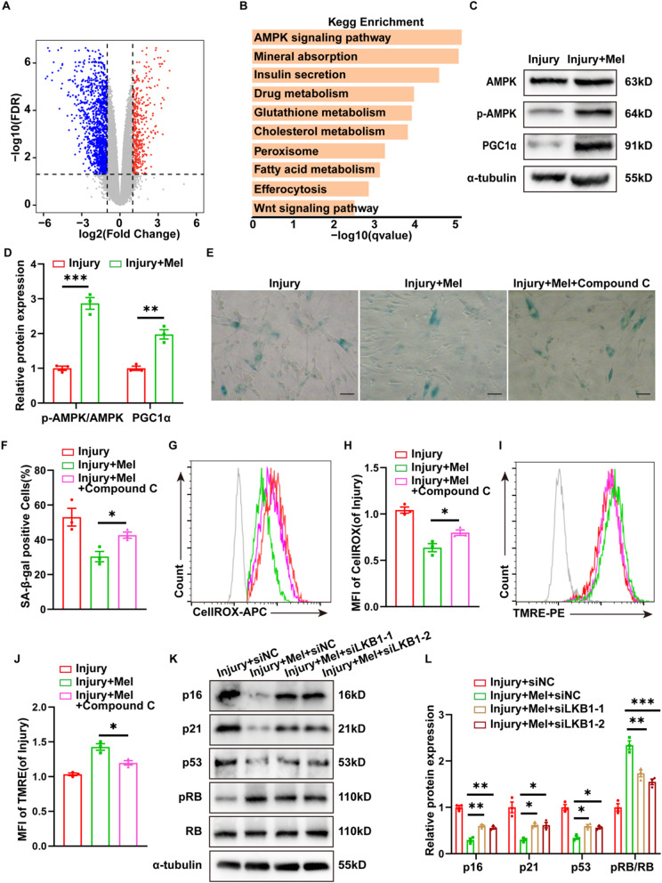
Results: In single-cell RNA sequencing analysis, we discovered the upregulation of senescence-related pathways in FAPs following injury. Immunofluorescence staining revealed the co-localization of FAPs and senescent markers in injured muscles. We established the CTX injury model and observed a reduction in the number of FAPs post-injury, accompanied by the manifestation of a senescent phenotype. Melatonin treatment was found to attenuate the injury-induced senescence of FAPs. Further co-culture experiments revealed that melatonin facilitated the restoration of FAPs' capacity to promote myoblast differentiation. Through GO and KEGG analysis, we found that the administration of melatonin led to the upregulation of AMPK pathway in FAPs, a pathway associated with antioxidant stress response. Finally, drug administration experiments corroborated that melatonin enhances skeletal muscle regeneration and repair by alleviating FAP senescence in vivo.

Conclusion: In this study, we first found FAPs underwent senescence and redox homeostasis imbalance after injury. Next, we utilized melatonin to enhance FAPs regenerative and repair capabilities by activating AMPK signaling pathway. Taken together, this work provides a novel theoretical foundation for treating skeletal muscle injury.

Keywords: FAPs; Melatonin; Senescence; Skeletal muscle injury.


Conflict of interest statement

Declarations. Ethics approval and consent to participate: All animal experiments were performed in accordance with approved protocols by the Institutional Animal Care and Use Committee (IACUC) of Shenzhen LingFu TopBiotech. Co., LTD. and were reported in line with the ARRIVE guidelines 2.0. (1) Title of the approved project: The Role of Oxidative Stress-Mediated Fibro-Adipogenic Progenitors Senescence in Skeletal Muscle Regeneration and Repair; (2) Name of the institutional approval committee: IACUC of Shenzhen LingFu TopBiotech. Co., LTD.; (3) Approval number: TOPGM-IACUC-2023-0200; (4) Date of approval: November 11th, 2023. Consent for publication: All authors have confirmed their consent for publication. Conflict of interest: The authors assert that they do not have any conflicts of interest.

Figures

Fig. 1
Fig. 1
FAPs exhibited a senescence phenotype following acute skeletal muscle injury. A: Cell clusters of the sc-RNA-seq data from database GSE214892(GEO Accession viewer (nih.gov). Tibialis anterior muscle were collected from C57BL/6 mice 5 days between control groups (50µL PBS injection) and injury groups (50µL 10µM CTX injection) (n = 5 mice for each group). B: The bubble plot showed the expression of distinctive marker genes in eight cell types. The dot size indicates the percentage of gene expression, and the dot color indicates the average expression level. C: The tSNE plot of different cell subtypes in (A) D: Density profiles of senescent fractions in (A) E: GESA analysis of the p53 pathway and the oxidative stress pathway in (A) F: Violin plot of senescence-related genes for FAPs in (A). G-H: The level of serum CK(G), LDH(H) from C57BL/6 mice 3 days between control(50µL PBS injection)and injury groups(50µL 10µM CTX injection) (n = 6 mice for each group). I-J: The Relative MFI of p16(I) and p21(J) in tibialis anterior muscle. K-L: Representative immunofluorescence images of the senescent FAPs (PDGFRα positive and p16/p21 positive cells) in tibialis anterior muscle. Scale bar = 20 μm. M-N: Masson trichrome staining (mice, n = 6) and quantification analysis of muscle fibrosis level in tibialis anterior muscle. Scale bar = 25 μm. The data above are presented as mean ± SEM of three independent experiments, P-values are calculated between two groups was performed using an unpaired t-test (ns, not significant; *P < 0.05; **P < 0.01; ***p < 0.001.)
Fig. 2
Fig. 2
In vitro validation of acute skeletal muscle injury-induced FAP senescence and redox imbalance. A: Flow cytometry sort the FAPs(CD31 CD45 PDGFRα+ Sca-1+ cells)from tibialis anterior muscle which were collected from C57BL/6 mice 3 days between control groups (50µL PBS injection) and injury groups (50µL 10µM CTX injection) (n = 6 mice for each group). B: The percentage of FAPs among nucleated cells in (A). C-D:SA-β-Gal images(n = 3) and quantification analysis showed the number of SA-β-Gal-positive cells in (A). Scale bar: 50 μm. E-F: Western Blot analysis(n = 3) of the proteins associated with cell senescence (p16, p21, p53, p-RB and RB) in (A). G-H: Flow cytometry and quantification analyses of CellROX fluorescence(n = 3) indicated the level of ROS in (A). I-J: Flow cytometry and quantification analyses of TMRE fluorescence(n = 3) indicated the level of mitochondrial membrane potential in (A). K: Quantification analyses of ATP content(n = 3) of (A). L: qRT-PCR analysis of the SASP genes (IL-1α, IL-6, CXCL15 and MMP-14) in (A). The data above are presented as mean ± SEM of three independent experiments, P-values are calculated between two groups was performed using an unpaired t-test (ns, not significant; *P < 0.05; **P < 0.01; ***p < 0.001.)
Fig. 3
Fig. 3
Melatonin maintained redox homeostasis and alleviated FAP senescence. A: Schematic diagram of FAPs in vitro experiment. Flow cytometry sort FAPs from tibialis anterior muscle which were collected from C57BL/6 mice 3 days between control groups (50µL PBS injection) and injury groups (50µL 10µM CTX injection), then the FAPs of Control and the Injury group were treated with ethanol (vehicle, 5 days) and the FAPs of Injury + Mel groups were treated with melatonin (100µM, 5 days). B-C: SA-β-Gal images(n = 3) and quantification analysis showed the number of SA-β-Gal-positive cells in FAPs of (A). Scale bar: 50 μm. D-E: Western Blot analysis(n = 3) of the proteins associated with cell senescence (p16, p21, p53, p-RB and RB) in FAPs of (A). F: Quantification analyses of ATP content(n = 3) of FAPs in (A) G-H: Flow cytometry and quantification analyses of CellROX fluorescence(n = 3) indicated the levels of ROS in FAPs of (A). I-J: Flow cytometry and quantification analyses of TMRE fluorescence(n = 3) indicated the levels of mitochondrial membrane potential in FAPs of (A). The data above are presented as mean ± SEM of three independent experiments, P-values were calculated between three groups were performed using one-way analysis of variance (anova) followed by the Tukey multiple-comparison test. (ns, not significant; *P < 0.05; **P < 0.01; ***p < 0.001.)
Fig. 4
Fig. 4
FAP senescence diminished their capacity to promote the differentiation of C2C12. A: Schematic diagram of FAPs-C2C12 coculture system. Flow cytometry sorted FAPs from tibialis anterior muscle which were collected from C57BL/6 mice 3 days between control groups (50µL PBS injection) and injury groups (50µL 10µM CTX injection), then the FAPs of Control and the Injury group were treated with ethanol (vehicle, 5 days) and the FAPs of Injury + Mel groups were treated with melatonin (100µM, 5 days). FAPs were cultured in top compartment and the C2C12 were cultured in the bottom. The FAPs and C2C12 were coculture for 7days. Indicators of differentiation were analyzed by WB and IF. Created with BioRender.com. B-C: Representative immunofluorescence images and quantification analyses displayed the differentiated C2C12 in (A). Scale bar = 50 μm. D-E: Western Blot analysis(n = 3) of the proteins associated with differentiation degree (Myosin, MyoD, Myogenin and Pax-7) in C2C12 in (A). The data above are presented as mean ± SEM of three independent experiments, P-values are calculated between three groups were performed using one-way analysis of variance (anova) followed by the Tukey multiple-comparison test. (ns, not significant; *P < 0.05; **P < 0.01; ***p < 0.001.)
Fig. 5
Fig. 5
Melatonin acts via AMPK activation. A-B: GO and KEGG pathway analyses of FAPs from database GSE214008. FAPs were sorted from tibialis anterior muscle which were collected from C57BL/6 mice and treated with vehicle or melatonin for 24 h in vitro. C-D: Western Blot analysis of the proteins (n = 3) associated with AMPK signal pathway (p-AMPK, AMPK, PGC1α) in FAPs from tibialis anterior muscle which were collected from C57BL/6 mice 3 days after CTX(50µL, 10µM) injection, then the FAPs of Injury group were treated with ethanol (vehicle, 5 days) and the FAPs of Injury + Mel groups were treated with melatonin(100µM, 5 days). E-F: SA-β-Gal images(n = 3) and quantification analysis showed the number of SA-β-Gal-positive cells in FAPs from tibialis anterior muscle which were collected from C57BL/6 mice 3 days after CTX(50µL, 10µM) injection, then the FAPs of Injury group were treated with ethanol (vehicle, 5 days), the FAPs of Injury + Mel groups were treated with melatonin(100µM, 5 days) and the FAPs of Injury + Mel + Compound C groups were treated with melatonin(100µM, 5 days) and compound C (4 µM, 24 h). G-H: Flow cytometry and quantification analyses of CellROX fluorescence(n = 3) indicated the level of ROS in FAPs in Injury, Injury + Mel, Injury + Mel + Compound C groups. I-J: Flow cytometry and quantification analyses of TMRE fluorescence(n = 3) indicated the level of mitochondrial membrane potential in FAPs in Injury, Injury + Mel, Injury + Mel + Compound C groups. K-L: Western Blot analysis of the proteins associated with cell senescence (p16, p21, p53, p-RB and RB) in FAPs from tibialis anterior muscle which were collected from C57BL/6 mice 3 days after CTX(50µL, 10µM) injection, then the FAPs were transfected with siNC or siLKB1-1 or siLKB1-2. The FAPs of Injury + siNC groups were treated with ethanol (vehicle, 5 days), and the FAPs of groups among Injury + Mel + siNC, Injury + Mel + siLKB1-1, Injury + Mel + siLKB1-2 were treated with melatonin (100µM, 5 days). The data above are presented as mean ± SEM of three independent experiments, P-values are calculated between two groups was performed using an unpaired t-test, and multiple-group statistical analysis was performed using one-way analysis of variance (anova) followed by the Tukey multiple-comparison test. (ns, not significant; *P < 0.05; **P < 0.01; ***p < 0.001.)
Fig. 6
Fig. 6
Antioxidant intervention in FAPs restored muscle function. A: Diagram of in vivo experimental modalities for melatonin treatment of skeletal muscle injury. The PBS (50µL) and CTX (50µL, 10µM) were injected into the tibialis anterior muscle of C57BL/6 mice for 3 days. Then the Control and Injury + Eth groups were injected intraperitoneally of Ethanol lasting for 15 days and the Injury + Mel groups were injected intraperitoneally of 20 mg/kg/day melatonin lasting for 15 days. Created with BioRender.com. B-C: Representative immunofluorescence (mice, n = 6) images displayed the senescent FAPs (PDGFRα positive and p16/p21 positive cells) among Control, Injury and Injury + Mel group. Scale bar = 20 μm. D-G: Sirius Red (mice, n = 6) and Masson staining (mice, n = 6) and quantification analysis indicated the muscle fibrosis level among Control, Injury and Injury + Mel group. Scale bar: 25 μm. H-I: PAS staining (mice, n = 6) and quantification analysis indicated the muscle glycogen reserve capacity among Control, Injury and Injury + Mel group. The data above are presented as mean ± SEM of three independent experiments, P-values are calculated between two groups was performed using an unpaired t-test, and multiple-group statistical analysis was performed using one-way analysis of variance (anova) followed by the Tukey multiple-comparison test. (ns, not significant; *P < 0.05; **P < 0.01; ***p < 0.001.)

Similar articles

References

    1. Cossu G, Birchall M, Brown T, De Coppi P, Culme-Seymour E, Gibbon S, Hitchcock J, Mason C, Montgomery J, Morris S, et al. Commission: stem cells and regenerative medicine. Lancet. 2018;391(10123):883–910. - PubMed
    1. Yoshimoto Y, Oishi Y. Mechanisms of skeletal muscle-tendon development and regeneration/healing as potential therapeutic targets. Pharmacol Ther. 2023;243:108357. - PubMed
    1. Chazaud B. Inflammation and skeletal muscle regeneration: leave it to the macrophages! Trends Immunol. 2020;41(6):481–92. - PubMed
    1. Espino-Gonzalez E, Dalbram E, Mounier R, Gondin J, Farup J, Jessen N, Treebak JT. Impaired skeletal muscle regeneration in diabetes:from cellular and molecular mechanisms to novel treatments. Cell Metabol. 2024;36(6):1204–36. - PubMed
    1. Zhang H, Ryu D, Wu Y, Gariani K, Wang X, Luan P, D’Amico D, Ropelle ER, Lutolf MP, Aebersold R, et al. NAD⁺ repletion improves mitochondrial and stem cell function and enhances life span in mice. Science. 2016;352(6292):1436–43. - PubMed