

Strategies for promoting neurovascularization in bone regeneration
- PMID: 40025573
- PMCID: PMC11874146
- DOI: 10.1186/s40779-025-00596-1
Strategies for promoting neurovascularization in bone regeneration
Abstract
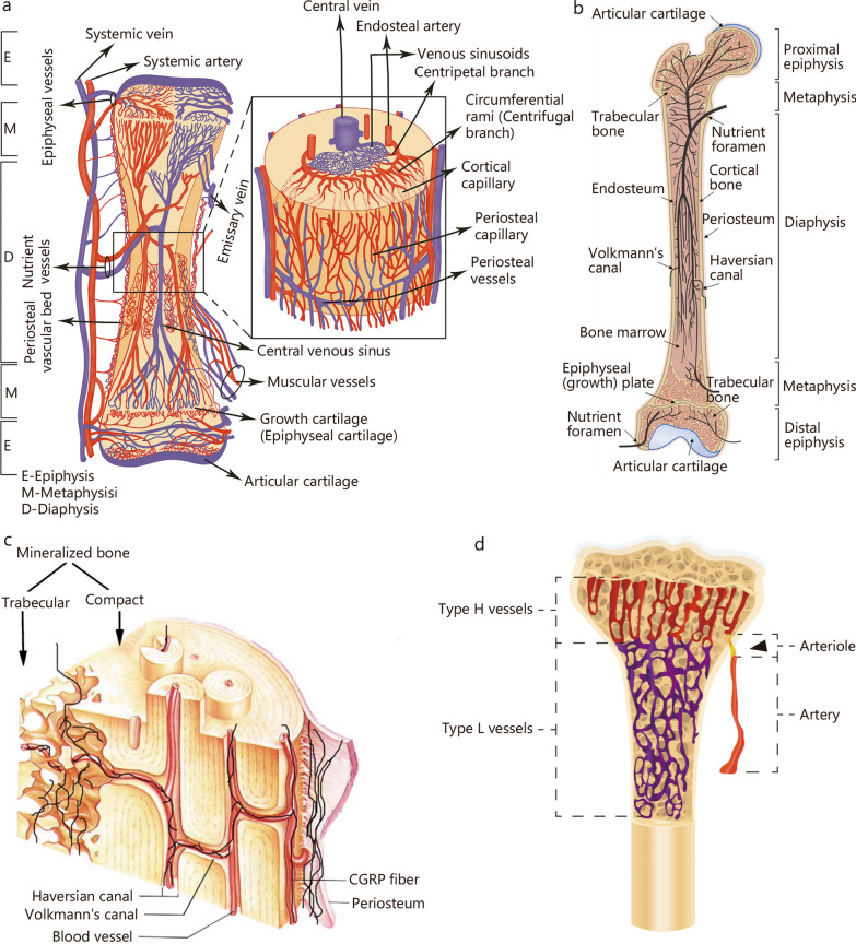
Bone tissue relies on the intricate interplay between blood vessels and nerve fibers, both are essential for many physiological and pathological processes of the skeletal system. Blood vessels provide the necessary oxygen and nutrients to nerve and bone tissues, and remove metabolic waste. Concomitantly, nerve fibers precede blood vessels during growth, promote vascularization, and influence bone cells by secreting neurotransmitters to stimulate osteogenesis. Despite the critical roles of both components, current biomaterials generally focus on enhancing intraosseous blood vessel repair, while often neglecting the contribution of nerves. Understanding the distribution and main functions of blood vessels and nerve fibers in bone is crucial for developing effective biomaterials for bone tissue engineering. This review first explores the anatomy of intraosseous blood vessels and nerve fibers, highlighting their vital roles in bone embryonic development, metabolism, and repair. It covers innovative bone regeneration strategies directed at accelerating the intrabony neurovascular system over the past 10 years. The issues covered included material properties (stiffness, surface topography, pore structures, conductivity, and piezoelectricity) and acellular biological factors [neurotrophins, peptides, ribonucleic acids (RNAs), inorganic ions, and exosomes]. Major challenges encountered by neurovascularized materials during their clinical translation have also been highlighted. Furthermore, the review discusses future research directions and potential developments aimed at producing bone repair materials that more accurately mimic the natural healing processes of bone tissue. This review will serve as a valuable reference for researchers and clinicians in developing novel neurovascularized biomaterials and accelerating their translation into clinical practice. By bridging the gap between experimental research and practical application, these advancements have the potential to transform the treatment of bone defects and significantly improve the quality of life for patients with bone-related conditions.
Keywords: Biomaterials; Blood vessels; Bone; Nerve; Neurovascularization.
© 2025. The Author(s).
Conflict of interest statement
Declarations. Ethics approval and consent to participate: Not applicable. Consent for publication: Not applicable. Competing interests: The authors declare that they have no competing interests.
Figures


Similar articles
-
Engineering vascularized and innervated bone biomaterials for improved skeletal tissue regeneration.Mater Today (Kidlington). 2018 May;21(4):362-376. doi: 10.1016/j.mattod.2017.10.005. Epub 2017 Nov 4. Mater Today (Kidlington). 2018. PMID: 30100812 Free PMC article.
-
Hydrogel-Based Systems in Neuro-Vascularized Bone Regeneration: A Promising Therapeutic Strategy.Macromol Biosci. 2024 May;24(5):e2300484. doi: 10.1002/mabi.202300484. Epub 2024 Feb 5. Macromol Biosci. 2024. PMID: 38241425 Review.
-
[Research progress on bone repair biomaterials with the function of recruiting endogenous mesenchymal stem cells].Zhongguo Xiu Fu Chong Jian Wai Ke Za Zhi. 2024 Nov 15;38(11):1408-1413. doi: 10.7507/1002-1892.202407101. Zhongguo Xiu Fu Chong Jian Wai Ke Za Zhi. 2024. PMID: 39542635 Free PMC article. Review. Chinese.
-
Biomaterial-mediated strategies targeting vascularization for bone repair.Drug Deliv Transl Res. 2016 Apr;6(2):77-95. doi: 10.1007/s13346-015-0236-0. Drug Deliv Transl Res. 2016. PMID: 26014967 Free PMC article. Review.
-
Progress in Biomaterials-Enhanced Vascularization by Modulating Physical Properties.ACS Biomater Sci Eng. 2025 Jan 13;11(1):33-54. doi: 10.1021/acsbiomaterials.4c01106. Epub 2024 Nov 30. ACS Biomater Sci Eng. 2025. PMID: 39615049 Review.
Cited by
-
From neuromodulation to bone homeostasis: therapeutic targets of nerve growth factor in skeletal diseases.Front Pharmacol. 2025 Aug 11;16:1614542. doi: 10.3389/fphar.2025.1614542. eCollection 2025. Front Pharmacol. 2025. PMID: 40860871 Free PMC article. Review.
-
Advancements in two-dimensional nanomaterials for regenerative medicine in skeletal muscle repair.Mater Today Bio. 2025 Jun 2;33:101924. doi: 10.1016/j.mtbio.2025.101924. eCollection 2025 Aug. Mater Today Bio. 2025. PMID: 40528839 Free PMC article. Review.
-
An Electrospun DFO-Loaded Microsphere/SAIB System Orchestrates Angiogenesis-Osteogenesis Coupling via HIF-1α Activation for Vascularized Bone Regeneration.Polymers (Basel). 2025 May 31;17(11):1538. doi: 10.3390/polym17111538. Polymers (Basel). 2025. PMID: 40508781 Free PMC article.
-
Recapitulating the bone extracellular matrix through 3D bioprinting using various crosslinking chemistries.Front Bioeng Biotechnol. 2025 Jun 5;13:1506122. doi: 10.3389/fbioe.2025.1506122. eCollection 2025. Front Bioeng Biotechnol. 2025. PMID: 40539097 Free PMC article. Review.
References
-
- Armiento AR, Hatt LP, Sanchez Rosenberg G, Thompson K, Stoddart MJ. Functional biomaterials for bone regeneration: a lesson in complex biology. Adv Funct Mater. 2020;30(44):1909874.
-
- Jia Z, Xu X, Zhu D, Zheng Y. Design, printing, and engineering of regenerative biomaterials for personalized bone healthcare. Prog Mater Sci. 2023;134:101072.
-
- de Melo PD, Habibovic P. Biomineralization-inspired material design for bone regeneration. Adv Healthc Mater. 2018;7(22):e1800700. - PubMed
Publication types
MeSH terms
Substances
Grants and funding
- LCA202204/Foundation of National Clinical Research Center for Oral Diseases
- 2024GH-YBXM-19/Key Research and Development Projects of Shaanxi Province
- LX2023-306/Clinical New Technology Program of Air Force Medical University
- 2019M653969/China Postdoctoral Science Foundation
- 82101069/National Natural Science Foundation of China
LinkOut - more resources
Full Text Sources
Miscellaneous
