. 2025 Feb 28:19:11779322251315922.
doi: 10.1177/11779322251315922. eCollection 2025.

A "Dock-Work" Orange: A Dual-Receptor Biochemical Theory on the Deterrence Induced by Citrusy Aroma on Elephant Traffic Central to a Conservation Effort

Affiliations

A "Dock-Work" Orange: A Dual-Receptor Biochemical Theory on the Deterrence Induced by Citrusy Aroma on Elephant Traffic Central to a Conservation Effort

Dilantha Gunawardana. Bioinform Biol Insights. .

Abstract

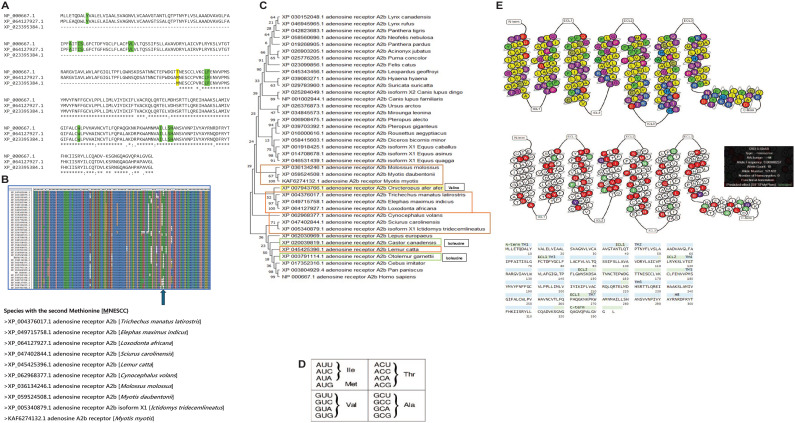
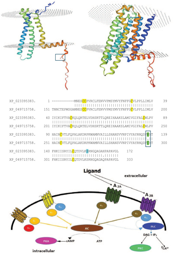
Conservation of elephants requires physical, chemical, and biological approaches to ensure the protection of these gargantuan pachyderms. One such approach is using orange plants (as biofencing) for the repellence of elephants, which precludes catastrophic events related to the encroachment of elephants into human habitats. Elephants have sensitive olfactory discrimination of plant volatile compounds for foraging and other behavior using G-protein-coupled receptors (GPCRs). However, 2 such receptors are the A2A and A2B receptors mediating olfaction elicited by a host of ligands, including limonene, the main volatile compound in citrus plants, which is hypothesized to be the chief repelling agent. Bioinformatics at the protein and mRNA levels (BLAST/Multiple Sequence Alignments) were employed to explore the multiple expression products of A2B receptors, namely full-length and truncated proteins produced by isoform mRNAs translated from multiple methionines, while the comparison of the limonene-binding pockets of human and elephant A2B receptors and prediction servers [Netphos 3.1; Protter] was used to focus, respectively, on the contacts limonene binding entails and the post-translational modifications that are involved in cell signaling. Finally, the link between limonene and antifeedant activity was explored by considering limonene content on trees that are preferentially foraged or avoided as part of the feeding behavior by elephants. The African bush elephant (Loxodonta africana) possesses a full-length A2A receptor but unlike most mammals, expresses a highly truncated A2B receptor isoform possessing only transmembrane helices 5, 6, and 7. Truncation may lead to higher traffic and expression of the A2B receptor in olfactory interfaces/pathways and aid stronger activation. In addition, all residues in the putative limonene-binding cleft are perfectly conserved between the human and African bush elephant A2B receptors, both full length and truncated. Shallow activation sites require micromolar affinity and fewer side-chain interactions, which is speculated to be the case for the truncated A2B receptor. An N-terminal extremity N-glycosylation motif is indicative of membrane localization of the truncated A2B receptor following accurate folding. A combination of truncation, indels, substitutions, and transcript isoforms are the attributed roles in the evolution of the L. africana A2B receptor, out of which limonene receptivity may be the key. It is also inferred how limonene may act as a dietary repellent/antifeedant to a generalist herbivore, with the documented limonene content being absent in some dietary favorites including the iconic Sclerocarya birrea.

Keywords: A2A receptors; A2B receptors; citrus; conservation; elephants; limonene.


Conflict of interest statement

The author(s) declared no potential conflicts of interest with respect to the research, authorship, and/or publication of this article.

Figures

Figure 1.
Figure 1.
Phylogeny of A2A receptor protein sequences using the Neighborhood Joining method. The sequences were first aligned using the ClustalW tool, sampled by 1000 bootstrap replications, prior to drawing the phylogenetic tree. The level of bootstrap support is indicated at the nodes of the phylogenetic tree.
Figure 2.
Figure 2.
Phosphorylated residues predicted using Netphos 3.1 for the XP_023395383.1 protein from L. africana. The upper panel depicts the putative phosphorylated residues as a function of protein sequence (X-Axis) from the N-terminal end to the C-terminus, while the Y-axis showcases the probability of phosphorylation over a threshold of 0.5. Serines are highlighted in red and threonines in green in the lower panel. The Netphos 3.1 server uses serine, threonine, and tyrosine phosphorylation sites using families of neural networks, drawing from experimentally verified phosphorylation sites.
Figure 3.
Figure 3.
Schematic illustration of (A) the linear and (B) the structural simplification of a GPCR. This was reproduced with permission from a specific research study where this illustration appeared.
Figure 4.
Figure 4.
The side chains of the limonene binding site mapped from the human A2B receptor as provided in the work by Patel et al., showcased here in analogy to the counterparts from the truncated A2B receptor where the analogous residues appear. The illustration is a modified picture of the one provided in the work by Patel et al., showcasing the side chains that are relevant and their positional IDs.
Figure 5.
Figure 5.
(A) The NES N-glycosylation site indicated in the sequence of the A2B protein from L. africana and an illustration of the receptor using Protter. (B) The N-glycosylation sites in structure of the A2A protein from L. africana illustrated using Protter.
Figure 6.
Figure 6.
Local sequence alignment of A2A [XP_064128479.1] and the full-length A2B receptor [XP_064127927.1] from L. africana to showcase similarities and divergences. In green is an insertion in the A2B receptor and in yellow is a region of weak conservation surrounding the initiator methionine of the truncated protein.
Figure 7.
Figure 7.
(A) 2 types of transcripts (mRNA isoforms) that are generated from the A2B receptor gene differing in their 5’ UTRs. The start codon is shown in a box while the differing 5’ UTRs are shown in a shaded blue rectangle. In addition, all 3 transcript isoforms are aligned in Supplementary Figure 1; (B) The alignment of the truncated A2B receptor mRNA with the corresponding A2B receptor protein sequence. Important conserved residues in the Kozak sequence are shown as pink residues. The start codon is colored yellow and the stop codon is colored gray.
Figure 8.
Figure 8.
(A) Sequence alignment of the human A2B receptor [NP_000667.1] with the counterparts from L. africana. The predicted limonene-binding residues are highlighted in green. Note that there is 100% conservation of the limonene-binding residues between the 3 sequences. Furthermore, the internal methionine from where the translation is proposed to begin in the truncated A2B receptor is showcased in yellow. Of relevance, there is no second methionine in the human A2B receptor [In Yellow]; (B) Sequence alignment of A2B receptor protein sequences. The species armed with the second methionine (the translated site of the A2B truncated receptor from L. africana) are shown underneath as relevant species; (C) Phylogeny of A2B receptor protein sequences using the Maximum Likelihood method. The sequences were first aligned using the ClustalW tool, sampled by 500 bootstrap replications, prior to drawing the phylogenetic tree. However, 10 out of the 39 sequences containing the second methionine residue (Figure 8[B]) are found within the orange boxes pointing to lineage-specific evolution. Moreover, 2 isoleucines as found in 2 distinct species, which the authors infer to be a stepping stone for the conversion into an internal methionine, are shown in green. (D) Codons encoding Threonine, Methionine, Valine, Isoleucine, and Alanine; (E) [Top] The color-coded biochemical properties of amino acids in the human A2B receptor arranged by GPCR residue numbering by the Ballesteros and Weinstein system. [Middle] The naturally occurring variation of the human A2B receptor showcasing the propensity of isoleucine 141 in being mutated into a methionine, hinting that a highly truncated mutant missense protein can be produced by the I → M mutation at this location (Allele frequency—0.00008237), drawing similarities to the highly truncated A2B receptor from L. africana. [Bottom] The sequence of the human A2B receptor showcases the residues classified according to structural localities. The isoleucine to methionine mutation appears to be a common adaptation to producing truncated proteins (Figure 8C).
Figure 9.
Figure 9.
(A) Secondary structure prediction of the region (60 residues in total) spanning the codon triplet encoding the second methionine. The initiator codon coding for the second methionine is shown in a red arrow. A secondary structural complex with a composite hairpin-loop structure is shown in a square, which is assumed to be a regulatory structural element upstream of the start codon. The web server employed was the RNA-Fold Web Server; (B) Secondary structure prediction of the divergent regions found further upstream of the start codon in the 2 mRNA transcripts, XM_023539615 and XM_023539616. In yellow are the consecutive hairpin-loop structures that distinctively identify XM_023539616 and underlined are the deleted residues in the XM_023539616 transcript compared with XM_023539615. The divergent region is demarcated in Figure 8(B). The web server employed was the RNA-Fold Web Server; (C) 4 consecutive codons coding for a region inclusive of the 3-residue N-glycosylated motifs in the A2B (top) and A2A (bottom) receptors of L. africana. It is inferred that the NESC coding region of the A2B receptor is an insertion.
Figure 10.
Figure 10.
(Top) Homology models of the A2B truncated receptor from L. africana (Left) and the full-length counterpart from Elephus maximus indicus (Right). The template used for the construction of the homology models was P29276.1.A (Adenosine receptor A2B from [Rattus norvegicus]). Ser-157 in the A2B truncated receptor and Cys-298 in the full-length A2B receptor of Elephus maximus indicus are shown in boxes. The Swiss-Model server was used to build the homology models. (Middle) The pairwise sequence alignment of A2B receptors of the African (XP_023395383.1) and Asian (Elephas maximum indicus—XP_049715758.1) elephants. A single (only) amino acid change between the 2 protein sequences is shown in green and Ser-157 is highlighted in cyan. (Bottom). Schematic illustration of the 4 types of adenosine receptors and their influence on downstream signaling events. This is a modified image from van Calker et al. showcasing the simplified architecture and signaling interactions.

Similar articles

References

    1. Bester T, Schmitt MH, Shrader AM. The deterrent effects of individual monoterpene odours on the dietary decisions of African elephants. Anim Cogn 2023;26:1049–1063. 10.1007/s10071-023-01755-4 - DOI - PMC - PubMed
    1. Nevo O, Schmitt MH, Ayasse M, Valenta K. Sweet tooth: elephants detect fruit sugar levels based on scent alone. Ecol Evol. 2020;10:11399-11407. doi:10.1002/ece3.6811 - DOI - PMC - PubMed
    1. Rizvanovic A, Amundin M, Laska M. Olfactory discrimination ability of Asian elephants (Elephas maximus) for structurally related odorants. Chem Senses. 2013;38:107-118. doi:10.1093/chemse/bjs106 - DOI - PubMed
    1. Niimura Y, Matsui A, Touhara K. Extreme expansion of the olfactory receptor gene repertoire in African elephants and evolutionary dynamics of orthologous gene groups in 13 placental mammals. Genome Res. 2014;24:1485-1496. doi:10.1101/gr.170828.113 - DOI - PMC - PubMed
    1. Patel M, Narke D, Kurade M, et al.. Limonene-induced activation of A2A adenosine receptors reduces airway inflammation and reactivity in a mouse model of asthma. Purinergic Signal. 2020;16:415-426. doi:10.1007/s11302-020-09734-0 - DOI - PMC - PubMed

LinkOut - more resources