. 2025 Feb 12:46:569-581.
doi: 10.1016/j.bioactmat.2024.09.027. eCollection 2025 Apr.

Synergistic restoration of spinal cord injury through hyaluronic acid conjugated hydrogel-polydopamine nanoparticles combined with human mesenchymal stem cell transplantation

Affiliations

Synergistic restoration of spinal cord injury through hyaluronic acid conjugated hydrogel-polydopamine nanoparticles combined with human mesenchymal stem cell transplantation

Yanbing Kao et al. Bioact Mater. .

Abstract

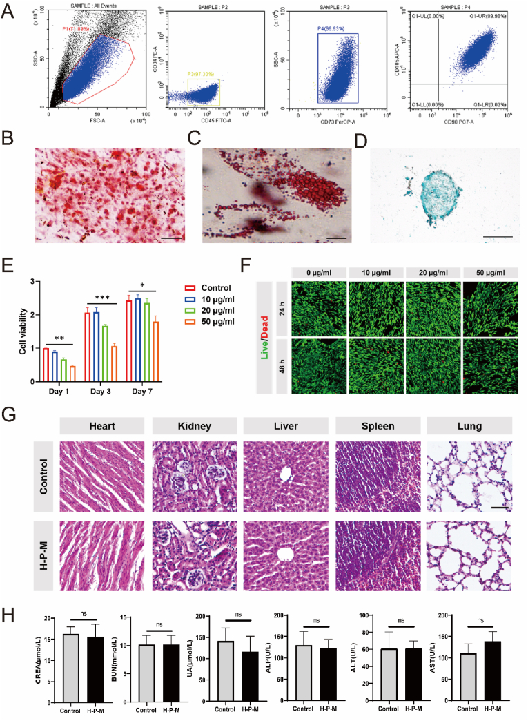
Spinal cord injury (SCI) is a devastating disease with limited treatment options due to the restricted regenerative capacity of the central nervous system. The accumulation of reactive oxygen species (ROS) and inadequate endogenous neural stem progenitor cells (eNSPCs) in the lesion site exacerbates neurologic deficits and impedes motor function recovery. We have developed a temperature-responsive hyaluronic acid conjugated hydrogel-polydopamine nanoparticles (PDA NPs) combined with human mesenchymal stem cell (hMSCs) transplantation, denoted as H-P-M hydrogel. Microglia cells treated with PDA NPs have been shown to reduce intracellular ROS levels by 65 % and suppress the expression of inflammatory cytokines such as IL-1β (decreased by 35 %) and IL-6 (decreased by 23 %), thus mitigating the microglia's inflammatory response. Additionally, our results have demonstrated that the H-P-M hydrogel combined with hMSCs transplantation can recruit eNSPCs to the injury site as evidenced by utilizing Nestin lineage tracer mice. The RNA-seq has unveiled the potential of the H-P-M hydrogel to facilitate eNSPCs neuronal differentiation through the MAPK pathway. Furthermore, these differentiated neurons are integrated into local neural circuits. Together, it suggests that the H-P-M hydrogel synergistically improves the SCI niche. It serves as catalysts inducing 5-HT axon regeneration and improving BMS score after SCI through the modulation of the ROS milieu and the promotion of neuronal differentiation from eNSPCs, thereby presenting a promising strategy for SCI repair.

Keywords: Endogenous neural stem progenitor cells; Human mesenchymal stem cells; Polydopamine nanoparticles; Spinal cord injury.


Conflict of interest statement

The authors declare that they have no known competing financial interests or personal relationships that could have appeared to influence the work reported in this paper.

Figures

Image 1
Graphical abstract
Fig. 1
Fig. 1
Schematic diagram of the preparation and use of the multifunctional hydrogel scaffold for SCI repair.
Fig. 2
Fig. 2
Preparation and characteristics of PDA NP-decorated hydrogel. (A) Schematic diagram of the gelation of the thermosensitive PDA NP-decorated HA hydrogel. (B) Transmission electron microscopy (TEM) images of PDA NPs. Scale bar: 200 nm. (C) Nuclear magneto-hydrogen spectroscopy (NMR) of HA-DA. (D) The vibration of the FTIR shows the absorption peaks of HA and HA-DA molecules. (E) The gelation of HA-DA loaded with or w/o PDA NPs at 37 °C. (F) Scanning electron microscopy (SEM) images of HA-DA hydrogel (The white arrow indicates the presence of the PDA NPs). Scale bar: 10 μm. (G) Storage modulus (G′) and loss modulus (G″) of HA-DA, H-P and H-P-M hydrogels. (H) The direct view of hydrogel adhesion to spinal cord tissues.
Fig. 3
Fig. 3
The biocompatibility and biotoxicity of PDA NPs. (A) Identification of surface markers of CD34, CD45, CD73, CD90 and CD105 in hUC-MSCs through flow cytometry. (B) The capacity of osteogenic differentiation by Alizarin Red staining. Scale bar: 100 μm. (C) The capacity of adipogenic differentiation by Oil Red staining. Scale bar: 25 μm. (D) Chondrogenic differentiation capacity by Alcian Blue staining. Scale bar: 100 μm. (E) Cell viability of hUC-MSCs at 1, 3 and 7 day (s) after treatment with different concentrations of PDA NP was assessed using the CCK8 assay. (F) The Live/Dead staining of hUC-MSCs at 1 and 2 day (s) post-treatment with different concentrations of PDA NPs was performed. Live cells are indicated by calcein staining (green), and dead cells by PI staining (red). Scale bar: 100 μm. (G) HE staining images of the heart, kidney, liver, spleen and lung, 8 weeks after hydrogel implantation after in mice. Scale bar: 50 μm. (H) The indicators (CERA/BUN/UA/ALP/ALT/AST) of the liver and kidney function 8 weeks after H-P-M hydrogel implantation in mice (n = 5). Data was presented as mean ± SEM. Results were analyzed by two-way ANOVA or t-test. Statistical significance: ∗p < 0.05, ∗∗p < 0.01, ∗∗∗p < 0.001.
Fig. 4
Fig. 4
Validation of H-P-M hydrogel to reduce ROS and promote NSPC neuronal differentiation in vitro. (A) Scavenging activity of ROS in inflammatory BV2 cells induced by LPS and treatment with PDA NPs at different concentrations (0 μg/ml, 10 μg/ml, 20 μg/ml, 50 μg/ml) of for 1 day, ROS (green), DAPI (blue). Scale bar: 50 μm. (B)The ROS level was detected by DCFH-DA in BV2 cells induced by LPS. The mRNA expression of (C) IL-1β (n = 3) and (D) IL-6 (n = 3) in BV2 cells induced by LPS. (E) Schematic diagram of co-culture of mouse NSPCs and H-P-M hydrogel. (F) The mRNA expression of Tuj1, MAP2 and GFAP after 7 days of co-culture with or w/o H-P-M hydrogel. (G) Representative images of GFAP and Tuj1 expression of NSPCs co-cultured with or w/o H-P-M hydrogel for 7 days. Scale bar: 50 μm. (H) The quantification of (G). (I and J) Western blot images showing the protein expression of GFAP and Tuj1 of NSPCs co-cultured with or w/o H-P-M hydrogel for 7 days and quantitative analysis. Data was presented as mean ± SEM. Results were analyzed by using t-test. Statistical significance: ∗p < 0.05, ∗∗p < 0.01, ∗∗∗p < 0.001.
Fig. 5
Fig. 5
H-P-M hydrogel induced 5-HT axon regeneration and promoted motor function recovery in SCI mice. (A) Schematic diagram of spinal cord transection surgery and hydrogel injection. (B) Representative images of HE-stained spinal cord section in different treatment groups. Scale bar: 500 μm. (C) Representative images of 5-HT staining of spinal cord section at 8 weeks after hydrogel injection. Scale bar (upper right): 500 μm. Scale bar (left and lower right): 50 μm. (D) The BMS scores in different treatment groups after SCI (Sham n = 8, SCI n = 8, HA n = 7, H-P n = 9, H-M n = 8, H-P-M n = 9). (E) Representative images of MEPs in different treatment groups at 8 weeks after hydrogel injection. (F) Representative images of footprints in different treatment groups at 8 weeks after hydrogel injection. Data was presented as mean ± SEM. Data were analyzed using two-way ANOVA. Statistical significance: ∗p < 0.05, ∗∗p < 0.01, ∗∗∗p < 0.001.
Fig. 6
Fig. 6
H-P-M hydrogel recruited eNSPCs to the injury site and facilitated neuronal differentiation for SCI repair. (A) Results of DEGs (FDR<0.05) were displayed by a volcano plot. (B) The significantly enriched biological processes (BPs) from upregulated DEGs were presented. (C) Heat map of key genes involved in neuronal differentiation (Tubb3, Map2, NeuN and Chat) and synapse formation (Syp and Snap25). (D) Representative fluorescence images showing Ki67 staining of spinal cord section of Nestin lineage tracer mice after SCI. The eNSPCs were labeled with tdTomato (red). Nuclei were labeled with DAPI (blue) in each group. Scale bar: 20 μm. (E) Representative fluorescence images showing the recruitment of eNSPCs labeled with tdTomato (red) at the lesion site in SCI mice. Nuclei were labeled with DAPI (blue) in each group. Scale bar: 400 μm. (F–G) Quantitation of Nestin+ eNSPCs and Ki67+ & Nestin+ proliferating eNSPCs around injury site at 8 weeks after hydrogel injection. (H)Representative fluorescence images showing Tuj1 and GFAP staining of spinal cord section at 8 weeks after hydrogel injection. The eNSPCs were labeled with tdTomato (red). Nuclei were labeled with DAPI (blue) in each group. Scale bar: 40 μm. (I) Representative fluorescence images showing Tuj1 and Syn1 staining of spinal cord section at 8 weeks after hydrogel injection. The eNSPCs were labeled with tdTomato (red). Nuclei were labeled with DAPI (blue) in each group. Scale bar: 200 μm. (J) The Western blot images and quantification showed the protein expression of JNK, ERK, p-JNK and p-ERK. Data was presented as mean ± SEM. Results were analyzed by t-test. Statistical significance: ∗p < 0.05, ∗∗p < 0.01, ∗∗∗p < 0.001.
Fig. 7
Fig. 7
H-P-M hydrogel could reduce the release of pro-inflammatory factors and inflammatory response of microglia in SCI mice. (A) The significantly enriched biological processes (BPs) from downregulated DEGs were presented. (B) GSEA of the acute inflammatory response gene set. (C) The expression of key genes involved in inflammatory response including iNOS, IL-1β and IL-6. (D) Representative fluorescence images and quantification showing Arg1 (red) and IBA1 (green) staining of spinal cord sections in each group at 1 week after hydrogel injection. Nuclei were labeled with DAPI (blue) in each group. Scale bar: 200 μm. (E) Representative fluorescence images and quantification showing iNOS (red) and IBA1 (green) staining of spinal cord sections in each group at 1 week after hydrogel injection. Nuclei were labeled with DAPI (blue) in each group. Scale bar: 200 μm. Data was presented as mean ± SEM. Results were analyzed by one-way ANOVA with the Holm-Sidak test. Statistical significance: ∗p < 0.05, ∗∗p < 0.01, ∗∗∗p < 0.001.

References

    1. Fan B., et al. Progression in translational research on spinal cord injury based on microenvironment imbalance. Bone Res. 2022;10:35. doi: 10.1038/s41413-022-00199-9. - DOI - PMC - PubMed
    1. Sofroniew M.V. Dissecting spinal cord regeneration. Nature. 2018;557:343–350. doi: 10.1038/s41586-018-0068-4. - DOI - PubMed
    1. Fan B., et al. Microenvironment imbalance of spinal cord injury. Cell Transplant. 2018;27:853–866. doi: 10.1177/0963689718755778. - DOI - PMC - PubMed
    1. Zhu B., et al. Schwann cell-derived exosomes and methylprednisolone composite patch for spinal cord injury repair. ACS Nano. 2023;17:22928–22943. doi: 10.1021/acsnano.3c08046. - DOI - PubMed
    1. Li L., et al. A MnO2 nanoparticle-dotted hydrogel promotes spinal cord repair via regulating reactive oxygen species microenvironment and synergizing with mesenchymal stem cells. ACS Nano. 2019;13:14283–14293. doi: 10.1021/acsnano.9b07598. - DOI - PubMed

LinkOut - more resources