[Preprint]. 2025 Feb 20:2025.02.20.639226.
doi: 10.1101/2025.02.20.639226.

In vitro reconstitution of minimal human centrosomes

Affiliations

In vitro reconstitution of minimal human centrosomes

Manolo U Rios et al. bioRxiv. .

Update in

Abstract

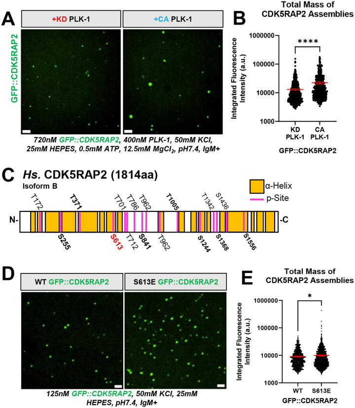
CDK5RAP2/CEP215 is a key pericentriolar material (PCM) protein that recruits microtubule-nucleating factors at human centrosomes. Using an in vitro reconstitution system, we show that CDK5RAP2 is sufficient to form micron-scale scaffolds around a nanometer-scale nucleator in a PLK-1-regulated manner. CDK5RAP2 assemblies recruited and activated gamma tubulin ring complexes (γ-TuRCs) which, in the presence of α/β tubulin, generated microtubule asters. We found that F75 in CDK5RAP2 is partially needed to recruit γ-TuRC yet is indispensable for γ-TuRC activation. Furthermore, our system recapitulated key features of centrosome-amplified cancer cells. CDK5RAP2 scaffolds selectively recruited the molecular motor KifC1/HSET, which enhanced concentration of α/β tubulin, microtubule polymerization, and clustering of the assemblies. Our results highlight the specificity and selectivity of in vitro generated CDK5RAP2 scaffolds and identify a minimal set of components required for human centrosome assembly and function. This minimal centrosome model offers a powerful tool for studying centrosome biology and dysfunction in human health and disease.


Figures

FIGURE 1.
FIGURE 1.. Purified human CDK5RAP2 is sufficient to form micron scale scaffolds.
A) Linear representation of Homo Sapiens (Hs.) CDK5RAP2/CEP215 Isoform B, human colon cancer DLD-1 cell expressing CDK5RAP2-mNeonGreen (mNG) and purified GFP::CDK5RAP2 isoform B from SF9 insect cells. Yellow blocks in linear CDK5RAP2 diagram represent alpha-helical regions predicted by AlphaFold2. CM1 (a.a. 51–100) and CM2 (a.a. 1715–1814) domains are indicated by red bars. The CM1 domain contains F75. B) Purified GFP::CDK5RAP2 combined with 9% PEG at various concentrations (10nM, 60nM, 100nM and 120nM). Scale bar, 5 μm. C) Quantification of panel 1B (mean +/− 95% C.I.; 10nM (n=1092 assemblies), 60nM (n=3845 assemblies), 100nM (n=5366 assemblies), 120nM (n=4821 assemblies). D) GFP::CDK5RAP2 assemblies were nucleated by adding <11 pM IgM raised against the CM2 domain of CDK5RAP2 (IgM+). Addition of buffer with no IgM (IgM−) or an anti-myosin heavy chain (Control IgM) did not nucleate micron-scale GFP::CDK5RAP2 assemblies. Scale bar, 5 μm. E) Quantification of panel 1D (mean +/− 95% C.I.; IgM+ (n=166 assemblies), IgM− (n=23 assemblies), Ctl IgM (n=20 assemblies)). P values from One-way ANOVA followed by Tukey’s multiple comparisons test. F) Assembly of full length (FL) GFP::CDK5RAP2 into micron-scale scaffolds (left panel). GFP::CDK5RAP2 lacking the CM2 domain does not form micron-scale assemblies (right panel). This domain (last 200 a.a.) is recognized by the IgM. Scale bar, 5 μm. G) Quantification of panel 1F. All data points represent individual GFP::CDK5RAP2 assemblies (mean +/− 95% C.I.; (FL (n=166 assemblies), ΔCM2 (n=131 assemblies)) P value from a student t-test.
FIGURE 2.
FIGURE 2.. Phospho-regulation of in vitro CDK5RAP2 scaffolds.
A) GFP::CDK5RAP2 assemblies generated in the presence of purified kinase dead (KD) or constitutively active (CA) human PLK-1. Scale bar, 5 μm. B) Quantification of panel 2A. Data points represent individual GFP::CDK5RAP2 assemblies (mean +/− 95% C.I.; KD PLK-1 condition (n=604 assemblies), CA PLK-1 condition (n=511 assemblies)). Significant differences were assessed using a student t-test. C) Identified PLK-1 phosphorylation sites (p-Sites) in GFP::CDK5RAP2 incubated with human PLK-1 plus ATP·MgCl2. Bold p-sites are also found in PhosphoSite Plus. S613 (red) is phosphorylated by PLK-1 in vivo and in vitro. Control reactions consisted of pre-dephosphorylated GFP::CDK5RAP2 plus PLK-1 but no ATP·MgCl2. D) In vitro assemblies of pre-dephosphorylated GFP::CDK5RA2 and pre-dephosphorylated GFP::CDK5RAP2(S613E). Scale bar, 5 μm. E) Quantification of total mass of CDK5RAP2 assemblies in panel D. Each data point represents individual GFP::CDK5RAP2 assemblies (mean +/− 95% C.I.; WT (n=1337 assemblies), S613E (n=2463 assemblies)). Significant differences were assessed using a student t-test.
FIGURE 3.
FIGURE 3.. Microtubule nucleating activity of CDK5RAP2 scaffolds with γ-TuRCs
A) 3-Component (3C) reactions were assembled with purified GFP::CDK5RAP2 (WT or F75A), mBFP::γ-TuRCs, and HiLyte647-labeled α/β tubulin. Scale bar, 3μm B) Quantification of γ-TuRC partitioning into GFP::CDK5RAP2 assemblies (mean +/− 95% C.I.; WT, n=95; F75A, n=117). Significant differences were assessed using a student t-test. C) Quantification of α/β tubulin partitioning into GFP::CDK5RAP2 assemblies (mean +/− 95% C.I.; WT, n=95; F75A, n=117). Significant differences were assessed using a student t-test. D) Time-lapse imaging of microtubule growth from GFP::CDK5RAP2 scaffolds + mBFP::γ-TuRCs. Z-projections were obtained every 2 min for 30 min at 37°C. Scale bar, 3μm.
FIGURE 4.
FIGURE 4.. Selective HSET recruitment, microtubule-nucleating effects, and clustering of CDK5RAP2 scaffolds in vitro.
A) Selective recruitment of purified mCherry::HSET (full-length), mCherry::HSET lacking its intrinsically disordered region (ΔIDR, missing a.a. 2–138), or mCherry into micron-scale GFP::CDK5RAP2 assemblies. Scale bar, 1μm. B) Quantification of panel 4A (mean +/− 95% C.I.; HSET(FL), n=293 scaffolds; HSET(ΔIDR), n=294 scaffolds; mCherry, n=285 scaffolds. Each data point represents a CDK5RAP2 scaffold. Significant differences were assessed using One-way ANOVA followed by Tukey’s multiple comparisons test. HSET(FL) n=293 scaffolds, HSET(ΔIDR) n=294 scaffolds, mCherry n=285 scaffolds. C) Z-projection of 4-Component (4C) asters consisting of 167nm GFP::CDK5RAP2, 33nM mBFP::γ-TuRCs, 33nM mCherry::HSET and 13μM HiLyte-647-labeled α/β tubulin. Buffer consists of 50mM KCl, 25mM HEPES, pH7.4, IgM+. D) Quantification of γ-TuRC partitioning in 3-component (3C) vs 4-component (4C) asters (mean +/− 95% C.I.; 3C, n=95; 4C, n=133). Significant differences were assessed using a student t-test. E) Quantification of α/β tubulin partitioning in 3-component (3C) vs 4-component (4C) asters. (mean +/− 95% C.I.; 3C, n=95; 4C, n=133). Significant differences were assessed using a student t-test. F) Z-projection of clusters formed by 4C asters in the presence of 0 or 0.66mM ATP plus MgCl2. Arrowheads indicate microtubule contacts between GFP::CDK5RAP2 scaffolds. Scale bar, 5μm.

References

    1. Andersen J.S., Wilkinson C.J., Mayor T., Mortensen P., Nigg E.A., and Mann M.. 2003. Proteomic characterization of the human centrosome by protein correlation profiling. Nature. 426:570–574. - PubMed
    1. Brito C., Serna M., Guerra P., Llorca O., and Surrey T.. 2024. Transition of human γ-tubulin ring complex into a closed conformation during microtubule nucleation. Science. 383:870–876. - PubMed
    1. Buchman J.J., Tseng H.-C., Zhou Y., Frank C.L., Xie Z., and Tsai L.-H.. 2010. Cdk5rap2 Interacts with Pericentrin to Maintain the Neural Progenitor Pool in the Developing Neocortex. Neuron. 66:386–402. - PubMed
    1. Cabral G., Laos T., Dumont J., and Dammermann A.. 2019. Differential Requirements for Centrioles in Mitotic Centrosome Growth and Maintenance. Developmental Cell. 50:355–366.e356. - PubMed
    1. Chavali P.L., Chandrasekaran G., Barr A.R., Tátrai P., Taylor C., Papachristou E.K., Woods C.G., Chavali S., and Gergely F.. 2016. A CEP215–HSET complex links centrosomes with spindle poles and drives centrosome clustering in cancer. Nature Communications. 7:11005. - PMC - PubMed

Publication types

LinkOut - more resources