. 2025 Feb 1;36(1):102472.
doi: 10.1016/j.omtn.2025.102472. eCollection 2025 Mar 11.

Enhanced HDR-mediated correction of heterozygous COL7A1 mutations for recessive dystrophic epidermolysis bullosa

Affiliations

Enhanced HDR-mediated correction of heterozygous COL7A1 mutations for recessive dystrophic epidermolysis bullosa

John M T Hunt et al. Mol Ther Nucleic Acids. .

Abstract

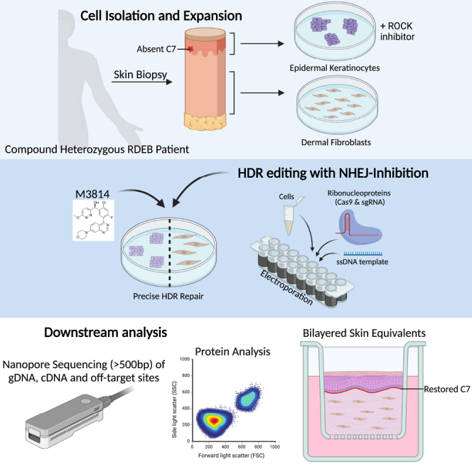
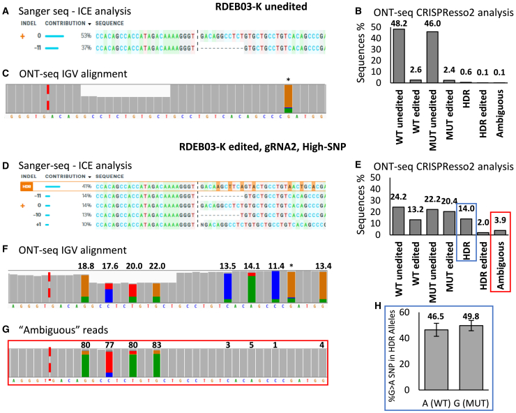
Gene editing facilitated by homology-directed repair (HDR) holds great potential for treating monogenetic disorders such as recessive dystrophic epidermolysis bullosa (RDEB). However, low efficiency and variability between loci must be overcome for its widespread adoption into personalized therapies. To address these challenges, we developed a highly efficient and versatile gene editing strategy for RDEB that incorporates the small molecule inhibitor M3814 to enhance HDR. We focused on three RDEB causative COL7A1 mutations not previously targeted by existing gene therapies. Editing was achieved using Cas9-nuclease ribonucleoproteins with short single-stranded DNA donor templates, and outcomes were assessed with an Oxford Nanopore Technology sequencing analysis pipeline. We demonstrate precise genomic HDR rates of up to 75% of alleles in primary RDEB keratinocytes and 32% in fibroblasts. This approach restored collagen VII expression in up to 80% of keratinocytes within a bulk-edited population and resulted in correct collagen VII deposition in a 3D skin model. Additionally, at one locus we show that a dual Cas9-nickase strategy is less effective than Cas9-nuclease and prone to large on-target deletions. Our results demonstrate a significant advancement in the efficiency and consistency of HDR editing, potentially paving the way for more effective personalized gene therapies.

Keywords: CRISPR-Cas9; MT: RNA/DNA Editing; Oxford Nanopore Technology sequencing; dual-nickase; epidermolysis bullosa; gene therapy; homology-directed repair; skin engineering.


Conflict of interest statement

The authors declare no conflict of interests.

Figures

None
Graphical abstract
Figure 1
Figure 1
HDR-mediated editing strategy for three COL7A1 mutations (A) Annotation of COL7A1 mutations (transcript GenBank: NM_000094.3) from three RDEB donors, with targeted alleles highlighted in red. (B) Immunohistochemistry analysis of skin punch biopsy from one healthy and three RDEB donors, showing DAPI-stained nuclei in blue and C7 in red. Scale bar (bottom right), 100 μM. (C–E) HDR-based editing reagent design for three heterozygous COL7A1 mutations. The approximate mutation locations within COL7A1 exons are shown. Both WT and MUT alleles are depicted for each locus. For RDEB01 and RDEB03 (C and D), deletion mutations are denoted with dashed lines. For RDEB05 (E), the substitution mutation is marked in bold and indicated by a gray asterisk. Black lines represent gRNA binding sites, with cut sites shown as black arrow heads and allele specificity is indicated. HDR templates are displayed below, showing mutation correction as underlined blue bases, gRNA blocking mutations as red bases (with asterisks), and bridging SNPs as green bases (with asterisks).
Figure 2
Figure 2
ONT-seq enables robust analysis of on-target editing events (A–C) Analysis of bulk DNA from unedited RDEB03 keratinocytes. (A) ICE analysis of Sanger sequencing data. Allele frequencies are displayed, where 0 and −11 represent the WT and MUT (c.8698_8708del) alleles, respectively. The gRNA cut site is marked by a vertical dashed line. (B) Allele frequencies reported as percentages from CRISPResso2 analysis of ONT-seq data. Categories include: WT unedited, WT edited, MUT unedited, MUT edited, HDR, HDR edited, and ambiguous. WT refers to the heterozygous allele, while MUT corresponds to the allele containing the c.8698_8708del mutation. Ambiguous reads align equally to multiple alleles and are given a distinct category. (C) Raw ONT-seq reads are aligned to the reference genome and visualized using the IGV. The histogram summarizes the depth of alignment for each base, with deletions represented by a proportional decrease in gray bar height and substitutions that exceed a 10% frequency threshold highlighted by a change in color. The Cas9 cut site is denoted by a dashed red line, and a frequent nanopore sequencing error is marked with an asterisk. (D–G) Analysis of bulk DNA from RDEB03 keratinocytes edited with gRNA2 (non-allele-specific) and the Full-SNP HDR template. (D) As described in (A), with HDR events highlighted in orange. (E) As described in (B). (F) As described in (C), with HDR-induced silent mutations highlighted, and their frequencies indicated above. (G) Reads classified as ambiguous by CRISPResso2 in panel E (boxed in red) were extracted, aligned to the reference genome, and visualized in IGV. (H) Reads that were classified as HDR by CRISPResso2 in (E) (boxed in blue) were extracted and the allelic frequency of a nearby heterozygous SNP (c.8780G>A) present in cis with the WT allele were calculated (n = 4). Adenine (A) is on the WT allele, and guanine (G) on the MUT allele.
Figure 3
Figure 3
High rates of on-target HDR-mediated gene editing is achieved in skin cells derived from donors with RDEB Keratinocytes and fibroblasts, derived from three donors with RDEB, were gene edited with Cas9-nuclease and HDR templates as indicated. HDR rates were measured by ONT-seq with CRISPResso2 analysis. (A) Calculated HDR rates for the MUT allele and (B) total indel rates are shown from bulk RDEB03-Ks edited with one of two sgRNAs as shown, the RDEB03 high-SNP template, with or without M3814. Data are presented as mean ± SD (n = 3, ∗p < 0.05, ∗∗p < 0.01, ∗∗∗p ≤ 0.001). (C) HDR rates in RDEB03 fibroblasts (RDEB03-Fs) edited with one of two sgRNAs and the RDEB03 high-SNP template and with or without M3814 (n = 2 for gRNA1, n = 3 for gRNA2). (D) Screen capture of CRISPResso2 allele alignment showing indel distribution in RDEB03-Ks edited with RDEB03 gRNA2 and the high-SNP template, with or without M3814. This alignment only includes reads which align closest to the c8698_8708del allele (MUT-unedited and MUT-edited) (see Figure 2). The reference allele is at the top, with indels below in decreasing frequency (displayed to <1%). The gRNA cut site is marked by a vertical dashed black line. Substitutions are in bold, insertions are outlined in red, and deletions are marked as dots. (E) Calculated HDR rates for the MUT allele in bulk keratinocytes derived from three RDEB donors comparing three HDR templates for each donor. The data are presented as an average percentage rate for each HDR-template, with individual donor rates indicated (RDEB03 n = 2, RDEB01 and RDEB05 n = 1). (F) HDR rates in RDEB03 fibroblasts edited with three different HDR templates (n = 2). (G) CRISPResso2 analysis output from low passage (P2) RDEB03 keratinocytes edited with gRNA2 and low-SNP HDR template (n = 1).
Figure 4
Figure 4
COL7A1 transcription and C7 protein analysis in HDR-edited RDEB cells (A) COL7A1 transcript analysis: ONT-seq data of COL7A1 transcripts aligned to the reference genome, displayed as coverage tracks in IGV. Rows from top to bottom: Transcripts from RDEB03-Ks edited with either high-SNP or low-SNP HDR templates, unedited RDEB03-Ks (UN), and normal human keratinocytes (NHKs). The target 11bp deletion (c.8698_8708del) is indicated with a black asterisk, the green asterisk indicates the non-target mutation (c.8780G>A) on the opposite allele. An aberrant splice site is marked by a red arrow. (B) Immunocytochemistry analysis of C7 (in red) in unedited and edited RDEB03-Ks and NHKs as labeled. RDEB03-Ks were edited with low-SNP template with M3814. Nuclei are stained with DAPI (blue). Nuclear FOXM1 staining (green) marks potential progenitor cells. Merged images are shown on the right-hand panel, where cyan represents merged DAPI (blue) and FOXM1 (green) nuclear stains. Scale bar (bottom right), 100 μM. On the far right, images are enlarged with white arrows indicating cells co-stained for FOXM1 and C7. (C) Flow cytometry analysis of C7 expression in HDR-edited RDEB03-Ks. Plots show C7 expression on the x axis and side scatter (SSC-A) on the y axis. The percentage of C7-positive cells is indicated in red text. (Top row) NHKs, unedited RDEB03-Ks (UN), and high-SNP edited RDEB03-Ks without M3814. (Bottom row) RDEB03-Ks edited with low-SNP, med-SNP, and high-SNP HDR templates with M3814. (D) Linear correlation (R2 = 0.9782) between HDR efficiency as assessed by ONT-seq/CRISPResso2 (x axis) and the percentage of C7-positive cells as assessed by flow cytometry (y axis). The y intercept set to 0. (E) The average mean fluorescence intensity (MFI) of the intracellular C7 signal above the baseline of unedited RDEB03 cells in NHKs (n = 3), RDEB03-Ks edited with the Low-SNP (n = 3), Med-SNP (n = 1), and High-SNP templates (n = 3).
Figure 5
Figure 5
Edited RDEB cells demonstrate normal deposition of C7 to the BMZ in 3D SEs Representative images are shown from sections derived from 3D SEs (fibroblasts and keratinocytes) generated from unedited RDEB03 cells (A–C), edited RDEB03 cells (D–F) or NHCs (G–I). H&E staining is shown in (A, D, and G). Immunohistochemistry staining of C7 (red) and nuclei stained with DAPI (blue) is shown in (B, E, and H). C7 alone is shown in (C, F, and I). Scale bars, 100 μM.
Figure 6
Figure 6
Evaluation of dual-nickase editing in RDEB03 keratinocytes (A) The dual-nickase based HDR-editing strategy for mutation c.8698_8708del present in RDEB03. Deleted bases are depicted with dashed lines. Black lines represent gRNA binding sites, with cut sites shown as black arrow heads. HDR templates are displayed below, showing mutation correction as underlined blue bases, gRNA blocking mutations as red bases (with asterisks), and bridging SNPs as green bases (with asterisks). (B) Representative ONT-seq data of dual-nickase RDEB03-Ks edited with or without M3814 as indicated, aligned to the reference genome and visualized using IGV. HDR templates and rates of genomic HDR, as assessed by ONT-Seq/Crispresso2 analysis, are shown. HDR-induced SNPs are indicated by colored lines on the IGV plots. A large 350-bp deletion is marked by a horizontal black arrow. The target 11 bp deletion (c.8698_8708del) is indicated with a black asterisk, and a non-target mutation (c.8780G>A) in cis with the WT allele is marked with a red asterisk. (C) Dual-nickase genomic HDR rates in bulk edited RDEB03-Ks edited with or without M3814, shown as mean ± SD (n = 3, p = 0.03). (D) Dual-nickase genomic HDR rates comparing three HDR templates with M3814. Percentages for perfect HDR (HDR), imperfect HDR, and total editing efficiency as shown (calculated as total HDR + indels, n = 2). (E) ONT-Seq/Crispresso2 analysis of cDNA derived from dual-nickase RDEB03-Ks edited with the High2-SNP template with or without M3814 (n = 1). (F) Immunocytochemistry analysis of C7 (red) in unedited (UN) and dual-nickase edited (edited) RDEB03-Ks and normal human keratinocytes (NHKs) as labeled. Nuclei are stained with DAPI (blue). Merged images are shown on the right-hand panel. Scale bar, 100 μM. (G) Flow cytometry analysis of C7 expression in unedited RDEB03-Ks (RDEB03 UN - left), RDEB03-Ks edited with High2-SNP with M3814 (middle), and NHKs (right). Plots show C7 on the x axis and side scatter (SSC-A) on the y axis. The percentage of C7-positive cells is indicated in red text.

References

    1. Bardhan A., Bruckner-Tuderman L., Chapple I.L.C., Fine J.D., Harper N., Has C., Magin T.M., Marinkovich M.P., Marshall J.F., McGrath J.A., et al. Epidermolysis bullosa. Nat. Rev. Dis. Prim. 2020;6:78. doi: 10.1038/S41572-020-0210-0. - DOI - PubMed
    1. Tang J.Y., Marinkovich M.P., Lucas E., Gorell E., Chiou A., Lu Y., Gillon J., Patel D., Rudin D. A systematic literature review of the disease burden in patients with recessive dystrophic epidermolysis bullosa. Orphanet J. Rare Dis. 2021;16:175. doi: 10.1186/S13023-021-01811-7. - DOI - PMC - PubMed
    1. Bischof J., Hierl M., Koller U. Emerging Gene Therapeutics for Epidermolysis Bullosa under Development. Int. J. Mol. Sci. 2024;25 doi: 10.3390/IJMS25042243. - DOI - PMC - PubMed
    1. Doudna J.A., Charpentier E. The new frontier of genome engineering with CRISPR-Cas9. Science. 2014;346 doi: 10.1126/SCIENCE.1258096. - DOI - PubMed
    1. Lee A.B.C., Tan M.H., Chai C.L.L. Small-molecule enhancers of CRISPR-induced homology-directed repair in gene therapy: A medicinal chemist’s perspective. Drug Discov. Today. 2022;27:2510–2525. doi: 10.1016/J.DRUDIS.2022.06.006. - DOI - PubMed

LinkOut - more resources