. 2025 Feb 1;28(2):111937.
doi: 10.1016/j.isci.2025.111937. eCollection 2025 Feb 21.

NFAT single-deficient murine T cells reduce the risk of aGvHD while controlling cytomegalovirus infection

Affiliations

NFAT single-deficient murine T cells reduce the risk of aGvHD while controlling cytomegalovirus infection

Nadine Hundhausen et al. iScience. .

Abstract

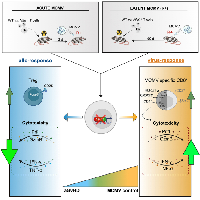
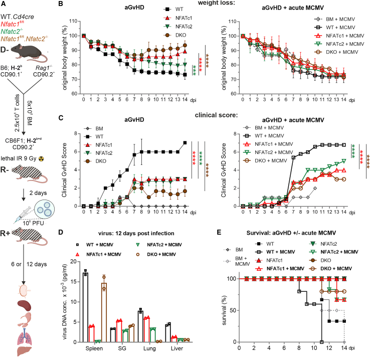
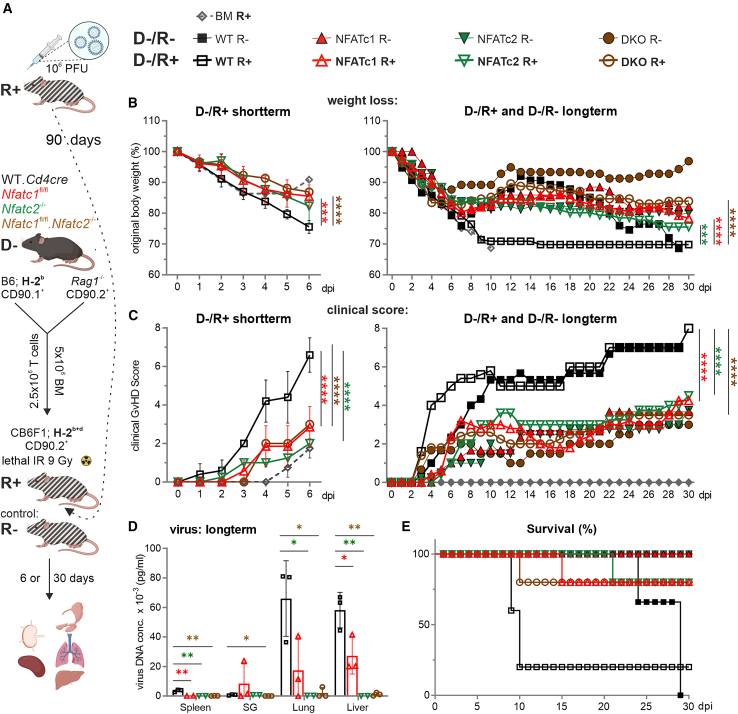
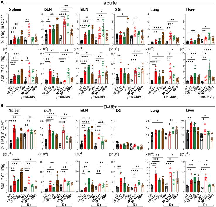
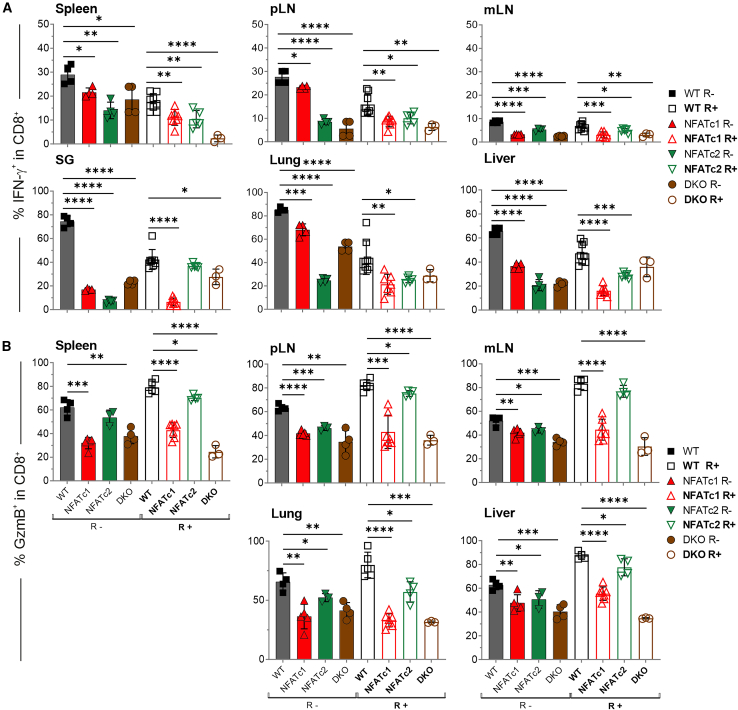
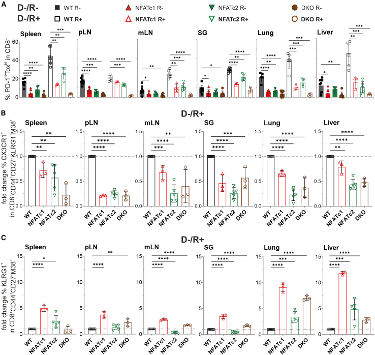
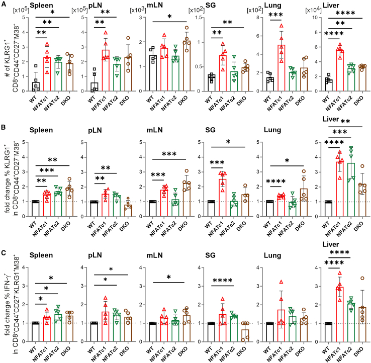
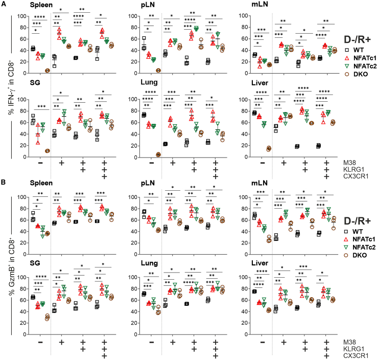
NFAT is a family of transcription factors whose activation is inhibited by calcineurin inhibitors (CNIs). In allogeneic hematopoietic stem cell transplantation (allo-HCT), CNIs are employed to prevent and treat graft-versus-host disease (GvHD). Unfortunately, control of cytomegalovirus (CMV), which exacerbates clinical outcomes, is simultaneously lost. Since single NFAT deficiency in T cells ameliorates GvHD in our major mismatch model, we investigated whether protection is maintained during CMV infection. Reassuringly, NFAT-deficient T cells still improved GvHD upon acute CMV infection and after allo-HCT in latently CMV-infected mice, showing reduced proinflammatory and cytotoxic potential. In sharp contrast, CMV-specific NFAT-deficient CD8+ inflated memory T cells expanded more and with higher levels of interferon gamma (IFN-γ) and GzmB expression, effectively controlling CMV. Notably, NFAT-deficient inflated memory T cells could migrate to non-lymphoid tissues and fight CMV. Therefore, CMV infection does not interfere with the protective effect of NFAT inhibition to attenuate GvHD while allowing an anti-CMV response.

Keywords: Biological sciences; Immunology; Microbiology; Natural sciences; Virology.


Conflict of interest statement

The authors declare no competing interests.

Figures

None
Graphical abstract
Figure 1
Figure 1
Co-transfer of single-deficient NFATc1 and NFATc2 or double-deficient NFATc1c2 T cells in a haploidentical major mismatch model limits signs of aGvHD even after an acute MCMV infection (A) Experimental in vivo set up of the induction of aGvHD in a haploidentical mouse model due to an H-2b → H-2b+d transfer with WT or NFAT-deficient T cells along with BM, preceded by lethal irradiation (9 Gy) of CB6F1 recipients. Two days post-transplantation, mice were acutely infected with 1 × 106 PFU of WT-MCMV Smith strain. Six or twelve days post-infection, ex vivo analyses were conducted on spleen, peripheral lymph nodes (pLNs), mesenteric lymph nodes (mLNs), salivary gland (SG), lungs, and liver. (B) Weight changes were determined over 14 days post-irradiation (dpi) in mice without (aGvHD) or with an acute MCMV infection (aGvHD + acute MCMV). Mice were evaluated every day and weight loss was calculated considering day 0 weight as 100%; n ≥ 5. (C) Clinical scores were determined over 14 days. (B and C) Two-way ANOVA and Tukey’s multiple comparisons test (∗∗∗p < 0.001, ∗∗∗∗p < 0.0001), mean ± SEM, n ≥ 5, two independent experiments. (D) Virus DNA concentration of acutely infected mice 14 dpi in indicated organs was determined by RT-PCR. (E) Survival over 14 days.
Figure 2
Figure 2
NFAT-deficient T cells can still provide protection when transferred into latently infected recipients (A) Experimental in vivo set up. Latent infected CB6F1 recipient (90 days prior transplantation) with WT-MCMV Smith strain. Acute GvHD was initiated by transferring H-2b donor T cells together with BM cells into H-2b+d recipients, preceded by lethal irradiation (9 Gy) of CB6F1 recipients. Six or thirty days post-transplantation, ex vivo analyses were conducted on various tissues. Symbols representing studied genotypes in only aGVHD (D−/R−) and latently infected CB6F1 recipients (D−/R+). (B) Weight loss and (C) clinical scores of GvHD-induced mice in latently infected recipients were monitored daily for short (6 dpi) or long term (30 dpi). Two-way ANOVA and Tukey’s multiple comparisons test (∗∗∗p < 0.001, ∗∗∗∗p < 0.0001), mean ± SEM, n ≥ 5, two independent experiments. (D) Virus DNA concentration 30 dpi, determined by RT-PCR, mean ± SD, n = 3, unpaired Student’s t test (∗p < 0.05, ∗∗p < 0.005). (E) Survival was monitored daily for 30 days.
Figure 3
Figure 3
Independent from MCMV infection, NFAT-deficient Tregs are significantly enriched during GvHD (A) Frequency and absolute cell numbers—calculated back to all cells of the particular organ—of donor CD90.1+CD4+CD25+Foxp3+ T cells 8 days post-transplantation in non-infected or acutely infected (MCMV+) recipients (spleen, pLN, mLN, salivary gland [SG], lungs, and liver). Student’s two-tailed t test (∗p < 0.05, ∗∗p < 0.005, ∗∗∗p < 0.001, ∗∗∗∗p < 0.0001), mean ± SEM, n ≥ 4, three independent experiments. (B) Flow cytometric analyses of indicated organs harvested 6 days post-transplantation from mice without or with latent MCMV infection (R+). Percentage of CD25+Foxp3+ Tregs within donor CD90.1+CD4+ T cells and absolute cell numbers. (A and B) Student’s two-tailed t test (∗p < 0.05, ∗∗p < 0.005, ∗∗∗p < 0.001, ∗∗∗∗p < 0.0001), mean ± SEM, n ≥ 3, two independent experiments.
Figure 4
Figure 4
The reduction of proinflammatory cytokines and cytotoxic molecules in NFAT-deficient CD8+ T cells during aGvHD remains significant regardless of a concurrent MCMV infection (A and B) Flow cytometric analyses of recipient mice without (R−) and with latent MCMV infection (R+) were conducted 6 days post-transplantation. (A) Frequency of donor CD90.1+CD8+IFN-γ+ T cells in the depicted organs, n ≥ 3. (B) Frequency of donor CD90.1+CD8+GzmB+ T cells. (A and B) Student’s two-tailed t test (∗p < 0.05, ∗∗p < 0.005, ∗∗∗p < 0.001, ∗∗∗∗p < 0.0001), mean ± SEM, n ≥ 3, two independent experiments.
Figure 5
Figure 5
NFAT deficiency reduces CD8+ T cell exhaustion while promoting terminal differentiation of MCMV-specific CD8+ T cells after allo-HCT (A) Percentage of PD-1hiTOXhi in CD8+ T cells in only aGVHD and latently infected CB6F1 recipients (+MCMV) 6 days post-transplantation, n ≥ 3. (B) Percentages of the chemokine receptor CX3CR1+ and (C) type II transmembrane protein receptor KLRG1+ within MCMV-specific donor CD90.1+CD8+M38+ T cells was determined through tetramer staining and subsequent flow cytometry analysis. Fold change was calculated relative to WT. (A, B and C) Student’s two-tailed t test (∗p < 0.05, ∗∗p < 0.005, ∗∗∗p < 0.001, ∗∗∗∗p < 0.0001), mean ± SEM, n ≥ 3, two independent experiments.
Figure 6
Figure 6
NFAT deficiency in CD8+ T cells is sufficient to increase the frequency of IFN-γ+ MI T cells (A–C) Flow cytometric analyses of allotransplanted WT CD4+ T cells and WT or NFAT-deficient CD8+ T cells (1:1 ratio) 6 days post-transplantation. (A) Absolute numbers and (B) fold change of the percentage of KLRG1+ within MCMV-specific donor CD90.1+CD8+CD44+CD27M38+ T cells was determined through tetramer staining and subsequent flow cytometry analysis. Fold change was calculated specifically on WT. (C) Frequency of IFN-γ+ within MCMV-specific donor CD90.1+CD8+CD44+CD27M38+ T cells (in vitro restimulated). (A, B and C) Student’s two-tailed t test (∗p < 0.05, ∗∗p < 0.005, ∗∗∗p < 0.001, ∗∗∗∗p < 0.0001), mean ± SEM, n = 5.
Figure 7
Figure 7
After allo-HCT in latently MCMV-infected mice, NFAT-deficient MI T cells present with a higher frequency of IFN-γ+ and GzmB+ cells Percentage of (A) IFN-γ+ and (B) GzmB+ cells of either M38 or MCMV-specific M38+, M38+KLRG1+, and M38+KLRG1+CX3CR1+ CD8+ T cells after 6 days post-transplantation and in vitro restimulation. (A and B) Student’s two-tailed t test (∗p < 0.05, ∗∗p < 0.0051, ∗∗∗p < 0.001, ∗∗∗∗p < 0.0001), mean ± SEM, n = 3.

Similar articles

References

    1. Ferrara J.L.M., Levine J.E., Reddy P., Holler E. Graft-versus-host disease. Lancet. 2009;373:1550–1561. - PMC - PubMed
    1. Zeiser R., Blazar B.R. Acute Graft-versus-Host Disease - Biologic Process, Prevention, and Therapy. N. Engl. J. Med. 2017;377:2167–2179. doi: 10.1056/NEJMra1609337. - DOI - PMC - PubMed
    1. Vaeth M., Bäuerlein C.A., Pusch T., Findeis J., Chopra M., Mottok A., Rosenwald A., Beilhack A., Berberich-Siebelt F. Selective NFAT targeting in T cells ameliorates GvHD while maintaining antitumor activity. Proc. Natl. Acad. Sci. USA. 2015;112:1125–1130. doi: 10.1073/pnas.1409290112. - DOI - PMC - PubMed
    1. Vaeth M., Feske S. NFAT control of immune function: New Frontiers for an Abiding Trooper. F1000Res. 2018;7:260. doi: 10.12688/f1000research.13426.1. - DOI - PMC - PubMed
    1. Vaeth M., Schliesser U., Müller G., Reissig S., Satoh K., Tuettenberg A., Jonuleit H., Waisman A., Müller M.R., Serfling E., et al. Dependence on nuclear factor of activated T-cells (NFAT) levels discriminates conventional T cells from Foxp3+ regulatory T cells. Proc. Natl. Acad. Sci. USA. 2012;109:16258–16263. doi: 10.1073/pnas.1203870109. - DOI - PMC - PubMed

LinkOut - more resources