. 2024 Dec 4;7(3):101294.
doi: 10.1016/j.jhepr.2024.101294. eCollection 2025 Mar.

Molecular MRI of T-cell immune response to cryoablation in immunologically hot vs. cold hepatocellular carcinoma

Affiliations

Molecular MRI of T-cell immune response to cryoablation in immunologically hot vs. cold hepatocellular carcinoma

Jessica Gois Santana et al. JHEP Rep. .

Abstract

Background & aims: Increasing enthusiasm around integrating locoregional therapy with systemic immunotherapy in primary liver cancer underscores the need for non-invasive imaging biomarkers. In this study, we aimed to establish advanced molecular MRI tools for monitoring T-cell responses to cryoablation in murine models, distinguishing between immunologically "hot" and "cold" hepatocellular carcinoma (HCC).

Methods: Immunocompetent 7-10-week-old C57BL/6J and BALB/cJ mice (n = 18 each) received carbon tetrachloride for 12 weeks to induce cirrhosis. Intrinsically immunogenic Hepa1-6 ("hot") and non-immunogenic TiB75 ("cold") cells were orthotopically implanted into C57BL/6 or BALB/c mice, respectively, to generate focal HCC lesions. After one week, animals were randomly assigned to (A) partial cryoablation (pCryo) (1.2 mm cryoprobe, -40 °C) or (B) no treatment (n = 8 per group and tumor type). Gadolinium 160 (160Gd)-labeled CD8+ antibody was administered intravenously either 1 week after tumor induction (control) or 1-week post (pCryo) (treatment). T1-weighted MRI scans were performed using a 9.4 T MRI scanner. Radiological-pathological correlation included imaging mass cytometry and immunohistochemistry.

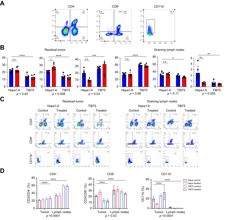
Results: pCryo-treated Hepa1-6 tumors displayed peritumoral ring enhancement on T1-weighted MRI with 160Gd-CD8, correlating with imaging mass cytometry signal patterns. Untreated Hepa1-6 tumors lacked such enhancement. Radiological-pathological correlation confirmed significantly increased tumor-infiltrating CD8+ T lymphocytes in pCryo Hepa1-6 tumors compared with untreated tumors (p <0.001), and a stronger local response compared with systemic lymph nodes (p = 0.0415). Increased T-lymphocyte infiltration was not observed in TiB75 tumors, as indicated by MRI and histopathology.

Conclusion: pCryo induced increased T-cell infiltration in Hepa1-6 tumors compared to TiB75 tumors. T1-weighted MRI, following 160Gd-CD8 antibody administration, reproducibly detected the ablation-induced changes. These findings encourage further investigation of MRI-based molecular imaging biomarkers to assess immune responses to local tumor therapies.

Impact and implications: This study successfully established reliable MR-based molecular imaging tools to visualize CD8+ anti-tumor specific T-cell infiltration following partial cryoablation (pCryo) in murine tumor models. The study's significance lies in advancing our understanding of immune responses within induced cirrhosis and distinguishing between "hot" and "cold" tumor phenotypes. The findings not only build upon previous proof-of-principle data but also extend this technology to include different immune cell types in hepatocellular carcinoma. The study reveals that pCryo may exert specific effects on the tumor microenvironment, augmenting the anti-tumor immune response in immunogenic tumors while displaying a weaker local effect in non-immunogenic tumors.

Keywords: Cryoablation; Immune Response; Immuno-metabolic interplay; In vivo Imaging; Magnetic Resonance Imaging.


Conflict of interest statement

The authors declare no conflicts of interest that pertain to this work. Please refer to the accompanying ICME disclosure forms for further details.

Figures

Image 1
Graphical abstract
Fig. 1
Fig. 1
Schematic view of establishing orthotopic HCC model in cirrhotic background. C57BL/6J and BALB/cJ mice (n = 18 each) were subjected to escalating doses of oral CCl4 three times per week for a total of 12 weeks. The dosing schedule consisted of 0.875 ml/kg (1st dose, week 1), 1.75 ml/kg (2nd to 9th dose, week 1 – 4), 2.5 ml/kg (10th to 23rd dose, week 4 – 8), and 3.25 ml/kg (after week 8). Upon completion of the CCl4 regimen, 1.5 x 10⁶ Hepa1-6 or TiB75 cells were orthotopically implanted in their respective syngeneic mouse strains, C57BL/6J and BALB/cJ. Animals were sacrificed and tumor tissues were histopathologically evaluated 7 days post-tumor implantation. CCl4, carbon tetrachloride; HCC, hepatocellular carcinoma.
Fig. 2
Fig. 2
Illustrative representation of experimental design for MR-based imaging of T lymphocytes in immunophenotypically diverse HCC mouse models. (A) Establishment of immunologically “hot” and “cold” syngeneic murine HCC models in toxin-induced underlying cirrhosis. (B) Animals were randomly assigned to receive pCryo on day 7 post- HCC tumors in both syngeneic models. Systemic administration of 2.4 mg/kg 160Gd-CD8 (n = 8/each) was given 7 days post-pCryo. (C) 24 h after systemic administration of 160Gd-CD8, animals were subjected to T1-weighted MRI, revealing hyperintensity in the peritumoral rim and successful in vivo labeling of tumor-infiltrating CD8+ T cells. Radiological-pathological correlation was confirmed with immunohistochemistry and imaging mass cytometry. 160Gd, gadolinium 160; CCl4, carbon tetrachloride; HCC, hepatocellular carcinoma; NK, natural killer; pCryo, partial cryoablation; Treg, regulatory T.
Fig. 3
Fig. 3
Syngeneic murine HCC tumor in cirrhotic background. (A) Gross pathology of CCl4-treated mice showing anterior images of orthotopic HCC tumor in a cirrhotic liver (from Hepa1-6-tumor-bearing mouse). (B) Histopathological evaluation of cirrhosis assessed with Masson’s trichome. (C) Quantification as the ratio of the fibrosis area to the total sample area, expressed in pixels in Masson’s trichome staining. Comparison of two variables was performed with an unpaired Student’s t test with Welch’s correction. ∗p <0.01, ∗∗∗p <0.0001. Statistical analyses were performed using GraphPad Prism version 10.0.0.
Fig. 4
Fig. 4
Partial cryoablation enhances cytotoxic CD8+ T-cell response in immunogenic tumor microenvironment. (A) IHC staining of CD8+ marker at 7 days post-tumor implantation (control, left column) and at 7 days post-pCryo (treated, right column) in both immunogenic (top row) and non-immunogenic (bottom row) HCC cell lines. The boxes represent selected area for 20X magnification (B) CD8+ stain quantification in residual tumor tissue. Percentage of positive CD8+ = Npositive/Ntotal. Comparison of two variables was performed with an independent non-parametric Mann-Whitney U or Kruskal-Wallis test. ∗p <0.01, ∗∗∗p <0.0001. Statistical analyses were performed using GraphPad Prism version 10.0.0. IHC, immunohistochemistry; HCC, hepatocellular carcinoma; NL, normal liver; pCryo, partial cryoablation; T, residual tumor.
Fig. 5
Fig. 5
Local and systemic immunomodulatory effects of partial cryoablation are tumor type dependent. Immune cell populations in draining lymph nodes and in the tumor microenvironment. (A) Representative flow cytometry staining for CD3+/CD4+, CD3+/CD8+ co-expression in CD90 gate, and CD11b+ cells (spleen sample). (B) Percentages of CD3+/CD4+, CD3+/CD8+, and CD11b+ cells in tumor and lymph node samples from Hepa1-6 and TiB75 tumor-bearing mice, without (control, blue color) and with cryoablation (treated, red color). (C–D) Comparison of immune cell population per group in both tumor and lymph node tissues. p values represent significance of interaction between treatment and tissue type. Data are represented as mean ± SEM, with differences assessed using two-way ANOVA followed by Sidak's multiple comparisons test. Statistical significance: ∗p <0.01, ∗∗∗p <0.001, ∗∗∗∗p <0.0001.
Fig. 6
Fig. 6
In vivo molecular imaging of peritumoral infiltrating cytotoxic T cells using 160Gd–labeled CD8+ antibodies. Radiological-pathological correlation of in vivo labeling of tumor-infiltrating cytotoxic T cells in both immunogenic (top row) and non-immunogenic (bottom row) HCC models. Immunohistochemistry assessment of CD8+ before and after pCryo (two left columns) shows enhanced T-cell tumor infiltration in immunogenic HCC model. A baseline T1-weighted coronal MRI scan of both tumor types (∗)7 days post-pCryo with 0.1 mmol/kg intravenous Dotarem contrast (third column). Peritumoral rim enhancement (arrows) on T1-weighted coronal MRI scan (repetition time, 1,500 ms; echo time, 5.46 ms) obtained 24 h after systemic administration of 160Gd-labeled CD8+ antibody (fourth column) indicates peritumoral cell infiltrate in immunogenic HCC tumors, as observed histologically. Bright field image from ex vivo IMC from animals (fifth column) after ablation of specific in vivo labeling of infiltrating T cells by 160Gd-CD8+. 160Gd, gadolinium 160; HCC, hepatocellular carcinoma; IMC, imaging mass cytometry; LRT, locoregional treatment (pCryo); pCryo, partial cryoablation; T, tumor.
Fig. 7
Fig. 7
160Gd metal concentration in the peritumoral rim by imaging mass cytometry. (A) Correlation of CD8+ positive signal on IHC and spatial localization of 160Gd metal distribution using IMC. (B) Histopathological analysis of ex vivo IMC and IHC from 7 days pCryo animals following 24 h post-160Gd-CD8+ antibody intravenous injection. Bright field image from IMC (left column) and IHC staining of CD8+ marker (right column). The boxes in upper row of B indicates the area of magnification for the bottom row. 160Gd, gadolinium 160; IHC, immunohistochemistry; IMC, imaging mass cytometry; pCryo, partial cryoablation.

References

    1. Chidambaranathan-Reghupaty S., Fisher P.B., Sarkar D. Hepatocellular carcinoma (HCC): epidemiology, etiology and molecular classification. Adv Cancer Res. 2021;149:1–61. - PMC - PubMed
    1. Leong T.Y., Leong A.S. Epidemiology and carcinogenesis of hepatocellular carcinoma. HPB (Oxford) 2005;7(1):5–15. - PMC - PubMed
    1. Chen Y.S., Lian L.F., Xu Y.H., et al. [Association of glycosylated hemoglobin level at admission with outcomes of intracerebral hemorrhage patients] Zhonghua Liu Xing Bing Xue Za Zhi. 2019;40(11):1445–1449. - PubMed
    1. Noor M.T., Manoria P. Immune dysfunction in cirrhosis. J Clin Transl Hepatol. 2017;5(1):50–58. - PMC - PubMed
    1. Cardarelli-Leite L., Hadjivassiliou A., Klass D., et al. Current locoregional therapies and treatment strategies in hepatocellular carcinoma. Curr Oncol. 2020;27(Suppl 3):S144–S151. - PMC - PubMed

LinkOut - more resources