Review
. 2025 May;266(1):9-25.
doi: 10.1002/path.6407. Epub 2025 Mar 3.

Cellular origins of mucinous ovarian carcinoma

Affiliations
Review

Cellular origins of mucinous ovarian carcinoma

Nicola S Meagher et al. J Pathol. 2025 May.

Abstract

Mucinous ovarian carcinoma (MOC) is a rare histotype of epithelial ovarian cancer. Its origins are obscure: while many mucinous tumours in the ovary are metastases from the gastrointestinal tract, MOC can occur as an ovarian primary; however, the cell of origin is not well established. In this review we summarise the pathological, epidemiological, and molecular evidence for the cellular origins of MOC. We propose a model for the origins of the various tumours of the ovary with mucinous differentiation. We distinguish Müllerian from gastrointestinal-type mucinous differentiation. A small proportion of the latter arise from teratoma and a distinct terminology has been proposed. Other gastrointestinal mucinous tumours are associated with Brenner tumours and arise from their associated benign lesions, Walthard nests. The remaining mucinous tumours develop either through mucinous metaplasia in established Müllerian tumours or with even greater plasticity through gastrointestinal metaplasia of epithelial or mesothelial ovarian inclusions. This model remains to be validated and mechanistically understood and we discuss future research directions. © 2025 The Author(s). The Journal of Pathology published by John Wiley & Sons Ltd on behalf of The Pathological Society of Great Britain and Ireland.

Keywords: cell of origin; mucinous; ovarian cancer.


Figures

Figure 1
Figure 1
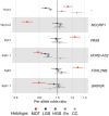
Top loci associated with the mucinous histotype identified in GWAS studies. The SNPs at loci 19q13.1, 2q13, and 3q23 are unique to mucinous ovarian tumours (MOT, including mucinous ovarian carcinoma (MOC) and mucinous borderline ovarian tumours, MBT); while others are shared to some extent with the other histotypes such as: LGS (low‐grade serous ovarian carcinoma); HGS (high‐grade serous ovarian carcinoma); En (endometrioid ovarian carcinoma); CC (clear‐cell ovarian carcinoma). Odds ratio shown is for the minor allele. The candidate genes shown are those with the highest combined score for the gene, transcriptome and chromatin analyses performed by Dareng et al [45]. 17q SNPs for MOC were not included in that analysis.
Figure 2
Figure 2
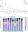
Mutations in MOC. (A) Frequency of genetic events in mucinous borderline tumours (MBT) and mucinous ovarian carcinoma (MOC). Each dot represents the frequency of the genetic event in one of the studies listed in Table 1. Not all studies had data for all genes. Amp = amplified, HD = homozygous deletion, mut = mutated; mutated only for other genes. Box and whiskers show median, quartiles and 1.5× interquartile range. (B) Frequency of KRAS variant types in selected cancers from TCGA Pan‐Cancer [79], mucinous ovarian carcinoma (MOC) [5] and low‐grade serous ovarian carcinoma (LGSC)/serous borderline ovarian tumours (SBT) [80, 81, 82]. Numbers of variants are given above each bar.
Figure 3
Figure 3
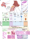
Potential cells of origin for MOC. (A) Anatomic depiction of the gynaecologic tract, showing relevant cell types. (B) A subset of mucinous tumours at the ovary may develop from germ cells, with evidence for GI differentiation to arise via a teratoma intermediate. These become teratoma‐associated low‐grade or high‐grade ovarian mucinous neoplasms (LOMN and HOMN respectively). (C) Similarly, some mucinous tumours may arise from Brenner tumours, from cells of origin in Walthard nests (whose origin is itself unknown). (D) Entrapped fallopian tube (FT) epithelium (or possibly ovarian surface ‘epithelium’ (OSE), sub‐OSE stem cells or hilar stem cells form an inclusion cyst within the ovary, which may undergo metaplasia to include mucinous‐type cells (see H below). (E) Endometriosis can form in the ovary or elsewhere, and comprises endometrial‐type epithelium. (F) Key immunohistochemical markers related to each cell type from B–E. (G) Progression models for mucinous and mucinous‐associated tumours of the ovary. LOMN and HOMN form from teratomas; mucinous metaplasia occurring in Brenner tumours, FTE, inclusion cysts or benign cystadenomas leads to a benign gastrointestinal type epithelium that further evolves into mucinous borderline tumours (MBT) and MOC, driven by genetic events in KRAS and/or CDKN2A. Mutations in these genes in Müllerian‐type benign epithelium (e.g. inclusion cyst or serous cystadenoma with no mucinous metaplasia) leads to serous borderline tumours (SBT) and low‐grade serous carcinomas. The inclusion cyst pathway could therefore be a shared origin with serous tumours and could also result in rare mixed serous/mucinous tumours (Ser + Muc). Mucinous metaplasia could also occur in a benign tumour arising from endometriosis, leading to the ‘seromucinous’ type, more correctly an endometrioid tumour with mucinous differentiation (En + Muc). These tumours may have KRAS mutations but are also likely to carry mutations in ARID1A and/or PIK3CA. Of course, endometriosis with mutations in these latter two genes also leads to endometrioid ovarian carcinomas (EnOC). (H) Example of mucinous metaplasia of the FT showing the mucinous component lacks WT1 staining but maintains PAX8. (I) Example of benign cystadenoma, comprising both serous and mucinous‐type epithelia, the mucinous component lacks ER staining. Created with BioRender.com

References

    1. Lee KR, Scully RE. Mucinous tumors of the ovary: a clinicopathologic study of 196 borderline tumors (of intestinal type) and carcinomas, including an evaluation of 11 cases with ‘pseudomyxoma peritonei’. Am J Surg Pathol 2000; 24: 1447–1464. - PubMed
    1. Kurman RJ, Shih IM. The origin and pathogenesis of epithelial ovarian cancer: a proposed unifying theory. Am J Surg Pathol 2010; 34: 433–443. - PMC - PubMed
    1. Talia KL, Parra‐Herran C, McCluggage WG. Ovarian mucinous and seromucinous neoplasms: problematic aspects and modern diagnostic approach. Histopathology 2022; 80: 255–278. - PubMed
    1. McCluggage WG. Immunohistochemistry in the distinction between primary and metastatic ovarian mucinous neoplasms. J Clin Pathol 2012; 65: 596–600. - PubMed
    1. Cheasley D, Wakefield MJ, Ryland GL, et al. The molecular origin and taxonomy of mucinous ovarian carcinoma. Nat Commun 2019; 10: 3935. - PMC - PubMed

Publication types