. 2025 Dec;17(1):2474256.
doi: 10.1080/19490976.2025.2474256. Epub 2025 Mar 3.

Extracellular vesicles of Limosilactobacillus fermentum SLAM216 ameliorate skin symptoms of atopic dermatitis by regulating gut microbiome on serotonin metabolism

Affiliations

Extracellular vesicles of Limosilactobacillus fermentum SLAM216 ameliorate skin symptoms of atopic dermatitis by regulating gut microbiome on serotonin metabolism

Hyejin Choi et al. Gut Microbes. 2025 Dec.

Abstract

Atopic dermatitis (AD) is a globally prevalent chronic inflammatory skin disorder. Its pathogenesis remains incompletely understood, resulting in considerable therapeutic challenges. Recent studies have highlighted the significance of the interaction between AD and gut microbiome. In this study, we investigated the effects of probiotic-derived extracellular vesicles on AD. Initially, we isolated and characterized extracellular vesicles from Limosilactobacillus fermentum SLAM 216 (LF216EV) and characterized their composition through multi-omics analysis. Gene ontology (GO) and pathway analysis classified LF216EV proteins into biological processes, molecular functions, and cellular components. Importantly, specific abundance in linoleic, oleic, palmitic, sebacic, and stearic acids indicating upregulated fatty acid metabolism were observed by metabolomic analysis. Furthermore, featured lipid profiling including AcylGlcADG and ceramide were observed in LF216EV. Importantly, in an atopic dermatitis-like cell model induced by TNFα/IFNγ, LF216EV significantly modulated the expression of immune regulatory genes (TSLP, TNFα, IL-6, IL-1β, and MDC), indicating its potential functionality in atopic dermatitis. LF216EV alleviated AD-like phenotypes, such as redness, scaling/dryness, and excoriation, induced by DNCB. Histopathological analysis revealed that LF216EV decreased epidermal thickness and mast cell infiltration in the dermis. Furthermore, LF216EV administration reduced mouse scratching and depression-related behaviors, with a faster onset than the classical treatment with dexamethasone. In the quantitative real-time polymerase chain reaction (qRT-PCR) analysis, we observed a significant increase in the expression levels of htrb2c, sert, and tph-1, genes associated with serotonin, in the skin and gut of the LF216EV-treated group, along with a significant increase in the total serum serotonin levels. Gut microbiome analysis of the LF216EV-treated group revealed an altered gut microbiota profile. Correlation analysis revealed that the genera Limosilactobacillus and Desulfovibrio were associated with differences in the intestinal metabolites, including serotonin. Our findings demonstrate that LF216EV mitigates AD-like symptoms by promoting serotonin synthesis through the modulation of gut microbiota and metabolome composition.

Keywords: Atopic dermatitis; extracellular vesicle; gut microbiome; gut-skin axis; multi-omics analysis.


Conflict of interest statement

No potential conflict of interest was reported by the author(s).

Figures

Figure 1.
Figure 1.
Verification and quantification of LF216EV by SEM, TEM, and NTA analyses. (a) SEM image of LF216 and LF216-derived EV (LF16EV). Scale bar = 200 nm. (b) TEM image of LF216EV particles from LF216. Scale bar = 50 nm. (c) NTA analysis showing the particle range of LF216EV.
Figure 2.
Figure 2.
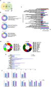
Characterization of LF216EV using proteomics and lipidomics. (a) Proteomic profiling of LF216 and LF216EV. Venn diagram displaying the number of LF216EV proteins identified in LF216. (b) functional classification of identified LF216EV proteins according to the cellular component, molecular function, and biological process. (c) gene ontology (GO) analysis of the identified LF216EV proteins. (d)lipidomic profiling of LF216 and LF216EV. (e) comparison of lipid profiles between LF216 and LF216EV. f relative comparison of the lipid content between LF216 and LF216EV.
Figure 3.
Figure 3.
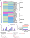
Characterization of LF216EV using metabolomics and miRNA sequencing analysis. (a) overview of the metabolic profiling of LF216 and LF216EV using heatmaps. (b) comparison of the fatty acid classes between LF216 and LF216EV. (c) analysis of the KEGG metabolic pathways related to significantly differential metabolites in LF216EV. (d) miRNA sequencing of LF216EV. Among a total of 771 identified miRNA, Dme-bantam and has-mir-9–1 are the most studied miRNAs among the annotated miRnas.
Figure 4.
Figure 4.
Evaluation of the function of LF216EV in an in-vivo model of C. elegans. (a) conditioning with LF216 and LF216EV for 24 h prolonged the lifespan of C. elegans CF512 fer-15(b26)II;fem-1(hc17) IV (fer-15;fem-1). (b) stimulating the immunity of C. elegans by pre-conditioning with LF216 and LF216EV for 24 h prolonged the lifespan of C. elegans exposed to S. aureus Newman. (c) stimulating the immunity of C. elegans by pre-conditioning with LF216 and LF216EV for 24 h prolonged the lifespan of C. elegans exposed to E. coli O157:H7 EDL933. d LF216EV induced the expression of the pmk-1 gene in C. elegans AY102 (p-vha-6::pmk-1::GFP +rol-6(su1006);pmk-1::GFP). Data are depicted as the mean values ± SEM of the samples. *p < 0.05, **p < 0.01, and ***p < 0.001 vs. The E. coli OP50 control.
Figure 5.
Figure 5.
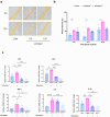
Confirming the functionality of LF216EV in ad-like cell models. (a, b) wound-healing effects of LF216EV in HaCaT cells. LF216EV significantly promoted wound healing of cells after 3 h of treatment. (c) effect of LF216EV on the expression of cytokines by HaCaT cells. HaCaT cells were pre-treated with LF216EV (109, 1010 particles/mL) and subsequently treated with TNF-α/IFN-γ for 24 h. TSLP, MDC, TNF-α, IL-6 and IL-1β mRNA expression of HaCaT cells, as detected by qRT-PCR. The mean of three independent experiments is represented by each bar. Data are depicted as mean values ± SEM of the samples. *p < 0.05, **p < 0.01, and ***p < 0.001 vs. The TNF-α/IFN-γ control.
Figure 6.
Figure 6.
LF216EV attenuates ad-like inflammation in a dncb-induced mouse model. (a) schematic diagram of the experiment. (c) Representative images of the dncb-treated skin area. (c) clinical score (redness, scaling/dryness, and excoriation) of each treatment group. (d) body weight of each treatment group. (e) H&E staining of the dorsal skin lesions from groups of mice and measurement of epidermal thickness. The black arrows indicate epidermal thickness. (f) TB staining and counting of mast cells of the dorsal skin lesions in a group of mice. Black arrows indicate mast cells infiltrating the skin. Scale bar = 50 µm. (g) measurement of spleen weight of each group. (h), i measurement of the total serum concentrations of IgE and histamine. j mRNA expression levels of inflammation-related genes (IFNγ, IL-13, IL-4, and TNF-α) using qRT-PCR. Data are depicted as mean values ± SEM of the samples. *p < 0.05, **p < 0.01, and ***p < 0.001 vs. The DNCB control.
Figure 7.
Figure 7.
LF216EV alleviates anxiety and depression-related behaviors in a mouse model of dncb-induced ad-like symptoms. (a) the scratch behavior of mice was observed for 10 min following sensitization. (b) LF216EV reduced the immobility time during the TST of mice. (c, d) LF216EV reduced anxiety- and depression-related behaviors in the OFT of mice. d LF216EV induced mouse activity in the OFT. Data are shown as mean values ± SEM of the samples. *p < 0.05, **p < 0.01, and ***p < 0.001 vs. The DNCB control.
Figure 8.
Figure 8.
Alteration of gut microbiota in a mouse model of ad-like symptoms by LF216EV. (a) alpha diversity of gut microbiota in each group using the Shannon index. (b, c) the similarity of the bacterial community structure of the gut microbiota using weighted and unweighted UniFrac distances. (d) phylum- and e genus-level gut microbiome compositions of treatment groups. (f) comparison between groups at eight representative genus levels. Data are shown as mean values ± SEM of the samples. *p < 0.05, **p < 0.01, and ***p < 0.001 vs. The DNCB control.
Figure 9.
Figure 9.
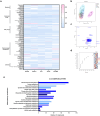
Alteration of gut metabolite changes in a mouse model of ad-like symptoms by LF216EV. (a) effects of LF216EV on the metabolites of gut microbiota in AD mice. Heat map illustrating the levels of gut metabolites. (b) Principal component analysis (PCA) of metabolites in AD mice. (c) differences in metabolites between treatments depicted using PCA loading plots. (d) variable importance in projection (VIP) score of metabolites of AD mice across treatment groups. (e) KEGG pathway classification of the increased metabolites in LF216EV.
Figure 10.
Figure 10.
LF216EV modulates the gut and skin serotonin genes to reduce ad-like symptoms in a mouse model. (a) measurement of the total serum concentrations of serotonin. (b) mRNA expression levels of serotonin receptor genes (htr2b and htr2c) in mice skin using qRT-PCR. (c) mRNA expression levels of serotonin related genes (htr2b, htr2c, sert, and tph-1) in mice intestine using qRT-PCR. Data are depicted as mean values ± SEM of the samples. *p < 0.05, **p < 0.01, and ***p < 0.001 vs. The DNCB control.
Figure 11.
Figure 11.
Schematic diagram. The schematic presentation shows the results of LF216EV ameliorating atopic dermatitis by regulating serotonin metabolism via gut microbial modulation. The analysis reveals that LF216EV induced changes in the composition and metabolites of the gut microbiota, particularly the composition of Limosilactobacillus and Desulfovibrio, and a significant increase in fumaric acid, lactulose, maltose, soyasapogenol B, and inosine. The altered composition of the gut microbiota and metabolome significantly increased the expression of serotonin-related genes SERT, TPH1, and HTR2C in the gut. Similarly, the HTR2C gene in skin tissue was upregulated. Notably, the altered gene expression attenuated the phenotype of a mouse model of atopic dermatitis and inflammation-induced scratching, anxiety, and depressive symptoms. Consequently, these effects contribute to the amelioration of atopic dermatitis and the improvement of atopic dermatitis-mediated psychiatric disorders. LF216EV, Limosilactobacillus fermentum SLAM 216 derived extracellular vesicles; SERT, serotonin transporter; TPH1, tryptophan hydroxylase 1; HTR2C, 5-hydroxytryptamine receptor 2C.

References

    1. Torres T, Ferreira E, Gonçalo M, Mendes-Bastos P, Selores M, Filipe P.. Update on atopic dermatitis. Acta medica portuguesa. 2019;32(9):606–28. doi:10.20344/amp.11963. - DOI - PubMed
    1. Lee S-Y, Lee E, Park YM, Hong S-J. Microbiome in the gut-skin axis in atopic dermatitis. Allergy, Asthma Immunol Res. 2018;10(4):354. doi:10.4168/aair.2018.10.4.354. - DOI - PMC - PubMed
    1. Petersen EB, Skov L, Thyssen JP, Jensen P. Role of the gut microbiota in atopic dermatitis: a systematic review. Acta dermato-venereologica. 2019;99:5–11. doi:10.2340/00015555-3008. - DOI - PubMed
    1. Reddel S, Del Chierico F, Quagliariello A, Giancristoforo S, Vernocchi P, Russo A, Fiocchi A, Rossi P, Putignani L, El Hachem M. Gut microbiota profile in children affected by atopic dermatitis and evaluation of intestinal persistence of a probiotic mixture. Sci Rep. 2019;9(1):4996. doi:10.1038/s41598-019-41149-6. - DOI - PMC - PubMed
    1. Song H, Yoo Y, Hwang J, Y-C Na, Kim HS. Faecalibacterium prausnitzii subspecies–level dysbiosis in the human gut microbiome underlying atopic dermatitis. J Allergy Clin Immunol. 2016;137(3):852–860. doi:10.1016/j.jaci.2015.08.021. - DOI - PubMed

MeSH terms

LinkOut - more resources