. 2025 Jun;292(12):3165-3179.
doi: 10.1111/febs.70048. Epub 2025 Mar 3.

Stabilization of the catalytically active structure of a molybdenum-dependent formate dehydrogenase depends on a highly conserved lysine residue

Affiliations

Stabilization of the catalytically active structure of a molybdenum-dependent formate dehydrogenase depends on a highly conserved lysine residue

Feilong Li et al. FEBS J. 2025 Jun.

Abstract

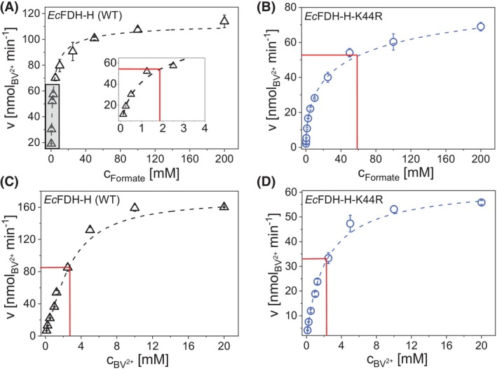
Molybdenum-dependent formate dehydrogenases (Mo-FDHs) reversibly catalyze the interconversion of CO2 and formate, and therefore may be utilized for the development of innovative energy storage and CO2 utilization concepts. Mo-FDHs contain a highly conserved lysine residue in the vicinity of a catalytically active molybdenum (Mo) cofactor and an electron-transferring [4Fe-4S] cluster. In order to elucidate the function of the conserved lysine, we substituted the residue Lys44 of Escherichia coli formate dehydrogenase H (EcFDH-H) with structurally and chemically diverse amino acids. Enzyme kinetic analysis of the purified EcFDH-H variants revealed the Lys-to-Arg substitution as the only amino acid exchange that retained formate oxidation catalytic activity, amounting to 7.1% of the wild-type level. Ultraviolet-visible (UV-Vis) spectroscopic analysis indicated that >90% of the [4Fe-4S] cluster was lost in the case of EcFDH-H variants -K44E and -K44M, whereas the cluster occupancy of the K44R variant decreased by merely 4.5%. Furthermore, the K44R substitution resulted in a slight decrease in its melting temperature and a significant formate affinity decrease, apparent as a 32-fold Km value increase. Consistent with these findings, molecular dynamics simulations predicted an increase in the backbone and cofactor mobility as a result of the K44R substitution. These results are consistent with the conserved lysine being essential for stabilizing the catalytically active structures in EcFDH-H and may support engineering efforts on Mo-FDHs to design more efficient biocatalysts for CO2 reduction.

Keywords: CO2 utilization; lysine; molybdenum‐dependent formate dehydrogenase; redox cofactor; site‐directed mutagenesis.


Conflict of interest statement

The authors declare no conflict of interest.

Figures

Fig. 1
Fig. 1
Sequence homology analysis and active‐site structure of the Mo‐dependent E. coli dehydrogenase H (EcFDH‐H). (A) Amino acid sequence alignment of EcFDH‐H and its homologous enzymes, including five molybdenum (Mo)/tungsten (W)‐dependent formate dehydrogenases [PDB identification codes: 1KQF (EcFDH‐N), 6SDR (DvFDH), 7BKB (MhFDH), 1H0H (DgFDH), and 6TG9 (RcFDH)] as well as three nitrate reductases (NARs) [PDB IDs: 2JIO (DdNAR), 2NYA (EcNAR), and 3O5A (CnNAR)] were performed using the Geneious Prime software tool (Biomatters; Auckland, New Zealand). The conserved [4Fe‐4S] cluster‐coordinated cysteine residues are numbered according to the amino acid sequence of EcFDH‐H. The lysine residue investigated in this study is highlighted in a red box, and the varying degrees of homology are indicated by different colors (yellow > blue). (B) Active site structure of EcFDH‐H (PDB ID: 1FDO) visualized using the PyMOL Molecular Graphics System (Version 1.7.2, Schrödinger; New York City, NY, USA). The enzyme‐bound [4Fe‐4S] cluster, amino acid residues, and the molybdopterin groups (MPT‐1 and MPT‐2) in EcFDH‐H are shown in stick representation, except the Mo ion and the water molecule shown in spheres. Yellow dotted lines between the [4Fe‐4S] cluster, residues Lys44, Asp179, and Ser180, a local water molecule and the MPT‐1 group indicate hydrogen bonding. The identity of iron, sulfur, carbon, nitrogen, oxygen, and molybdenum atoms is indicated by coloring in brown, yellow, gray, blue, red, and cyan, respectively.
Fig. 2
Fig. 2
Analyses of EcFDH‐H [wildtype (WT)] and its variants ‐K44R, ‐K44A, ‐K44M, ‐K44E, ‐K44Q, and ‐K44H as well as their E. coli production host strains JG‐X, FL008, FL009, FL010, FL011, FL012, and FL013, respectively. (A) Final optical density at λ = 600 nm (OD600) of E. coli strains after anaerobic cell cultivation in gas‐tight 2‐L laboratory bottles containing 1 L lysogeny broth (LB) medium supplemented with 4 g·L−1 glucose, 1 mm Na2MoO4 and 10 μm Na2SeO3. (B) Sodium dodecyl sulfate/polyacrylamide gel electrophoresis (SDS/PAGE) analysis of reference proteins (lane labeled M) and purified EcFDH‐H (WT) as well as its K44 substitution variants using 4–20% acrylamide gradient. (C) Enzyme yields obtained from lysed E. coli cells by Ni‐ion affinity chromatography under strictly anaerobic conditions. (D) Formate oxidation rates determined by spectrometry at λ = 555 nm and 25 °C under strictly anaerobic conditions in 50 mm phosphate buffer (pH 7.5) containing the purified enzymes at 1.5 μg·mL−1 [EcFDH‐H (WT)], 7.5 μg·mL−1 (‐K44R), and 30 μg·mL−1 (‐K44A, ‐K44E, ‐K44Q, ‐K44M, and ‐K44H) as well as 10 mm formate and 2 mm oxidized benzyl viologen (BV2+). Data are mean values of three independent experiments and the error bars indicate the standard deviation.
Fig. 3
Fig. 3
Ultraviolet–visible (UV–Vis) spectroscopic analysis of EcFDH‐H [wildtype (WT)] and its variants ‐K44R, ‐K44M, and ‐K44E. (A) UV–Vis absorption spectra of EcFDH‐H (WT) dissolved at a concentration of 2.5 mg·mL−1 in 25 mm 2‐(N‐morpholino)ethanesulfonic acid (MES) buffer (pH 6.0) containing 100 mm Na2SO4. The data sets were recorded using freshly purified samples (anaerobic) and samples exposed to air for 30 min and 60 min at 4 °C and 1200 rpm orbital shaking. Comparison of the UV–Vis spectra of the freshly purified enzyme solutions containing EcFDH‐H (WT) and its variants K44R, ‐K44M, and ‐K44E (B–D, respectively) at a concentration of 2–3 mg·mL−1. The enlarged regions containing spectroscopic data at λ = 300–500 nm are shown as insets and the shaded areas indicate the standard deviation of data measured from three independent experiments (n = 3). (E) Spectral signals at λ = 350–450 nm were obtained by baseline subtraction from the data shown in B–D.
Fig. 4
Fig. 4
Kinetic analysis of EcFDH‐H [wildtype (WT)] and its variant K44R. Effect of formate (A and B) or oxidized benzyl viologen (BV2+) (C and D) concentration on the activity of EcFDH‐H (WT) (black) and the K44R variant (light blue). The 200‐μL reaction mixture contains the tested EcFDH‐H (WT) and K44R enzyme samples at a concentration of 1.5 and 7.5 μg·mL−1, respectively, as well as, the respective co‐substrates at BV2+ [2 mm] and formate [10 mm]. The consumption of BV2+ was monitored for 60 s at 25 °C under strictly anaerobic conditions. The inset of Panel A displays the activity data recorded for EcFDH‐H (WT) at formate concentrations ranging between 0 and 4 mm (shaded area). Estimation of the kinetic parameters KmFormate and KmBV2+ were performed by fitting the data to the Michaelis–Menten equation (dashed line), in which red lines intersect the y‐axis at the apparent ½ V max and indicate the respective K m values as x‐axis intersections. Data are mean values of three independent experiments with error bars indicating the standard deviation.
Fig. 5
Fig. 5
The pH dependence of the catalytic activity of EcFDH‐H [wildtype (WT)] and its variant K44R. Formate oxidation activity of EcFDH‐H (WT) and variant K44R measured at pH 4.0–9.5 in 50 mm buffer (pH 3.0–6.0: citrate; pH 6.0–9.5: bis‐tris propane) prepared by addition of aqueous NaOH and H2SO4 solutions to avoid EcFDH‐H inhibition by chloride ions [55]. The displayed relative activity is determined by normalizing the measured activity at the respective pH to the maximum activity at pH 7.5. The formate oxidation activity of EcFDH‐H enzymes was quantified spectroscopically at λ = 555 nm inside an anaerobic cabinet. The reaction mixture contained the reaction substrates formate [10 mm] and oxidized benzyl viologen (BV2+) [2 mm] as well as either 1.5 μg·mL−1 EcFDH‐H (WT) or 7.5 μg·mL−1 EcFDH‐H‐K44R. The measurements were performed in triplicate and error bars indicate the standard deviation.
Fig. 6
Fig. 6
Circular dichroism (CD) analysis of EcFDH‐H [wildtype (WT)] and its variants ‐K44R, ‐K44M, and ‐K44E. The CD ellipticity was measured at 25 °C (A) and at temperatures varying between 25 and 55 °C (B–E). Signals were recorded from 300 μL samples containing 100 mm Na2SO4 and ~0.5 mg·mL−1 enzyme dissolved in 25 mm 2‐(N‐morpholino)ethanesulfonic acid (MES) buffer (pH 6.0). Samples were equilibrated for 60 s at the respective temperature in sealed QS‐110 quartz cuvettes prior to CD signal recording. Data are mean values of three independent experiments with error bars indicating the standard deviation.
Fig. 7
Fig. 7
Thermal stability analysis of EcFDH‐H [wildtype (WT)] and its variants ‐K44R, ‐K44E and ‐K44M. Melting curves of EcFDH‐H (WT) (A) and ‐K44R (B), ‐K44E (C) and ‐K44M (D) obtained by nonlinear least squares fitting of circular dichroism (CD) signals recorded at λ = 222 nm and different temperatures (Fig. 6) to the Boltzmann equation using T m as midpoint value x0 [56]. The displayed data are mean values calculated from three biological replicates (n = 3) with error bars indicating the standard deviation.
Fig. 8
Fig. 8
Molecular dynamics (MD) simulation of EcFDH‐H [wildtype (WT)] and its variants ‐K44R, ‐K44M, and ‐K44E. The system was simulated at a temperature of 45 °C using the Desmond MD engine (D. E. Shaw Research; New York City, NY, USA). The root mean square deviation (RMSD) of protein backbone‐Cα atoms (A) and the bound Mo‐bis‐pyranopterin guanine dinucleotide (Mo(MGD)2) cofactor (B) throughout the 150‐ns simulation period are displayed. Each enzyme variant is represented by a single RMSD data set (n = 1). Visualization of the active‐site structures of the EcFDH‐H variants ‐K44R (C–E), ‐K44M (F–H), ‐K44E (I–K) and EcFDH‐H (WT) was performed using the PyMOL Molecular Graphics System (Version 1.7.2, Schrödinger; New York City, NY, USA). The displacement of the molybdopterin group of Mo(MGD)2 cofactor proximal to site 44 in EcFDH‐H (MPT‐1) in the ‐K44R variant is marked with red arrows.

Similar articles

References

    1. Niks D & Hille R (2019) Molybdenum‐containing enzymes. In Metalloproteins: Methods and Protocols (Hu Y, ed.), pp. 55–63. Springer New York, New York, NY. - PMC - PubMed
    1. Hille R, Hall J & Basu P (2014) The mononuclear molybdenum enzymes. Chem Rev 114, 3963–4038. - PMC - PubMed
    1. Schwarz G, Mendel RR & Ribbe MW (2009) Molybdenum cofactors, enzymes and pathways. Nature 460, 839–847. - PubMed
    1. Leimkühler S (2020) The biosynthesis of the molybdenum cofactors in Escherichia coli . Environ Microbiol 22, 2007–2026. - PubMed
    1. Mendel RR & Leimkühler S (2015) The biosynthesis of the molybdenum cofactors. J Biol Inorg Chem 20, 337–347. - PubMed

MeSH terms

LinkOut - more resources