. 2025 Mar 20;207(3):e0041624.
doi: 10.1128/jb.00416-24. Epub 2025 Mar 3.

Broken beyond repair: TA system ParE toxins mediate effective gyrase inhibition without driving resistance

Affiliations

Broken beyond repair: TA system ParE toxins mediate effective gyrase inhibition without driving resistance

Chih-Han Tu et al. J Bacteriol. .

Abstract

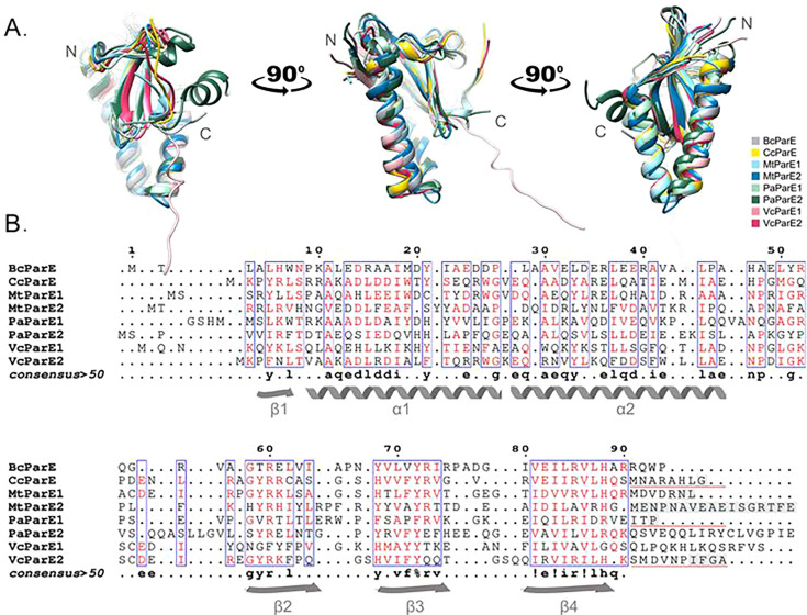
DNA gyrase is an essential bacterial-specific type IIA topoisomerase that corrects DNA overwinding during transcription and replication. Compounds capable of stabilizing gyrase-mediated double-strand DNA breaks are valuable antibacterials; however, these can trigger error-prone repair, potentially inducing DNA mutations leading to antimicrobial resistance. ParE toxin proteins, which belong to a family of type II toxin-antitoxin systems, inhibit DNA gyrase and promote the persistence of double-strand DNA breaks. However, it is unclear if the ParE-induced gyrase inhibition is equivalent for all ParE family members, or if any mutations arise and can accumulate to cause antibiotic resistance. Selected chromosomal ParE toxins were examined for toxicity to their native bacterial hosts, and the frequency of mutations and impact on susceptibility to selected antibiotics were assessed. Our results show that ParE toxins from Burkholderia cenocepacia, Mycobacterium tuberculosis, Pseudomonas aeruginosa, and Vibrio cholerae exert potent toxicities toward the native cells, whereas one tested ParE toxin from P. aeruginosa was not toxic. The contribution to toxicity of the ParE toxin C-terminal amino acid sequences was examined using two lab-generated chimeric ParE toxins; our results demonstrate that this region did not impact the toxicity level. Our study finds that the relative potency of individual ParE toxins correlates with increases in mutation frequency. While some ParE toxins induced limited collateral sensitivity to selected antibiotics, no increases in MIC values were found. Overall, this study demonstrates the relative toxicity of different ParE toxins. Importantly, the toxicity appears to result in loss of viability before productive resistance-inducing mutations can accumulate.

Importance: Toxin-antitoxin (TA) systems can halt growth or kill cells when the toxin protein engages with the host cell target. In the ParDE TA system, the toxin ParE inhibits DNA gyrase, resulting in loss of viability that phenocopies fluoroquinolone antibiotics. Our study demonstrates that ParE toxins increase the frequency of mutations, presumably by a mechanism similar to fluoroquinolone antibiotics. These increases scale to the resulting toxicity, and importantly, these mutations do not accumulate into productive antibacterial resistance. This suggests that ParE toxins are not intrinsic drivers of resistance and, if the molecular mechanism can be harnessed, could generate a new class of gyrase inhibitors.

Keywords: ParE toxin; antimicrobial susceptibility; bacterial cell viability; gyrase inhibition; mutation frequency; toxin antitoxin.


Conflict of interest statement

The authors declare no conflict of interest.

Figures

Fig 1
Fig 1
The BcParE toxin induces loss of viability that correlates with a dose-dependent increase in mutagenic frequency. (A) Induction of BcParE encoded in the rhamnose (rhm)-inducible pSCrha vector harbored in its native host B. cenocepacia J2315 causes stagnation and loss of viability more than 4-log values at timepoints equivalent to approx. 5 cell doublings (15 h timepoint with a 3 h doubling time). (B) Increasing stagnation or toxicity at the 15 h timepoint triggers increases in mutation frequency, with the highest impacts comparable to treatment with gyrase inhibitor ciprofloxacin (CIP). (C) In contrast, the BcParE toxin has modest impacts on the viability of E. coli MG1655. Viability data are presented as change from initial CFU/mL (dashed line); all data are displayed as standard error of the mean (SEM) calculated from three independent experiments. Mutation frequency of ParE-induced or CIP-treated cells was compared to that of the 0% induction cells using an unpaired two-tailed Student’s t-test: ***P < 0.001.
Fig 2
Fig 2
The two ParE toxins encoded in the genome of M. tuberculosis induce loss of viability to surrogate bacterial host E. coli MG1655 that correlates with a dose-dependent increase in mutagenic frequency. (A) Induction of MtParE1 and (C) MtParE2 from the arabinose (ara)-inducible pMindBAD vector causes a loss of viability to E. coli with a stronger effect noted for MtParE2. (B) Mutation frequency is increased for both MtParE1 and (D) MtParE2, with even mild induction producing frequencies in excess of the positive control gyrase-inhibiting ciprofloxacin (CIP) treatment. Viability data are presented as change from initial CFU/mL (dashed line); all data are displayed as standard error of the mean (SEM) calculated from three independent experiments. Mutation frequency of ParE-induced or CIP-treated cells was compared to that of the 0% induction cells using an unpaired two-tailed Student’s t-test: *P < 0.05, **P < 0.01, and ***P < 0.001.
Fig 3
Fig 3
The two ParE toxins encoded in the genome of V. cholerae exert toxicity and trigger corresponding increases in mutation frequencies. (A) Induction of VcParE1 and (D) VcParE2 cause dose-dependent loss of viability with a stronger effect mediated by VcParE2. The level of toxicity correlates directly with the increase in mutation frequency (B, E), with a much stronger effect seen for VcParE2 at more than a 4-log increase. Both toxins also display toxicity in E. coli MG1655 (C, F) although to a lower level than to the native host. Viability data are presented as change from initial CFU/mL (dashed line); all data are displayed as standard error of the mean (SEM) calculated from three independent experiments. Mutation frequency of ParE-induced or CIP-treated cells was compared to that of the 0% induction cells using an unpaired two-tailed Student’s t-test: *P < 0.05 and **P < 0.01.
Fig 4
Fig 4
One ParE toxin encoded in the genome of P. aeruginosa does not impact the viability of any bacterial strain tested, consistent with a lack of impact on the frequency of mutations, while the ParE2 toxin found in many strains of this bacterium is potently toxic yet has an attenuated impact on mutation frequency. (A) Induction of PaParE1 in either the PAO1 or PA14 (C) strains has no impact on viability or on mutation frequency (B, D). In contrast, the ParE2 toxin is potently toxic to the PAO1 strain (E) with values at 4 h below the limit of detection (dashed line), and this induction triggers (F) modest increases in mutation frequency. It is of note that the positive control treatment with ciprofloxacin (CIP) also causes only modest impacts to the mutation frequency. (G) PaParE2 also displays toxicity in E. coli MG1655, although to a lower level than to its native host. Viability data are presented as change from initial CFU/mL (dashed line); all data are displayed as standard error of the mean (SEM) calculated from three independent experiments except for PaParE1 in the PA14 strain, which has two independent replicates that are consistent with the PAO1 results. Mutation frequency of ParE-induced or CIP-treated cells was compared to that of the 0% induction cells using an unpaired two-tailed Student’s t-test: **P < 0.01 and ***P < 0.001.
Fig 5
Fig 5
Structural and sequence alignments of ParE toxins tested in the current work. (A) The three-dimensional structures of seven ParE toxins were superimposed: BcParE, AlphaFold model AF-B4EF23-F1-model_v4; CcParE, PDB 3KXE (50); MtParE1, PDB 8C24 (15); MtParE2, PDB 8C26 (15); PaParE1, PDB 6XRW (49); PaParE2, AlphaFold model AF-Q9I5J9-F1-model_v4; VcParE1, AlphaFold model AF-O68848-F1-model_v4; VcParE2, PDB 7R5A (35). Note that all crystallographic structures are pictured without the bound ParD antitoxin; further, the C-terminal regions are not complete in those structures (shaded gray boxes, panel B). (B) Structure-based aligned sequences with secondary structures indicated (ESPript [55]). Residues with a similarity score exceeding 50% (4 out of 8) are deemed highly similar and are colored in red and framed in blue; amino acids omitted from crystallographic structures due to disorder are indicated by gray shaded boxes. C-terminal sequences used for construction of chimeric ParE toxins are underlined in red. Consensus is listed below, % = F or Y, ! = I or V.
Fig 6
Fig 6
The amino acid sequence of ParE C-termini does not impact toxicity but can enhance expression. (A) E. coli cells transformed with a pET15b expression vector (control, gray line), or this vector harboring the coding sequence for PaParE1 (magenta line), PaParE1 with the C-terminus from the C. crescentus ParE toxin (+Cc, blue line) and with the C-terminus from VcParE2 (+Vc, green line) were induced and viability monitored (data presented as in Fig. 1). No significant differences in viability as a function of C-terminal sequence are observed. (B) The expression of each ParE toxin was assessed by electrophoresis, and based on the intensity of staining with Coomassie dye, the levels are comparable to improved with the changed C-terminal sequences.

References

    1. Jurėnas D, Fraikin N, Goormaghtigh F, Van Melderen L. 2022. Biology and evolution of bacterial toxin–antitoxin systems. Nat Rev Microbiol 20:335–350. doi:10.1038/s41579-021-00661-1 - DOI - PubMed
    1. Bardaji L, Añorga M, Echeverría M, Ramos C, Murillo J. 2019. The toxic guardians - multiple toxin-antitoxin systems provide stability, avoid deletions and maintain virulence genes of Pseudomonas syringae virulence plasmids. Mob DNA 10:7. doi:10.1186/s13100-019-0149-4 - DOI - PMC - PubMed
    1. Fernández-García L, Blasco L, Lopez M, Bou G, García-Contreras R, Wood T, Tomas M. 2016. Toxin-antitoxin systems in clinical pathogens. Toxins (Basel) 8:227. doi:10.3390/toxins8070227 - DOI - PMC - PubMed
    1. Guan J, Chen Y, Goh Y-X, Wang M, Tai C, Deng Z, Song J, Ou H-Y. 2024. TADB 3.0: an updated database of bacterial toxin-antitoxin loci and associated mobile genetic elements. Nucleic Acids Res 52:D784–D790. doi:10.1093/nar/gkad962 - DOI - PMC - PubMed
    1. Fraikin N, Van Melderen L. 2024. Single-cell evidence for plasmid addiction mediated by toxin-antitoxin systems. Nucleic Acids Res 52:1847–1859. 10.1093/nar/gkae018. - DOI - PMC - PubMed

MeSH terms

LinkOut - more resources