. 2025 Dec;14(1):2473165.
doi: 10.1080/2162402X.2025.2473165. Epub 2025 Mar 3.

MERTK inhibition improves therapeutic efficacy of immune checkpoint inhibitors in hepatocellular carcinoma

Affiliations

MERTK inhibition improves therapeutic efficacy of immune checkpoint inhibitors in hepatocellular carcinoma

Diana Llopiz et al. Oncoimmunology. 2025 Dec.

Abstract

Immunotherapy with immune checkpoint inhibitors (ICI) in hepatocellular carcinoma (HCC) patients only achieves response rates of 25%-30%, indicating the necessity of new therapies for non-responder patients. Since myeloid-related suppressive factors are associated with poor responses to ICI in a subgroup of HCC patients, modulation of these targets may improve response rates. Our aim was to characterize the expression of the efferocytosis receptor MERTK in HCC and to analyze its potential as a new therapeutic target. In HCC patients, MERTK was expressed by myeloid cells and was associated with poorer survival. In a murine HCC model with progressive myeloid cell infiltration, MERTK was detected in dendritic cells and macrophages with an activated phenotype, which overexpressed the checkpoint ligand PD-L1. Concomitant expression of PD-1 in tumor T-cells suggested the pertinence of combined PD-1/PD-L1 and MERTK blockade. In vivo experiments in mice showed that inhibition of MERTK improved the therapeutic effect promoted by anti-PD-1 or by ICI combinations currently approved for HCC. This effect was associated with enhanced tumor infiltration and superior activity of antigen presenting cells and effector lymphocytes. Our results indicate that MERTK may behave as a relevant target for immunotherapeutic combinations in those HCC patients with tumors enriched in a myeloid component.

Keywords: Hepatocellular carcinoma; MERTK; dendritic cells; immune checkpoint inhibitors; tumor-associated macrophages.


Conflict of interest statement

No potential conflict of interest was reported by the author(s).

Figures
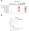
Figure 1.
Figure 1.
Expression of MERTK in human healthy and tumorous liver samples. (a) Expression of MERTK in HCC cell subsets according to TISCH2. (b) Overall survival of grade 3 HCC patients according to the expression of MERTK.
Figure 2.
Figure 2.
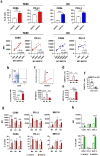
Evolution of myeloid cell subsets during murine PM299L HCC growth and expression of MERTK. (a) The presence of total leukocytes (among alive cells) and myeloid cells (in CD45+ cells) was analyzed by flow cytometry in PM299L HCC orthotopic tumors obtained at different time points. (b) Proportion of total leukocytes and myeloid cells in the liver of healthy mice (HL) (n = 5), and in tumor (TUM) (n = 15) and non-tumor (NTUM) (n = 8) samples. (c) Percentage of mertk-expressing cells in different myeloid subsets. (d) Contribution of each myeloid subset to total pool of MERTK+ cells. (e) Intensity of MERTK expression (measured as mean fluorescence index-MFI) in myeloid subsets. (f) MERTK score (product of the percentage of MERTK+ cells in CD45+ cells and the MFI of MERTK) of myeloid subsets. g. Correlation between tumor volume and the percentage of MERTK+ cells in total myeloid cells, DC and TAM (ns, non-statistically significant; *p < 0.05; **p < 0.01; ***p < 0.001; ****p < 0.0001).
Figure 3.
Figure 3.
Phenotype of TAM and DC according to MERTK expression in orthotopic PM299L HCC tumors. (a) Percentage of TAM and DC expressing CD86 and PD-L1 according to MERTK expression (n = 6–12 mice) (upper panels) and correlation of expression of these markers and MERTK, expressed as mean fluorescence intensity (MFI) (lower panels). (b) Identification of M0, M1 and M2 subsets within TAM. (c) Intensity of expression of MERTK (represented as MFI) in M0, M1 and M2 TAM. (d) Percentage of MERTK+ cells in M0, M1 and M2 TAM. (e) MERTK score of M0, M1 and M2 TAM. (f) Percentage of MERTK+ cells in KC according to CD38 expression. (g, h) MFI of CD86, PD-L1 and MHC-II (upper panels) and percentage of cells positive for these markers (bottom panels) in TAM (g) and in KC (h) was determined in MERTK+ and MERTK cells. (*p < 0.05; **p < 0.01; ***p < 0.001; ****p < 0.0001).
Figure 4.
Figure 4.
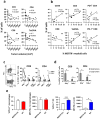
MERTK correlates with higher levels of antigen experienced T-cells and promotes PD-L1 expression. (a) The presence of lymphoid cells (in CD45+ cells) was analyzed in orthotopic PM299L HCC at different time points. (b) Correlation between percentage of CD45, CD4, CD8 and TetOVA+ cells, and of PD-1+ CD4 and PD-1+ CD8 cells, and the percentage of MERTK+ myeloid cells. (c) Tumor-infiltrating lymphocytes were stimulated with PMA/Ionomycin and cytokine production by PD-1+ and PD-1 CD8 and CD4 T cells was determined by flow cytometry. (d) Production of cytokines after PMA/Ionomycin by CD8 and CD4 T cells obtained from small and large tumors. (e) PD-L1 expression was analyzed in hepatic macrophages (left panels) and DC (right panels) of wild type (WT) C57BL/6J and MERTK KO mice (n = 5/group). (*p < 0.05; **p < 0.01; ***p < 0.001).
Figure 5.
Figure 5.
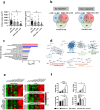
Simultaneous blockade of MERTK and PD-1 results in enhanced therapeutic efficacy. (a) C57BL/6J mice with orthotopic PM299L tumors were treated with control (ISO) or anti-PD-1 antibodies, RXDX-106, or the combination (RXDX-106 + anti-PD-1). They were sacrificed at day 18 to determine tumor volume and weight. (b) Number of up- and down-regulated genes in RNAseq experiments comparing tumor samples from isotype-treated mice (ISO) and treatment groups. (c) Pathway enrichment analyses according to reactome of genes upregulated by the combination vs ISO. (d) Protein–protein interaction networks of genes upregulated by the combination vs ISO according to Metascape. (e) Normalized expression of individual genes corresponding to immune functions and cell components in the four groups. (f) Analysis by flow cytometry of tumor infiltrating cells of mice (n = 7–9/group) corresponding to the different treatments. (*p < 0.05; **p < 0.01; ***p < 0.001; ****p < 0.0001).
Figure 6.
Figure 6.
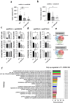
Combination of MERTK blockade with approved HCC immunotherapies improves their efficacy. C57BL/6J mice with orthotopic PM299L tumors were treated with control (ISO), immunotherapy (IT) based on blockade of PD-L1 + VEGFR (a), IT based on blockade of PD-L1 + CTLA-4 (b), or combination of these IT with RXDX-106, and tumor volume was determined. Flow cytometry analyses of tumor infiltrating cells obtained from mice (n = 5–8 mice/group) treated with PD-L1 + VEGFR (c) or PD-L1 + CTLA-4 (d) IT combinations. (e) Genes up- and down-regulated in mice treated with ISO, PD-L1 + CTLA-4 blockade (IT) or IT + RXDX-106 in RNAseq experiments using tumor samples (n = 4 mice/group) from each groups. (f) Pathway enrichment analyses of genes upregulated only by IT + RXDX-106.

References

    1. Finn RS, Qin S, Ikeda M, Galle PR, Ducreux M, Kim TY, Kudo M, Breder V, Merle P, Kaseb AO, et al. Atezolizumab plus bevacizumab in unresectable hepatocellular carcinoma. N Engl J Med. 2020;382(20):1894–12. doi:10.1056/NEJMoa1915745. - DOI - PubMed
    1. Abou-Alfa GK, Lau G, Kudo M, Chan SL, Kelley RK, Furuse J, Sukeepaisarnjaroen W, Kang Y-K, Van Dao T, De Toni EN, et al. Tremelimumab plus durvalumab in unresectable hepatocellular carcinoma. NEJM Evid. 2022;1(8):1–12. doi:10.1056/evidoa2100070. - DOI - PubMed
    1. Zhu AX, Abbas AR, de Galarreta MR, Guan Y, Lu S, Koeppen H, Zhang W, Hsu CH, He AR, Ryoo BY, et al. Molecular correlates of clinical response and resistance to atezolizumab in combination with bevacizumab in advanced hepatocellular carcinoma. Nat Med. 2022;28(8):1599–1611. doi:10.1038/s41591-022-01868-2. - DOI - PubMed
    1. Keskin DB, Anandappa AJ, Sun J, Tirosh I, Mathewson ND, Li S, Oliveira G, Giobbie-Hurder A, Felt K, Gjini E, et al. Neoantigen vaccine generates intratumoral T cell responses in phase Ib glioblastoma trial. Nature. 2019;565(7738):234–239. doi:10.1038/s41586-018-0792-9. - DOI - PMC - PubMed
    1. Zheng H, Peng X, Yang S, Li X, Huang M, Wei S, Zhang S, He G, Liu J, Fan Q, et al. Targeting tumor-associated macrophages in hepatocellular carcinoma: biology, strategy, and immunotherapy. Cell Death Discov. 2023;9(1). doi:10.1038/S41420-023-01356-7. - DOI - PMC - PubMed

MeSH terms