. 2025 May 1;31(9):1766-1782.
doi: 10.1158/1078-0432.CCR-24-2449.

XMT-2056, a HER2-Directed STING Agonist Antibody-Drug Conjugate, Induces Innate Antitumor Immune Responses by Acting on Cancer Cells and Tumor-Resident Immune Cells

Affiliations

XMT-2056, a HER2-Directed STING Agonist Antibody-Drug Conjugate, Induces Innate Antitumor Immune Responses by Acting on Cancer Cells and Tumor-Resident Immune Cells

Raghida A Bukhalid et al. Clin Cancer Res. .

Abstract

Purpose: Targeted tumor delivery may be required to potentiate the clinical benefit of innate immune modulators. The objective of the study was to apply an antibody-drug conjugate (ADC) approach to STING agonism and develop a clinical candidate.

Experimental design: XMT-2056, a HER2-directed STING agonist ADC, was designed, synthesized, and tested in pharmacology and toxicology studies. The ADC was compared with a clinical benchmark intravenously administered a STING agonist.

Results: XMT-2056 achieved tumor-targeted delivery of the STING agonist upon systemic administration in mice and induced innate antitumor immune responses; single dose administration of XMT-2056 induced tumor regression in a variety of tumor models with high and low HER2 expressions. Notably, XMT-2056 demonstrated superior efficacy and reduced systemic inflammation compared with a free STING agonist. XMT-2056 exhibited concomitant immune-mediated killing of HER2-negative cells specifically in the presence of HER2-positive cancer cells, supporting the potential for activity against tumors with heterogeneous HER2 expression. The antibody does not compete for binding with trastuzumab or pertuzumab, and a benefit was observed when combining XMT-2056 with each of these therapies as well as with trastuzumab deruxtecan ADC. The combination of XMT-2056 with anti-PD-1 conferred benefit on antitumor activity and induced immunologic memory. XMT-2056 was well tolerated in nonclinical toxicology studies.

Conclusions: These data provide a robust preclinical characterization of XMT-2056 and provide rationale and strategy for its clinical evaluation.


Conflict of interest statement

R.A. Bukhalid reports a patent for Mersana Therapeutics and holds patents and patent applications relevant to this work pending, issued, and licensed to various institutions. J.R. Duvall reports personal fees from Mersana Therapeutics outside the submitted work; in addition, J.R. Duvall has a patent for ADCs comprising STING agonists pending. K. Lancaster reports other support from Mersana Therapeutics outside the submitted work. K.C. Catcott reports other support from Mersana Therapeutics outside the submitted work and is employed by Mersana Therapeutics. N. Malli Cetinbas reports personal fees and other support from Mersana Therapeutics during the conduct of the study; in addition, N. Malli Cetinbas has a patent for Mersana Therapeutics pending and issued. J.D. Thomas reports personal fees from Mersana Therapeutics outside the submitted work; in addition, J.D. Thomas has a patent for Mersana Therapeutics and holds patents and patent applications relevant to this work pending. L. Xu reports a patent for W02021202984A1 issued and licensed to Mersana Therapeutics. D. Toader reports a patent for certain pending patent applications and granted patents pending. M. Damelin reports a patent for Mersana Therapeutics and holds patents and patent applications relevant to this work pending. T.B. Lowinger reports other support from Mersana Therapeutics outside the submitted work; in addition, T.B. Lowinger has various patents pending and issued to Mersana Therapeutics. No disclosures were reported by the other authors.

Figures

Figure 1.
Figure 1.
XMT-2056 elicits potent antigen-dependent and Fc-R–mediated activation of monocytes in cocultures. A, Schematic of the XMT-2056 mechanism, which includes HER2-dependent ADC uptake into tumor cells and Fcγ-R–expressing myeloid cells. B, Chemical structure of XMT-2056. C, Cytokine induction as measured by a multiplex Luminex assay from supernatants of fresh human WBCs treated for 6 (IFN-β) or 24 (CXCL10, IL-6, and TNF-a) hours. Bars represent mean value of n = 2 data points shown as symbols. D, Competition with trastuzumab by biolayer interferometry (Octet): trastuzumab was loaded onto the sensor chip, and HER2 ECD or HT19 antibody associations are indicated by blue arrows. Additional binding by HT19 indicates noncompetitive binding. E, Competition with trastuzumab by cell-based flow cytometry. HT19 hIgG1 or mIgG2a formats detected with either Alexa Fluor anti-hIgG1 (h647) or Alexa Fluor anti-mIgG2a (m647) secondary antibodies in the presence or absence of trastuzumab–mIgG2a. Each point represents the mean and SD (n = 3). F, Binding of XMT-2056 or HT19 antibody to HCC1954 cells showing fluorescence intensities measured by flow cytometry. Each point represents the mean and SD (n = 2). G, CXCL10 cytokine induction in HCC1954 monocultures treated for 24 hours with XMT-2056, nonbinding control ADC, HT19, or the free payload. Each point represents the mean and SD (n = 2). H, Binding of XMT-2056 or Fc-mutant XMT-2056, HT19, and nonbinding control ADC to SKOV3 cells showing fluorescence intensities measured by flow cytometry. Each point represents the mean and SD (n = 3). I and J, IRF3 reporter activity of THP1 cells in coculture with SKOV3 cells (I) or cultured on recombinant HER2 antigen-coated plates (J) treated for 24 hours with XMT-2056 or Fc-mutant XMT-2056, nonbinding control ADC, or STING agonist payload. Each point represents the mean and SD (n = 3). When noted, EC50 and Bmax values represent mean of two independent experiments. gMFI, geometric mean fluorescence intensity; RLU, relative light units. (A, Created in BioRender. Cetinbas, N. [2025], https://BioRender.com/s16c825; G, Created in BioRender. Cetinbas, N. [2025], https://BioRender.com/s16c825; I and J, Created in BioRender. Cetinbas, N. [2025], https://BioRender.com/s16c825.)
Figure 2.
Figure 2.
XMT-2056 induces killing of HER2-high and HER2-low cancer cells and induces concomitant immune-mediated killing of HER2-negative cancer cells in vitro. A and B, Cancer cell death induced by XMT-2056, nonbinding control ADC, or STING agonist payload, shown as percent viable SKBR3–NR (A) or MDA-MB-175-VII–NR (B) cells in PBMC coc ultures (84 hours time point). Each point represents the mean and SD (n = 3). C and D, Cytokine induction by XMT-2056, nonbinding control ADC, or STING agonist payload in supernatants of SKBR3–NR (C) or MDA-MB-175-VII–NR (D) cells in PBMC cocultures (24 hours time point). Each point represents the mean and SD (n = 3). Note that the y-axis scales vary. E, Schematic of concomitant antigen-dependent, immune-mediated killing of HER2-negative cancer cells. F, Flow cytometry of HER2 expression on SKBR3 and nuclear red expressing MDA-MB-231–NR. Each point represents the mean and SD (n = 4). G, Cancer cell death mediated by XMT-2056, nonbinding control ADC, or STING agonist payload, shown as percent viable MDA-MB-231–NR cells in coculture with PBMCs and HER2-positive SKBR3 at the indicated ratios or in the absence of SKBR3 (84 hours time point). Each point represents the mean and SD (n = 3). (E, Created in BioRender. Cetinbas, N. [2025], https://BioRender.com/k38t564.)
Figure 3.
Figure 3.
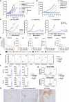
Intravenous administration of XMT-2056 elicits tumor-specific cytokine changes, immune cell tumor infiltration, and antitumor activity. A, Antitumor activity of XMT-2056 in SKOV3 xenograft model. Tumor-bearing CB.17 SCID mice were intravenously administered a single dose (black arrowhead) of XMT-2056, nonbinding control ADC, HT19 antibody, or STING agonist payload, or three doses (orange arrowheads) of diABZI STING agonist. Each point indicates the mean tumor volume and SEM (n = 10). The free payload dose of 0.128 mg/kg is equivalent to the payload dose of 3 mg/kg XMT-2056. B, Antitumor activity of XMT-2056 and Fc-mutant XMT-2056 at the indicated doses in the SKOV3 xenograft model. Each point represents the mean tumor volume and SEM (n = 10). C, Antitumor activity of XMT-2056 in SKOV3 after 3 weekly administrations of lower doses; the fractions of animals with partial response (PR) or complete response (CR) are indicated. D, Cytokines/chemokines measured in serum of SKOV3 tumor–bearing mice after a single intravenous injection of XMT-2056, nonbinding control ADC, or diABZI STING agonist using a 32-plex Luminex assay. Sampling was performed at 6, 12, 24, and 72 hours after administration. Each point represents the mean and SEM (n = 5). E, Mouse gene signature scores for tumors 12 hours after treatment as measured by NanoString analysis. DC, dendritic cell. F, Normalized counts for mouse mRNA or human mRNA of individual tumor cytokine/chemokines in the xenografts sampled at 12 hours. Each point represents the mean and SD (n = 2). G,CD68 and CD45 murine mRNA counts in tumors at the indicated time points. Each floating bar represents the mean and minimum/maximum values (n = 2). H, Representative IHC images of tumors collected 72 hours after treatment stained for the macrophage marker CD68. Scale bar, 100 µm.
Figure 4.
Figure 4.
XMT-2056 elicits antitumor activity in a broad range of tumor models. A, HER2 expression by IHC in tumor xenograft models (human HER2) and syngeneic tumors (rat HER2); see Supplementary Table S3 for quantitation. Scale bar, 20 µm. B–D, SCID beige mice bearing subcutaneous HCC1954 xenograft tumors (B) or CB.17 SCID mice bearing subcutaneous SNU-5 (C) or JIMT-1 (D) xenograft tumors were intravenously administered a single dose (black arrowhead) of XMT-2056, nonbinding control ADC, or HT19 antibody, or three doses of the diABZI STING agonist (orange arrowheads). Each point represents the mean tumor volume and SEM (n = 10). E, FVB/NJ immune-competent mice bearing syngeneic mBR9013 subcutaneous tumors derived from a spontaneous tumor in MMTV-ERBB2 FVB mouse expressing rat HER2 were intravenously administered a single dose of XMT-2056 surrogate ADC targeting rat HER2, nonbinding control ADC, or three doses of the diABZI STING agonist. Each point represents the mean tumor volume and SEM (n = 10). F, BALB/c immune-competent mice bearing syngeneic EMT-6 subcutaneous tumors that were engineered to express rat HER2 (EMT-6–rHER2) were treated as described in E. Each point represents the mean tumor volume and SEM (n = 10).
Figure 5.
Figure 5.
Observed benefit in combining XMT-2056 with other HER2-targeted agents. A–C, Combination of XMT-2056 and trastuzumab. CB.17 SCID mice bearing subcutaneous SKOV3 (A), JIMT-1 (B), or SNU-5 (C) xenograft tumors were administered in 3 weekly doses (A and B) or a single dose (C) of XMT-2056, nonbinding control ADC, trastuzumab, or the combinations indicated. ADCs were administered intravenously, whereas trastuzumab was administered intraperitoneally. Each point represents the mean tumor volume and SEM (n = 10). D, Combination of XMT-2056 and T-Dxd. CB.17 SCID mice bearing subcutaneous JIMT-1 xenograft tumors were intravenously administered XMT-2056, nonbinding control ADC, T-Dxd, or a combination of XMT-2056 and T-Dxd. STING agonist ADCs were administered as a single dose, whereas T-Dxd was administered twice, 1 week apart (red triangles). Each point represents the mean tumor volume and SEM (n = 10).
Figure 6.
Figure 6.
Combination of XMT-2056 with immune checkpoint inhibitor anti–PD-1 elicits tumor clearance and immunologic memory in the EMT-6–rHER2 syngeneic model. A, Normalized counts for PD-L1 human mRNA and mouse mRNA in SKOV3 xenograft tumors. B, Study design including efficacy and rechallenge. Tumor implantations are indicated by circles on the left and right flanks of the mice. C, BALB/c immune-competent mice bearing syngeneic EMT-6–rHER2 engineered tumors were treated with XMT-2056 surrogate ADC targeting rat HER2, anti-mouse PD-1 (clone RPM1-14), or the indicated combinations. The ADCs were administered as single doses, whereas the mouse anti–PD-1 was administered twice weekly for 2 weeks, as indicated by the red triangles. Each point represents the mean tumor volume and SEM (n = 10). D, Tumor-free animals from the XMT-2056 surrogate ADC (blue) and the combination (green) groups were implanted with EMT-6 parental cells on the left flank (opposite the original EMT-6–rHER2 implantation) and CT26 cells on the right flank. Age-matched untreated mice were included as controls. Right, tumor growth in individual mice in each group after implantation on day 0 plotted relative to age-matched naïve mice. CR, complete response; LF, left flank; RF, right flank. (B, Created in BioRender. Cetinbas, N. [2025], https://BioRender.com/k61r850.)

References

    1. Chen DS, Mellman I. Elements of cancer immunity and the cancer-immune set point. Nature 2017;541:321–30. - PubMed
    1. Trujillo JA, Sweis RF, Bao R, Luke JJ. T cell-inflamed versus non-T cell-inflamed tumors: a conceptual framework for cancer immunotherapy drug development and combination therapy selection. Cancer Immunol Res 2018;6:990–1000. - PMC - PubMed
    1. Kono H, Rock KL. How dying cells alert the immune system to danger. Nat Rev Immunol 2008;8:279–89. - PMC - PubMed
    1. Fuertes MB, Kacha AK, Kline J, Woo S-R, Kranz DM, Murphy KM, et al. . Host type I IFN signals are required for antitumor CD8+ T cell responses through CD8{alpha}+ dendritic cells. J Exp Med 2011;208:2005–16. - PMC - PubMed
    1. Akira S, Uematsu S, Takeuchi O. Pathogen recognition and innate immunity. Cell 2006;124:783–801. - PubMed