. 2025 Mar 11;122(10):e2419992122.
doi: 10.1073/pnas.2419992122. Epub 2025 Mar 3.

Recessive genetic contribution to congenital heart disease in 5,424 probands

Affiliations

Recessive genetic contribution to congenital heart disease in 5,424 probands

Weilai Dong et al. Proc Natl Acad Sci U S A. .

Abstract

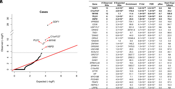
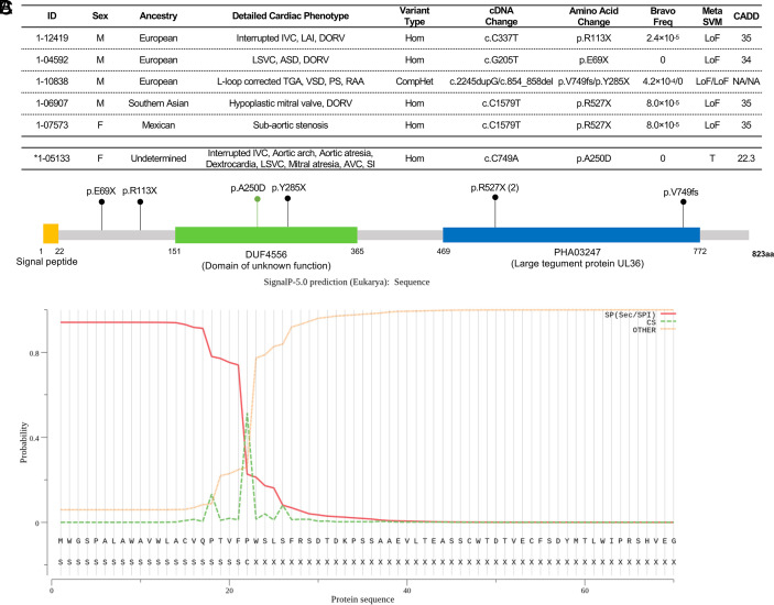
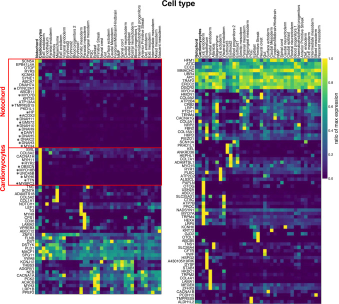
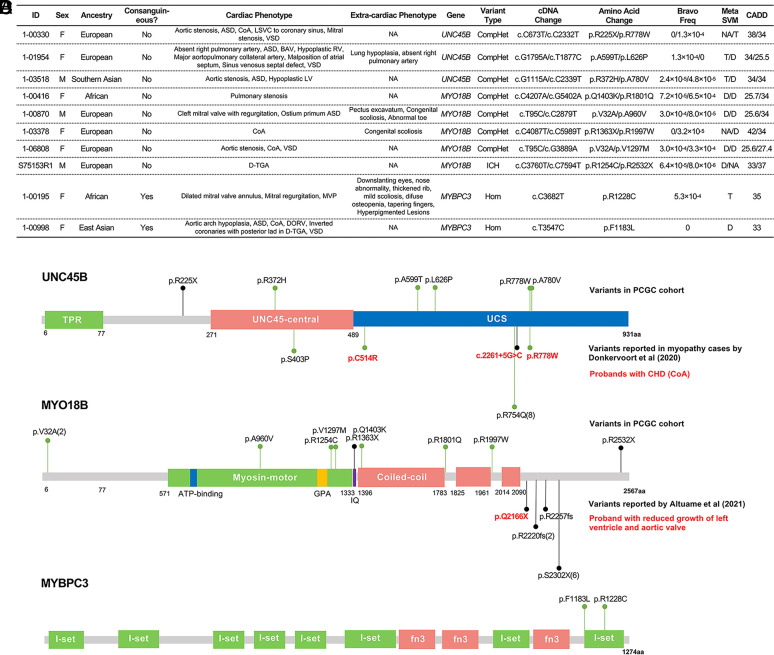
Variants with large effect contribute to congenital heart disease (CHD). To date, recessive genotypes (RGs) have commonly been implicated through anecdotal ascertainment of consanguineous families and candidate gene-based analysis; the recessive contribution to the broad range of CHD phenotypes has been limited. We analyzed whole exome sequences of 5,424 CHD probands. Rare damaging RGs were estimated to contribute to at least 2.2% of CHD, with greater enrichment among laterality phenotypes (5.4%) versus other subsets (1.4%). Among 108 curated human recessive CHD genes, there were 66 RGs, with 54 in 11 genes with >1 RG, 12 genes with 1 RG, and 85 genes with zero. RGs were more prevalent among offspring of consanguineous union (4.7%, 32/675) than among nonconsanguineous probands (0.7%, 34/4749). Founder variants in GDF1 and PLD1 accounted for 74% of the contribution of RGs among 410 Ashkenazi Jewish probands. We identified genome-wide significant enrichment of RGs in C1orf127, encoding a likely secreted protein expressed in embryonic mouse notochord and associated with laterality defects. Single-cell transcriptomes from gastrulation-stage mouse embryos revealed enrichment of RGs in genes highly expressed in the cardiomyocyte lineage, including contractility-related genes MYH6, UNC45B, MYO18B, and MYBPC3 in probands with left-sided CHD, consistent with abnormal contractile function contributing to these malformations. Genes with significant RG burden account for 1.3% of probands, more than half the inferred total. These results reveal the recessive contribution to CHD, and indicate that many genes remain to be discovered, with each likely accounting for a very small fraction of the total.

Keywords: congenital heart disease; exome-sequencing; genomics; human genetics.


Conflict of interest statement

Competing interests statement:M.Y. is a cofounder and consultant for Fabric Genomics Inc., M.Y. has stock in Fabric Genomics.

Figures

Fig. 1.
Fig. 1.
Enrichment of damaging RGs in cases and controls. (A) Q-Q plot comparing observed versus expected P values for damaging RGs in probands; genes with genome-wide significant enrichment of RGs are marked with a red arrow. The dashed line indicates the genome-wide significant p-value threshold of 2.6 × 10⁻⁶ (0.05/19,347). (B) Top 30 genes from the binomial test. Genes with genome-wide significant burden of RGs are in bold font; * denotes previously identified human recessive CHD genes. pRec: probability of being intolerant to biallelic LoF variants based on gnomAD dataset (doi: 10.1038/nature19057). Heart Expr % Rank: the percentile of gene expression in the mouse heart at embryonic day E14.5 (doi: 10.1038/nature12141).
Fig. 2.
Fig. 2.
RGs in C1orf127, a likely secreted protein. (A) RGs in probands with RGs in C1orf127 and associated phenotypes. HTX: heterotaxy; LVO: left ventricular outflow tract obstruction; CTD: conotruncal defect; IVC: inferior vena cava; LAI: left atrial isomerism; DORV: double-outlet right ventricle; LSVC: Persistent left superior vena cava; ASD: atrial septal defect; TGA: transposition of the great arteries; VSD: ventricular septal defect; PS: pulmonary stenosis; RAA: right aortic arch; AVC: atrioventricular canal; SI: situs inversus. * (below the dashed line), proband identified post hoc with heterotaxy and rare RG predicted as benign by MetaSVM but damaging by SIFT and Polyphen with CADD score > 20. (B) Location of C1orf127 variants on the C1ORF127 protein. C1ORF127 protein contains a likely N-terminal signal peptide, a DUF4556 domain, and a PHA03237 domain. The LoF variants are shown in black while missense variant is shown in green. (C) Signal peptide prediction in C1ORF127. The signal peptide was predicted using SignalP-5.0 (http://www.cbs.dtu.dk/services/SignalP/). The red line indicates the probability the sequence comprises a secretory signal peptide (Sec/SPI); the green line indicates the probability of a proteolytic cleavage site; the orange line shows the probability that the sequence does not encode a signal peptide. At the Bottom of the figure, the encoded amino-terminal amino acid sequence of C1ORF127 is shown. The cleavage site is predicted to be between amino acids 22 and 23, and is more than twice as likely as the next most likely site. The overall likelihood of the protein having a Sec signal peptide is calculated to be 0.9.
Fig. 3.
Fig. 3.
Single-cell expression of genes with ≥ 2 RGs during mouse gastrulation. The single-cell RNA-seq data of mouse gastrulation were acquired from Pijuan-Sala et al(33). The expression of each gene was normalized to the level of its expression in the cell/tissue type with highest expression (ratio of max expression). Genes were then clustered by their ratio of max expression in different cell/tissue types using the UPGMA hierarchy clustering algorithm. Only genes with available expression data were shown. *indicates genes with at least fivefold higher expression in notochord or cardiomyocytes compared to other tissues. The x-axis indicates the cell types, the y-axis shows the genes with ≥ 2 damaging RGs. The column on the Right is a continuation of the column on the Left.
Fig. 4.
Fig. 4.
RGs and phenotypes in UNC45B, MYO18B, and MYBPC3. (A) RGs in UNC45B, MYO18B, and MYBPC3 in probands with CHD. ASD; CoA: coarctation; LSVC: left superior vena cava; VSD: ventricular septal defect; BAV: bicuspid aortic valve; RV: right ventricle; LV: left ventricle; MVP: mitral valve prolapse; DORV; D-TGA: D-transposition of great artery. (BD) Location of variants in UNC45B (B), MYO18B (C), and MYBPC3 (D). Variants denoted above each gene were found in probands with CHD described in (A) while variants below each gene are previously reported in patients with RGs and cardiomyopathy and/or CHD. LoF variants are shown in black and missense variants are shown in green. Numbers in parentheses indicate the number of probands with the variant. (B) UNC45B contains the N-terminal TPR domain which interacts with Hsp90, a conserved central domain, and a C-terminal UCS domain which binds myosin. Variants reported in ref. are shown. Variants found in patients with CoA in both cohorts are labeled in red. (C) MYO18B contains an N-terminal myosin-motor domain, a myosin light chain binding IQ motif, and 3 C-terminal coiled-coil regions which might allow for dimerization. Variants found in our cohort are mostly missense and distribute across the encoded protein, RGs reported by Altuame et al (49) in patients with Klippel–Feil syndrome with cardiomyopathy are all LoF and cluster in the C-terminal end of the protein. One patient with p.Gln2166X variant reported reduced growth of the left ventricle and aortic valve. (D) MYBPC3 contains eight Ig-like C2 (I-set) domains and three fibronectin type-III (fn3) domains. Both of the variants in our cohort are missense and map to the last I-set domain, which is known to bind to the myosin rod (53).

References

    1. Zaidi S., Brueckner M., Genetics and genomics of congenital heart disease. Circ. Res. 120, 923–940 (2017). - PMC - PubMed
    1. G. B. D. Congenital Heart Disease Collaborators, Global, regional, and national burden of congenital heart disease, 1990–2017: A systematic analysis for the Global Burden of Disease Study 2017. Lancet Child Adole sc. H ealth 4, 185–200 (2020). - PMC - PubMed
    1. Wu W., He J., Shao X., Incidence and mortality trend of congenital heart disease at the global, regional, and national level, 1990–2017. Medicine (Baltimore) 99, e20593 (2020). - PMC - PubMed
    1. van der Linde D., et al. , Birth prevalence of congenital heart disease worldwide: A systematic review and meta-analysis. J. Am. Coll Cardiol. 58, 2241–2247 (2011). - PubMed
    1. van der Bom T., et al. , The changing epidemiology of congenital heart disease. Nat. Rev. Cardiol. 8, 50–60 (2011). - PubMed

Substances

Grants and funding

LinkOut - more resources