. 2025 Mar 11;122(10):e2414514122.
doi: 10.1073/pnas.2414514122. Epub 2025 Mar 3.

In the activation of HPV-specific human B cells HPV-VLP vaccines mimic membrane-associated antigens

Affiliations

In the activation of HPV-specific human B cells HPV-VLP vaccines mimic membrane-associated antigens

Charles Torgbor et al. Proc Natl Acad Sci U S A. .

Abstract

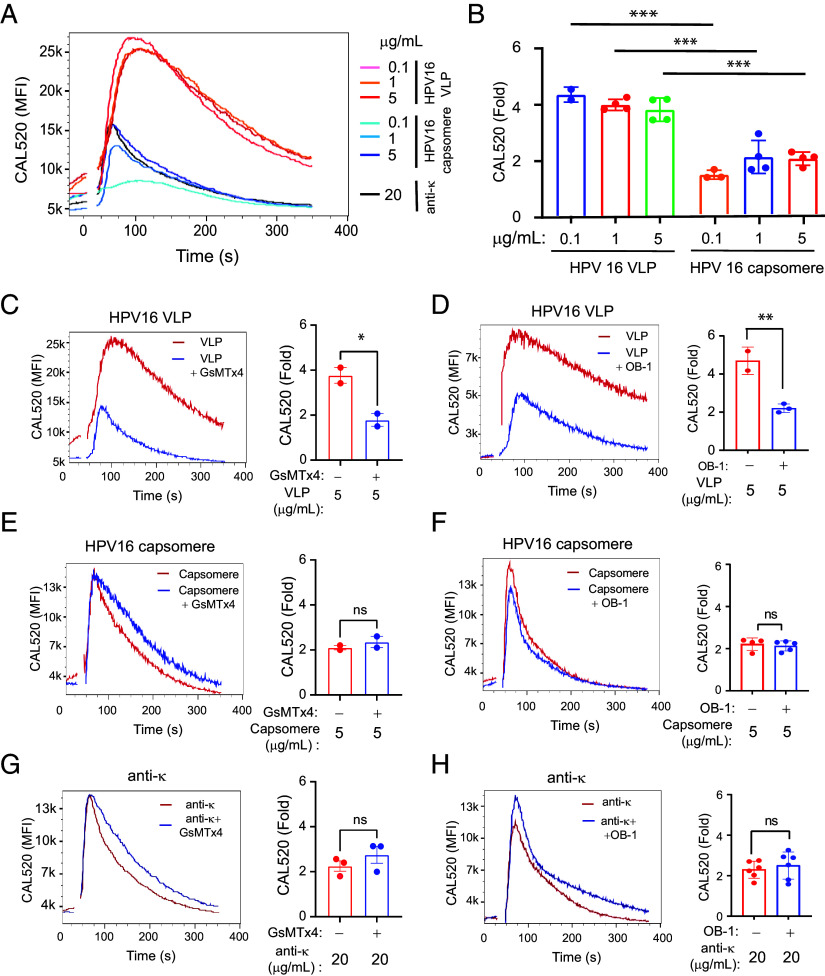
B cell responses to membrane-presented antigens appear to be strongly favored over soluble antigens in vivo suggesting that vaccines that mimic membrane-presented antigens may be highly efficacious. We recently demonstrated that human B cell responses to membrane-associated but not to soluble antigens in vitro depended on the expression and activity of the plasma membrane mechanosensitive ion channel, Piezo1. Here, we provide evidence that the efficacy of the current human papillomavirus virus-like particle (HPV VLP) vaccines may be due in part to their inherent ability to mimic Piezo1-dependent membrane presentation of antigens to B cells. We compared HPV-specific human B cell responses to HPV VLPs versus soluble HPV pentameric capsomeres and showed that although both induced calcium responses, only HPV VLP-induced responses were blocked by Piezo1 inhibitors. The kinetics of internalization of HPV-VLP and capsomeres into HPV-specific B cells were similar and neither required Piezo1 function as shown by small interfering RNA (siRNA)-mediated knockdown of Piezo. However, trafficking of HPV-VLPs into intracellular major histocompatibility complex (MHC) class II, lysosomal associated membrane protein 1 (LAMP1)+ antigen-processing compartments was Piezo1-dependent, whereas trafficking of capsomeres was not. In addition, a time course of intracellular trafficking suggested that colocalization of HPV-VLP with MHC classII was more stable over time as compared to capsomeres. Taken together these findings suggest that the ability of HPV-VLP vaccines to mimic Piezo1-dependent membrane antigen presentation may be exploited in the design of highly effective human vaccines.

Keywords: HPV-VLP vaccine; Piezo1; antigen presentation; human B cells.


Conflict of interest statement

Competing interests statement:The authors declare no competing interest.

Figures

Fig. 1.
Fig. 1.
HPV16 VLPs and HPV16 capsomeres bind exclusively to HVP16-specific B cells. (A) Flow cytometry plots show the binding of HPV16 VLP-AF647 and HPV16 VLP-AF488 to PBMCs alone (Left), WT Ramos (Middle), or A24 Ramos (Right). (B) Flow cytometry plots showing binding of either HPV16 VLP-AF488 and HPV16 VLP-AF647 (Left), HPV16 VLP-AF647 and capsomere-AF488 (Middle), or capsomere-AF647 and capsomere-AF488 (Right) to A24 Ramos diluted into PBMCs at a ratio of 1 A24 Ramos:100 PBMCs. (C) Representative flow cytometry plots of A24 Ramos diluted into PBMCs at a ratio of 1 A24 Ramos:100 PBMCs are gated on A24 Ramos labeled with fluorescent Abs specific for CD19, CD27, and CD10 (CD19+CD27brightCD10bright). A24 Ramos were preincubated without competitor or with the indicated unlabeled competitors HPV16 VLPs, BPV, or HPV58 VLPs, at concentrations of 4 or 20 µg/mL followed by incubation with HPV16 VLP-AF647 and HPV16 VLP-AF488. (D) A summary of the data as in C is given, representing replicates in three independent experiments. The data were analyzed by a Kruskal–Wallis test with a Dunn’s correction and P-values > 0.05 were considered not significant (ns). **P < 0.01. (E) Representative flow cytometry plots of A24 Ramos diluted into PBMCs at a ratio of 1 A24 Ramos:100 PBMCs and gated on A24 Ramos as in C. Cells were preincubated without competitor or with the indicated concentrations of unlabeled HPV16 capsomeres, followed by incubation with HPV16 VLP-AF647 and HPV16 VLP-488. (F) A summary of the data in E is shown, representing replicates in three independent experiments. The data were analyzed by a Kruskal–Wallis test with a Dunn’s correction. ns, P > 0.05, ***P < 0.001.
Fig. 2.
Fig. 2.
HPV16 VLPs induce more robust and prolonged calcium fluxes as compared to HPV16 capsomeres in a Piezo1-dependent manner. A24 Ramos cells were loaded with a calcium indicator dye, CAL520, and baseline calcium levels were recorded by flow cytometry. Upon addition of 0.1, 1, and 5 µg/mL of HPV16 VLPs, HPV16 capsomeres or 20 µg/mL goat F(ab′)2 anti-Igκ Abs (anti-κ) calcium fluxes were recorded with time. (A) Shown is a representative plot of the median CAL520 fluorescence intensities (MFI) over time at the indicated concentrations of HPV16 VLP or HPV16 capsomere or anti-κ. (B) Quantifications of the fold change in CAL520 MFI during BCR stimulation relative to the baseline at each indicated concentration of HPV16 VLPs or HPV16 capsomere are given as the mean ± SEM for 2-4 independent experiments. (CH) A24 Ramos B cells were either untreated or pretreated with GsMTx4 (5 µM) for 30 min or OB-1 (20 µM) for 1 h at 37 °C and calcium fluxes were recorded upon addition of HPV16 VLP (C and D), HPV16 capsomere (E and F), or anti-κ (G and H). Shown for each antigen/inhibitor combination are the representative kinetics plots of the calcium fluxes (Left) and the fold change (Right) as the mean ± SEM from 2 to 3 independent experiments. Statistical significance was determined by the two-tailed paired t-test. ns, P > 0.05, *P ≤ 0.05, **P ≤ 0.01, and ***P ≤ 0.001.
Fig. 3.
Fig. 3.
HPV16 VLP-induced B cell activation results in loss of viability that can be partially rescued by T cell help. A24 Ramos cells were cultured in the absence or presence of HPV16 capsomeres (10 µg/mL) or HPV16 VLPs (10 µg/mL) in the absence or presence of T cell help (Th) media for the indicated time. Shown are the graphs with mean ± SD from flow cytometry for: (A) the percentage of surface CD69-expressing cells; (B) the percent of viable cells (Viability) measured using a Live/Dead fixable viability dye. Each symbol is an individual replicate. Data are representative of two independent experiments, and statistical significance was determined by one-way ANOVA with Tukey’s multiple comparison test. ****P < 0.0001, ***P < 0.001, **P < 0.01, ns, not significant. ####P < 0.0001 between unstimulated and either HPV16 capsomeres or HPV16 VLPs-stimulated cells in A.
Fig. 4.
Fig. 4.
Piezo1 down-expression has no influence on both HPV16 VLPs and capsomeres-driven BCR down-modulation. A24 Ramos cells were transfected with 1 µM of either control or PIEZO1 siRNA using Amaxa nucleofection system as described in Method and cultured for 3 d at 37 °C in the 5% CO2 incubator. (A) siGlo-Red transfection indicator, a red fluorescent oligonucleotide duplex, was used in three different concentrations (0.5, 1.0, and 1.5 µM) for the optimization of siRNA transfection efficiency in A24 Ramos cells. (B and C) Representative flow cytometry plot for total Piezo1 expression stained by rabbit anti-Piezo1-Janelia Fluor 549 polyclonal antibodies (Piezo1-JF549) for either Control (Control)–or PIEZO1 siRNA (PIEZO1)-transfected cells (B). The level of Piezo1 expression in PIEZO1 siRNA transfected cells is given as a percent of Piezo1 expressed in Control cells (C). Shown is a bar graph with mean ± SD with dots, indicating data from three independent experiments. One-sample t test was performed for the statistical analysis. **P < 0.01. (D) Surface BCR downregulation was analyzed for HPV16 VLPs and capsomeres in Control and Piezo1 knock-down (KD) cells (Piezo1 KD-VLP and Piezo1 KD-Capsomere) by Control and PIEZO1 siRNA, respectively. HPV16 VLPs or capsomeres (5 µg/mL) were added to Control or Piezo1 KD cells, allowed to bind to the A24 BCR on ice for 30 min and then chased for the indicated time at 37 °C and fixed with 4% paraformaldehyde (PFA). Surface BCR was labeled with either goat IgG anti-Igµ heavy chain (IgM HC) or anti-Igκ light chain (Igκ LC) and analyzed for the percent of Piezo1 expression in control cells in surface level of BCR by flow cytometry. Ratios of surface IgM HC or Igκ LC gMFIs at each time point (T = t) relative to those on ice (T = 0) was calculated (T = t/T =0). Statistical significance (95% CI) was assessed with the “compareGrowthCurves” function from the statmod statistical modeling package for three independent experiments (26).
Fig. 5.
Fig. 5.
Piezo1 down-expression influences on the trafficking of HPV16 VLPs into the LAMP1+HLA-DR+ organelles. Control or Piezo1-KD A24 Ramos cells were allowed to settle on the chambered cover glass for 20 min at 37 °C and incubated with 5 µg/mL of Alexa Fluor 488-conjugated HPV16 VLPs (VLP) or HPV16 capsomeres (Capsomere) for 120 min at 37 °C in the CO2 incubator. The cells were fixed with 4% PFA, permeabilized with 0.1% Triton-X100, and labeled with mouse mAbs specific for LAMP1 and HLA-DR conjugated with Alexa Fluor 549 and Alexa Fluor 647, respectively. 3D surface and sectional views of single cell images were obtained by Z-stack confocal imaging using a ZEISS 880 confocal laser scanning microscope followed by 3D reconstruction using Imaris and Huygens software as described in Materials and Methods. (A and B) Shown are 3D surface views of HPV16 VLPs or capsomeres (Antigen, green), LAMP1 (red), or HLA-DR (Blue) or three-color merged images of the representative cells (A). Also shown are orthogonal views by XY, XZ, and YZ planes of three-color merged 3D images of the same cells shown in panel A to provide a clear image of the colocalization of the three markers in intracellular vesicles (B). Yellow arrowheads indicate the vesicles containing all three markers: antigen, HLA-DR, and LAMP1. Fluorescence intensity is not comparable between different cells. (Scale bar, 2 µm.) (CE) The ratio of antigens colocalized with LAMP1 or HLA-DR, or that of LAMP1 colocalized with HLA-DR was quantified among different experimental conditions by Manders overlap coefficient per cell between antigens and LAMP1 (C), between antigens and HLA-DR (D) or between LAMP1 and HLA-DR (E) using Huygens software. Over ninety cells per condition combined from two independent experiments were analyzed. Graphs are shown as box plots with median and Tukey whiskers. ****P < 0.0001, **P < 0.01, ns, not significant; Statistical significance was performed by the Kruskal–Wallis test in all four groups followed by Dunn’s multiple comparison tests between Control and Piezo1 KD cells treated with either HPV16 VLPs or HPV16 capsomeres, or between HPV16 VLPs and HPV16 capsomeres within Control or Piezo1 KD cells. (F) HPV16 VLPs or capsomeres colocalized with LAMP1 and HLA-DR double positive (LAMP1+HLA-DR+) organelles were quantified and compared between Control and Piezo1 KD cells as the percentage of HPV16 VLPs or capsomeres colocalized with LAMP1+HLA-DR+ vesicles by Manders overlap coefficient. Data are shown as box plots with median and Tukey whiskers for over 40 cells per condition from two independent experiments. Statistical significance was compared between VLP- and Capsomere-treated cells in Control siRNA-transfected cells, and between Control and Piezo1 KD cells in either VLP or Capsomere-treated cells by the two-tailed Mann–Whitney test *P < 0.05, ns, P > 0.05.

References

    1. Markowitz L. E., Schiller J. T., Human papillomavirus vaccines. J. Infect. Dis. 224, S367–S378 (2021). - PMC - PubMed
    1. Schiller J. T., Lowy D. R., Understanding and learning from the success of prophylactic human papillomavirus vaccines. Nat. Rev. Microbiol. 10, 681–692 (2012). - PMC - PubMed
    1. Bachmann M. F., et al. , The influence of antigen organization on B cell responsiveness. Science 262, 1448–1451 (1993). - PubMed
    1. Batista F. D., Neuberger M. S., Affinity dependence of the B cell response to antigen: A threshold, a ceiling, and the importance of off-rate. Immunity 8, 751–759 (1998). - PubMed
    1. Kato Y., et al. , Multifaceted effects of antigen valency on B cell response composition and differentiation in vivo. Immunity 53, 548–563.e8 (2020). - PMC - PubMed

MeSH terms