. 2025 Mar 11;122(10):e2417943122.
doi: 10.1073/pnas.2417943122. Epub 2025 Mar 3.

TEX38 localizes ZDHHC19 to the plasma membrane and regulates sperm head morphogenesis in mice

Affiliations

TEX38 localizes ZDHHC19 to the plasma membrane and regulates sperm head morphogenesis in mice

Yuki Kaneda et al. Proc Natl Acad Sci U S A. .

Abstract

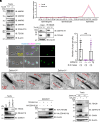
Sperm morphogenesis is a tightly regulated differentiation process, disruption of which leads to sperm malfunction and male infertility. Here, we show that Tex38 knockout (KO) male mice are infertile. Tex38 KO spermatids exhibit excess retention of residual cytoplasm around the head, resulting in abnormal sperm morphology with backward head bending. TEX38 interacts and colocalizes with ZDHHC19, a testis-enriched acyltransferase catalyzing protein S-palmitoylation, at the plasma membrane of spermatids. ZDHHC19 and TEX38 are each downregulated in mouse testes lacking the other protein. TEX38 stabilizes and localizes ZDHHC19 to the plasma membrane of cultured cells and vice versa, consolidating their interdependence. Mice deficient in ZDHHC19 or harboring a C142S mutation that disables the palmitoyltransferase activity of ZDHHC19 display phenotypes resembling those of Tex38 KO mice. Strikingly, ZDHHC19 palmitoylates ARRDC5, an arrestin family protein regulating sperm differentiation. Overall, our findings indicate that TEX38 forms a stable complex with ZDHHC19 at the plasma membrane of spermatids, which governs downstream S-palmitoylation of proteins essential for morphological transformation of spermatids.

Keywords: S-acylation; spermiation; sterility.


Conflict of interest statement

Competing interests statement:The authors declare no competing interest.

Figures
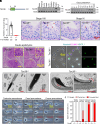
Fig. 1.
Fig. 1.
Tex38–/– male mice were infertile due to sperm head folding. (A) A transmembrane domain (TMD) in TEX38 was predicted using SMART (http://smart.embl-heidelberg.de/). (B) RT-PCR of Tex38 using RNAs of multiple adult mouse tissues. Tex38 is predominantly expressed in the testis. Actb was used as a loading control. (C) RT-PCR of Tex38 using RNAs obtained from postnatal day 0 to 35 mouse testes. (D) The numbers of pups per plug. WT or Tex38–/– male mice were individually mated with three WT females. (E) Histology of stage VIII and IX seminiferous tubules. Red arrowheads indicate unreleased sperm lining the lumen. (F) Histology of cauda epididymides. Yellow arrowheads indicate round cells, presumably degenerated early spermatids. (G) Immunostaining of IZUMO1 (magenta) and SYPL1 (green) in WT and Tex38 KO spermatozoa. IZUMO1 and SYPL1 was used as a marker for the acrosome and CD, respectively. Nuclei were visualized with Hoechst 33342 (blue). (H) Ultrastructural analysis of cauda epididymal spermatozoa by TEM. Retained residual cytoplasm was observed in the sperm head (red asterisks). The sperm plasma membrane does not fold inward at the head bending site (red arrowheads). (I) Testicular and caput and corpus epididymal spermatozoa are subdivided into three morphological categories; straight (Straight), partially bent (Partial), and completely bent (Complete). (J) Percentages of spermatozoa with the three types of head abnormalities in WT and Tex38−/− male mice (n = 3). Head folding was rarely observed in testicular spermatozoa, while a multitude of folded spermatozoa appeared in the corpus and cauda epididymides.
Fig. 2.
Fig. 2.
TEX38 interacts with ZDHHC19, ablation of which phenocopies Tex38–/– male mice. (A) Immunoblot analyses of representative TEX38-interacting proteins identified by co-IP/MS in WT and Tex38–/– testes. β-actin was used as a loading control. The black asterisk indicates nonspecific bands. (B) Expression patterns of Tex38 and Zdhhc19 in mouse spermatogenic cells based on a previously published scRNA-seq dataset (25). (C) Validation of the interaction between TEX38 and ZDHHC19 in testes. ZDHHC19 was coimmunoprecipitated with TEX38. β-actin was used as a loading control. The black asterisk indicates nonspecific protein bands. (D) The numbers of pups per plug. (E) Immunostaining of IZUMO1 (magenta) and SYPL1 (green) in WT and Zdhhc19 KO spermatozoa. IZUMO1 and SYPL1 was used as a marker for the acrosome and CD, respectively. Nuclei were visualized with Hoechst 33342 (blue). (F) Ultrastructural analysis of elongated spermatids by TEM. Red arrowheads indicate the cytoplasm of WT and Zdhhc19 KO spermatids. (G) Immunoblot analyses of TEX38 and ZDHHC19 in WT, Tex38–/–, and Zdhhc19–/– testes. IZUMO1 was used as a loading control. The black asterisk indicates nonspecific bands. (H) TEX38-1D4 and/or HA-ZDHHC19 were individually or simultaneously overexpressed in HEK293T cells. β-actin was used as a loading control. (I) acyl-MfTag exchange (AME) assay of proteins extracted from WT and Zdhhc19–/– testes. Palmitic acid was cleaved by NH2OH and replaced by the MfTag. CANX was used as a positive control as it contains two S-palmitoylation sites. β-actin was used as a loading control.
Fig. 3.
Fig. 3.
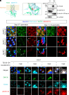
TEX38 and ZDHHC19 colocalize at the plasma membrane of spermatids. (A) The structure of the TEX38–ZDHHC19 heterodimer predicted by AlphaFold3. The DHHC domain of ZDHHC19 is colored in red. (B) Immunostaining of TEX38 and ZDHHC19 in testis cryosections. TEX38 and ZDHHC19 were localized to the plasma membrane of WT spermatids and to the Golgi apparatus (white arrowheads) near the proacrosome of the KO spermatids lacking the other protein. The nuclei and acrosome of spermatids were visualized with Hoechst 33342 (blue) and lectin-PNA (cyan), respectively. White asterisks indicate nonspecific signals observed in the manchette. (C) Fractionation analyses of testis proteins. CY and PM indicate cytosol and plasma membrane, respectively. Na+/K+-ATPase α1 was used as a positive control for the plasma membrane fraction while GAPDH was used as a positive control for the cytosol fraction. The black asterisk indicates nonspecific bands. (D) TEX38-1D4 and/or HA-ZDHHC19 were individually or simultaneously expressed in COS-7 cells for immunocytochemistry analyses. The nuclei and Golgi apparatus of COS-7 cells were visualized with Hoechst 33342 (blue) and an anti-GM130 antibody (Green), respectively.
Fig. 4.
Fig. 4.
The PAT activity of ZDHHC19 is important for sperm head morphogenesis. (A) The domain composition of ZDHHC19. Cysteine 142 in the DHHC domain is crucial for autoacylation. TMD indicates a transmembrane domain. (B) The complex of TEX38 and the autoacylated ZDHHC19 predicted by AlphaFold3. The DHHC domain of ZDHHC19 was colored in red. Palmitic acid is indicated in purple. (C) Schematics of generating Zdhhc19C142S/C142S mice by CRISPR/Cas9. White and black boxes indicate untranslated regions and protein coding regions, respectively. gRNAs are shown in purple. (D) Confirmation of the C142S KI mutation with direct sequencing of the PCR product amplified by Fw #3 and Rv #3 primers. (E) Morphology of spermatozoa obtained from the cauda epididymides of WT and Zdhhc19C142S/C142S mice. (F) Proteomic analyses unraveling potential substrates of ZDHHC19 in testes. Co-IP/MS analyses detected 130 proteins showing differential binding ability to WT and C142S mutant ZDHHC19 [FC (WT/C142S) > 3 = 82 proteins; FC (C142S/WT) > 3 = 48 proteins]. acyl-biotin exchange (ABE)/MS analyses detected 79 proteins that were higher palmitoylated in WT than Zdhhc19–/– testes [FC (WT/KO) > 3]. (G) Four top hits of ZDHHC19 substrates detected in both co-IP/MS and ABE/MS. All four proteins were previously identified as candidate palmitoylated proteins in testes (31). (H) AME assay of ZDHHC19-mediated palmitoylation in HEK293T cells. (I) AME assay of ZDHHC19-mediated palmitoylation in COS-7 cells.

References

    1. Miyata H., Shimada K., Kaneda Y., Ikawa M., Development of functional spermatozoa in mammalian spermiogenesis. Development 151, dev202838 (2024). - PubMed
    1. Clermont Y., Kinetics of spermatogenesis in mammals: Seminiferous epithelium cycle and spermatogonial renewal. Physiol. Rev. 52, 198–236 (1972). - PubMed
    1. O’Donnell L., Nicholls P. K., O’Bryan M. K., McLachlan R. I., Stanton P. G., Spermiation. Spermatogenesis 1, 14–35 (2011). - PMC - PubMed
    1. Breucker H., Schäfer E., Holstein A.-F., Morphogenesis and fate of the residual body in human spermiogenesis. Cell Tissue Res. 240, 303–309 (1985). - PubMed
    1. Agarwal A., Majzoub A., Parekh N., Henkel R., A schematic overview of the current status of male infertility practice. World J. Men’s Health 37, 308–322 (2019). - PMC - PubMed