. 2025 Dec;17(1):2469716.
doi: 10.1080/19490976.2025.2469716. Epub 2025 Mar 3.

Rhodococcus spp. interacts with human norovirus in clinical samples and impairs its replication on human intestinal enteroids

Affiliations

Rhodococcus spp. interacts with human norovirus in clinical samples and impairs its replication on human intestinal enteroids

Cristina Santiso-Bellón et al. Gut Microbes. 2025 Dec.

Abstract

Viral infections, particularly human norovirus (NoV), are a leading cause of diarrheal diseases globally. To better understand NoV susceptibility, it is crucial to investigate both host glycobiology and the influence of the microbiota. Histo-blood group antigens (HBGA) displayed on surfaces of host cells act as NoV receptors, while certain bacteria express HBGA-like substances, facilitating virus-bacteria interactions. To identify bacterial species interacting with NoV during infection, stool samples from children infected with NoV GII.4 were analyzed. The samples were subjected to bacteria separation using anti-NoV GII.4 polyclonal antibodies coupled to magnetic beads, followed by microbiota profiling through 16S rDNA sequencing. This approach identified the genus Rhodococcus as enriched in samples captured with anti-NoV antibodies compared to controls. Electron microscopy confirmed the binding of NoV GII.4 Sydney 2012 viral-like particles (VLP) to five Rhodococcus strains from different species, which expressed HBGA-like substances on their surfaces, particularly from A and B blood groups. In human intestinal enteroids, Rhodococcus erythropolis reduced NoV GII.4 Sydney 2012 infection levels under displacement, exclusion and competitive exclusion settings. Extracellular polysaccharides (EPS) isolated from Rhodococcus strains bound VLP from both GII.4 and GII.6 genotypes. Blocking antibodies targeting A and B epitopes reduced the binding of the EPS from R. erythropolis to GII.6 VLP, while enhanced binding to GII.4 VLP was observed when A and B epitopes were blocked. These findings revealed the interaction of Rhodococcus to NoV in an in vitro setting and open new avenues for developing innovative antiviral strategies to prevent and treat NoV infections through the HBGA-like substances present in their EPS.

Keywords: Human norovirus; Rhodococcus; gut microbiota; histo-blood group antigens; human intestinal enteroids; stool samples.


Conflict of interest statement

No potential conflict of interest was reported by the author(s).

Figures
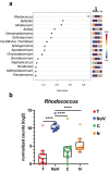
Figure 1.
Figure 1.
Bacteria interacting with NoV GII.4 Sydney 2012 in stool samples. (a) LDA of bacterial genera quantified from clinical samples with NoV. The LDA score of the 15 taxa with significant differences (p < 0.05 in Kruskal-Wallis test, adjusted by FDR) in a LEfse analysis is presented. Rhodococcus genus obtained the highest LDA score. The differences in color intensity correspond to higher or lower abundance of genera for the four bacterial populations. T, total bacteria present in stools before experiment; C, bacteria recognized by the IgG isotype control antibody; N, bacteria not recognized by the isotype control antibody nor the specific antibody; NoV, bacteria recognized by the anti-NoV GII.4 Sydney 2012 antibody. (b) Abundance of Rhodococcus (normalized counts) for each subsample obtained from the nine diarrhea samples. The counts of a sample that showed abnormally high Rhodococcus counts are displayed as outliers. ****p < 0.0001 for Sidak’s test after comparing NoV subsample to the rest of subsamples.
Figure 2.
Figure 2.
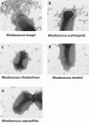
Interaction between Rhodococcus spp. And NoV GII.4 Sydney 2012 VLP. TEM images with negative staining of the adhesion between (a), Rhodococcus hoagie; (b), Rhodococcus erythropolis; (c), Rhodococcus rhodochrous; (d), Rhodococcus rhodnii, and. (e) Rhodococcus coprophilus and NoV GII.4 Sydney 2012 VLP (magnification at 21000X). The bars correspond to 200 nm.
Figure 3.
Figure 3.
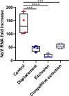
Effect of the presence of R. erythropolis on NoV infection in HIE. The viral RNA-fold increase from displacement, exclusion and competitive exclusion experiments are shown. In the three experimental settings, R. erythropolis (OD600 1) and 104 genome equivalents of NoV GII.4 Sydney 2012 isolate were added. After 2 h of incubation, the HIE monolayers were washed and NoV infection was let to proceed for further 24 h. Viral RNA was measured by rt-qPCR. ***p < 0.001, ****p < 0.0001 for Sidak’s test compared to control without R. erythropolis added.
Figure 4.
Figure 4.
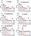
Rhodococcus spp. express compounds similar to blood group antigens on their surface. The ELISA results of recognition by specific monoclonal anti-hbga antibodies on surface substances of R. hoagii (a), R. erythropolis (b), R. rhodochrous (c), R. rhodnii (d), R. coprophilus (e) and E. coli (f) are shown. The image depicts the mean of triplicates ± SD. Statistical significance is shown with asterisks (*p < 0.05, **p < 0.01, ***p < 0.001, ****p < 0.0001; Sidak’s test) for each strain compared to the negative control, “None” (samples incubated only with the secondary antibody anti-mouse HRP).
Figure 5.
Figure 5.
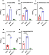
The EPS from Rhodococcus spp. bind to NoV GII.4 Sydney 2012 and NoV GII.6 VLP. Binding of the five isolated EPS to two NoV VLP (GII.4 Sydney 2012 and GII.6) and to rotavirus a DLP (RVA DLP) is shown. RVA DLP were used as negative control since they do not bind HBGA. Panel (a) shows the binding of Rhodococcus hoagie; panel (b), R. erythropolis; panel (c), R. rhodochrous; panel (d), R. rhodnii, and panel (e), R. coprophilus. The GII.6 VLP show a significant (**p < 0.01. Student’s t-test) higher binding to each EPS compared to GII.4 Sydney 2012 VLP.
Figure 6.
Figure 6.
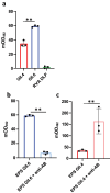
Blocking assay by anti-A and anti-B antibodies of R. erythtopolis EPS binding to NoV VLP. Biotinylated EPS (EPS-Biotin) from R. erythropolis was utilized in a binding assay with NoV GII.4 Sydney 2012 and NoV GII.6 VLP. Panel (a) shows that after biotinylation, the EPS still bound specifically to the NoV VLP but not to RVA DLP, being the binding to GII.6 VLP significantly (**p < 0.01. Student’s t-test) higher compared to GII.4 Sydney 2012 VLP. Panel (b) shows that anti-A and anti-B antibodies significantly (**p < 0.01. Student’s t-test) decreased the binding of the EPS to NoV GII.6. Panel (c) shows that incubation with anti-A and anti-B antibodies significantly (**p < 0.01. Student’s t-test) increased the binding of the EPS to NoV GII.4 Sydney 2012.

References

    1. World Health Organization (WHO) . The top 10 causes of death [Internet]. 2020. [accessed 2023 June 19]. https://www.who.int/news-room/fact-sheets/detail/the-top-10-causes-of-death.
    1. Centers for Disease Control and Prevention (CDC). Norovirus Burden and Trends [Internet]. 2023. [accessed 2023 June 19]. https://www.cdc.gov/norovirus/burden.html#worldwide.
    1. Bucardo F, Lindgren PE, Svensson L, Nordgren J, Sestak K.. Low prevalence of rotavirus and high prevalence of norovirus in hospital and community wastewater after introduction of rotavirus vaccine in Nicaragua. PLOS ONE. 2011;6(10):6. doi:10.1371/journal.pone.0025962. - DOI - PMC - PubMed
    1. Patel MM, Hall AJ, Vinjé J, Parashar UD.. Noroviruses: a comprehensive review. J Clin. 2009;44(1):1–19. doi:10.1016/j.jcv.2008.10.009. - DOI - PubMed
    1. Donaldson EF, Lindesmith LC, Lobue AD, Baric RS. Norovirus pathogenesis: mechanisms of persistence and immune evasion in human populations. Immunological Rev. 2008;225(1):190–211. doi:10.1111/j.1600-065X.2008.00680.x. - DOI - PubMed

Substances

LinkOut - more resources