. 2025 Mar 3;10(1):82.
doi: 10.1038/s41392-025-02184-0.

Ultra-high dose rate radiotherapy overcomes radioresistance in head and neck squamous cell carcinoma

Affiliations

Ultra-high dose rate radiotherapy overcomes radioresistance in head and neck squamous cell carcinoma

Hong-Shuai Li et al. Signal Transduct Target Ther. .

Abstract

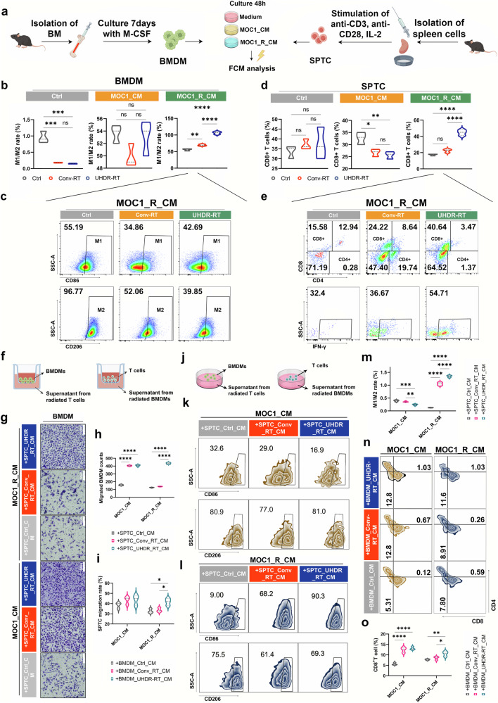
Radiotherapy (RT) resistance in head and neck squamous cell carcinoma (HNSCC) significantly hampers local control and patient prognosis. This study investigated the efficacy and molecular mechanisms of high-energy X-ray-based ultra-high dose rate radiotherapy (UHDR-RT) in overcoming RT resistance. The established RT-resistant HNSCC cell lines and animal models were subjected to UHDR-RT or conventional RT (Conv-RT) via a high-power rhodotron accelerator. Cellular assays assessed the malignant phenotype, viability, and degree of DNA damage, whereas in vivo evaluations focused on tumor proliferation and the tumor immune microenvironment (TiME). Transcriptome sequencing and Olink proteomics were employed to explore the underlying mechanisms involved. In vitro experiments indicated that UHDR-RT suppressed radioresistant cell proliferation and invasion, while promoting apoptosis and exacerbating DNA damage. In contrast, its efficacy in radiosensitive cells was comparable to that of Conv-RT. In vivo studies using patient-derived xenograft nude mice models demonstrated that UHDR-RT only partially reversed RT resistance. Transcriptomic and proteomic analyses of C57BL/6J mice models revealed the predominant role of TiME modulating in reversing radioresistance. Immunofluorescence and flow cytometry confirmed increased CD8+ T cells and an increased M1/M2 macrophage ratio post-UHDR-RT. Mechanistically, UHDR-RT activated CD8+ T cells, which stimulated M1 macrophages through paracrine IFN-γ signaling, thereby enhancing TiME activation. Furthermore, the activated M1 macrophages secreted CXCL9, which in turn reactivated CD8+ T cells, forming a feedforward loop that amplified TiME activation. This study elucidates the dual role of UHDR-RT in directly inducing DNA damage and modulating the TiME, highlighting its potential in treating radioresistant HNSCC.


Conflict of interest statement

Competing interests: The authors declare no competing interests.

Figures

Fig. 1
Fig. 1
Cellular experiments demonstrated that UHDR-RT reversed radioresistance and led to increased DNA damage. Compared with Conv-RT, UHDR-RT significantly inhibited CAL33_R cell proliferation (a, b), migration (c, d), and infiltration ability (e, f), increased DNA damage (gi), decreased the mitochondrial membrane potential (g, h, j), and increased the degree of cell death (g, h, k), apoptosis (g, h, l), and ICD (m). Ctrl control; Conv-RT conventional radiotherapy; UHDR-RT ultra-high dose rate radiotherapy; ICD immunogenic cell death. *p < 0.05; **p < 0.01; ***p < 0.001; ****p < 0.0001. The data are presented as the mean ± SEM (a, b, d, f, and im). Comparisons were performed using one-way ANOVA with Tukey’s test for multiple comparisons
Fig. 2
Fig. 2
Mouse experiments confirmed that UHDR-RT reversed radioresistance and led to TiME activation and DNA damage. C57BL/6J mouse experiments confirmed that in the MOC1_R group, tumors (n = 5) in the UHDR-RT group achieved better tumor control than those in the Conv-RT group (n = 5) (a, b). Ki-67 staining, TUNEL staining, and DNA damage IF of mouse tumor tissues revealed that UHDR-RT significantly increased DNA damage and apoptosis levels and decreased tumor proliferative activity in the MOC1_R group (cg). Transcriptome PCA revealed subgroups within the MOC1_R group (h), volcano plots revealed differential genes in the UHDR-RT and Conv-RT subgroups within the MOC1_R group (i), and KEGG analysis revealed an enriched signaling pathway for the UHDR-RT subgroup within the MOC1_R group (j). GSEA enrichment analysis revealed upregulated (k) and downregulated (l) pathways in the UHDR-RT subgroup. Ctrl control; Conv-RT conventional radiotherapy; UHDR-RT ultra-high dose rate radiotherapy; PCA principal component analysis. ns not significant; *p < 0.05; **p < 0.01; ***p < 0.001; ****p < 0.0001. The data are presented as the mean ± SEM (b and eg). Comparisons were performed using two-way (b) or one-way (eg) ANOVA with Tukey’s test for multiple comparisons
Fig. 3
Fig. 3
Abscopal effects and PDX experiments confirmed the crucial role of TiME activation in reversing resistance. Compared with Conv-RT (n = 5), UHDR-RT (n = 5) also significantly decreased the volume of contralateral tumors (a, b). Compared with Conv-RT, UHDR-RT significantly reduced the spleen burden in the MOC1_R group (c, d). Compared with Conv-RT, UHDR-RT (n = 5) reduced the PDX volume (n = 5) but not significantly (e, f). HE staining revealed typical squamous carcinoma histologic features, and TUNEL staining and DNA damage staining suggested that UHDR-RT significantly increased apoptosis and DNA damage levels (gi). The ring heat plot shows differential gene expression profiles between groups (j), and GSEA demonstrated different signaling pathway enrichment between the two groups (k). Ctrl control; Conv-RT conventional radiotherapy; UHDR-RT ultra-high dose rate radiotherapy; PDX patient-derived xenograft; HE staining hematoxylin‒eosin staining; TiME tumor immune microenvironment; ns not significant; *p < 0.05; **p < 0.01; ***p < 0.001; ****p < 0.0001. The data are presented as the mean ± SEM (b, d, f, and h, i). Comparisons were performed using two-way (b, f) or one-way (d, h, i) ANOVA with Tukey’s test for multiple comparisons
Fig. 4
Fig. 4
Integrative analysis of transcriptomics and proteomics confirmed the activation of the TiME. Olink proteome PCA revealed subgroup clustering of the MOC1_R group (a). Volcano plot showing significantly upregulated proteins in the UHDR-RT subgroup compared with the Conv-RT subgroup (b) and an interaction between these proteins (c). There is a cross-relationship between pathways enriched in proteome-differentiated proteins and pathways enriched in transcriptome-differentiated genes, and the cross-pathways (d) are strongly associated with intrinsic as well as adaptive immune responses (e). Transcriptomic data (f), and Olink data (g) revealed significant upregulation of CD8+ T-cell and M1-type macrophage surface markers in the UHDR-RT subgroup. Cybersort analysis revealed differences in TiME infiltration between the UHDR-RT subgroup and the Conv-RT subgroup (h). Ctrl control; Conv-RT conventional radiotherapy; UHDR-RT ultra-high dose rate radiotherapy; TiME tumor immune microenvironment; DEGs differentially expressed genes; DEPs differentially expressed proteins; IHC immunohistochemistry; IF immunofluorescence. *p < 0.05; **p < 0.01; ***p < 0.001; ****p < 0.0001. The data are presented as the median and interquartile range (h). The boxplots indicate median (center), 25th and 75th percentiles (bounds of box), and minimum and maximum (whiskers). Comparisons were performed using one-way ANOVA method
Fig. 5
Fig. 5
Significant aggregation of CD8+ T cells along with notable M1 polarization of macrophages led to TiME activation. A monochromatic IF assay suggested significant aggregation of CD8+ T cells, macrophages, and DCs in the UHDR-RT subgroup of the MOC1_R group (ae), and multicolor IF verified the colocalization of CD8+ T cells and macrophages as well as significant M1 polarization of macrophages (fh). The multi-color FCM scheme (i). The results of tumor and spleen multicolor FCM analysis were consistent with those of IF (jl). Ctrl control; Conv-RT conventional radiotherapy; UHDR-RT ultra-high dose rate radiotherapy; DC dendritic cells; TiME tumor immune microenvironment. *p < 0.05; **p < 0.01; ***p < 0.001; ****p < 0.0001. The data are presented as the mean ± SEM (ce) or median and interquartile range (j–l). Comparisons were performed using one-way (ce) ANOVA with Tukey’s test for multiple comparisons
Fig. 6
Fig. 6
Endogenous antibody blockade experiments in mice confirmed the crucial role of CD8+ T cells and macrophages. Subcutaneous injection of the MOC1_R cell line was used to construct a radiotherapy-resistant C57BL/6J mouse model, which received Conv-RT, UHDR-RT, or UHDR-RT combined with anti-PD-1, anti-CD8a or anti-CSF1R (a, b). Tumor growth curves of the different groups (c). Multicolor immunofluorescence demonstrated the colocalization of CD8+ T cells and macrophages in response to different treatments (df). FCM was used to determine the proportions of CD8+ T cells (g) and macrophages (h, i) in the different treatment groups. The proportions of CD8-, CD86- and CD206-positive cells (j, k) were calculated, and the M1/M2 ratio was computed (l). Ctrl control; Conv-RT conventional radiotherapy; UHDR-RT ultra-high dose rate radiotherapy. *p < 0.05; **p < 0.01; ***p < 0.001; ****p < 0.0001. The data are presented as the mean ± SEM (c). Comparisons were performed using two-way (c) or one-way (e, j, k) ANOVA with the Tukey’s test for multiple comparisons
Fig. 7
Fig. 7
In vitro experiments demonstrated the interplay between CD8+ T cells and macrophages induced by UHDR-RT. Independent in vitro irradiation assay procedure (a). BMDMs (b, c) and SPTCs (d, e) that were cultured with different CM were treated with different radiotherapy modalities and then subjected to FCM. Transwell assay design (f). Transwell staining results of BMDMs (g). Statistical analysis of the transwell assay results for BMDMs (h). Statistical analysis of the transwell assay results for SPTCs (i). Co-culture assay design diagram (j). FCM results of the co-culture assay for BMDMs (k, l). Statistical analysis of the FCM results of the co-culture assay for BMDMs (m). FCM results of the co-culture assay for SPTC (n). Statistical analysis of the FCM results of the co-culture assay for SPTCs (o). Ctrl control; Conv-RT conventional radiotherapy; UHDR-RT ultra-high dose rate radiotherapy; BMDMs bone marrow-derived macrophages; SPTCs spleen-derived T cells; CM tumor-conditioned medium; FCM flow cytometry. *p < 0.05; **p < 0.01; ***p < 0.001; ****p < 0.0001. The data are presented as the median and interquartile range (bd, h, i, m, o). Comparisons were performed using one-way ANOVA with Tukey’s test for multiple comparisons
Fig. 8
Fig. 8
The paracrine positive feedback loop between CD8+ T cells and macrophages induced by UHDR-RT. Venn diagram showing the intersecting genes/proteins obtained from transcriptome sequencing and proteomics analysis (a). IFN-γ and CXCL9 IF and IHC validation results (bd). IF confirmed the colocalization of M1 macrophages with CXCL9 and of CD8+ T cells with IFN-γ (e). ELISAs verified the significant upregulation of CXCL9 and IFN-γ in the supernatants of radiated BMDMs and SPTCs, respectively (fh). Mechanism diagram of UHDR-RT overcoming radioresistance. (i). Ctrl control; Conv-RT conventional radiotherapy; UHDR-RT ultra-high dose rate radiotherapy; BMDM bone marrow-derived macrophage; SPTC spleen-derived T cell; IHC immunohistochemistry; IF immunofluorescence; DSB double-strand break; CRT calreticulin; TiME tumor immune microenvironment; DC dendritic cell. *p < 0.05; **p < 0.01; ***p < 0.001; ****p < 0.0001. The data are presented as the median and interquartile range (c, d and g, h). Comparisons were performed using one-way ANOVA with Tukey’s test for multiple comparisons. This figure was completed on the Figdraw platform (https://www.figdraw.com/)

References

    1. Sung, H. et al. Global Cancer Statistics 2020: GLOBOCAN estimates of incidence and mortality worldwide for 36 cancers in 185 countries. CA Cancer J. Clin.71, 209–249 (2021). - PubMed
    1. Xia, C. et al. Cancer statistics in China and the United States, 2022: profiles, trends, and determinants. Chin. Med. J.135, 584–590 (2022). - PMC - PubMed
    1. Slater, N. N. et al. Reirradiation in head and neck squamous cell carcinoma; prognostic indicators, oncologic and functional outcomes. Am. J. Otolaryngol.45, 104482 (2024). - PubMed
    1. Wu, Y., Song, Y., Wang, R. & Wang, T. Molecular mechanisms of tumor resistance to radiotherapy. Mol. Cancer22, 96 (2023). - PMC - PubMed
    1. Baro, M., Lopez Sambrooks, C., Burtness, B. A., Lemmon, M. A. & Contessa, J. N. Neuregulin signaling is a mechanism of therapeutic resistance in head and neck squamous cell carcinoma. Mol. Cancer Ther.18, 2124–2134 (2019). - PMC - PubMed

MeSH terms