. 2025 Mar 3;15(1):7473.
doi: 10.1038/s41598-025-91751-0.

MUSE and PROPELLER DWI for ADC in parasagittal dura: insights from high-resolution and reduced-distortion DWI

Affiliations

MUSE and PROPELLER DWI for ADC in parasagittal dura: insights from high-resolution and reduced-distortion DWI

Yi-Jui Liu et al. Sci Rep. .

Abstract

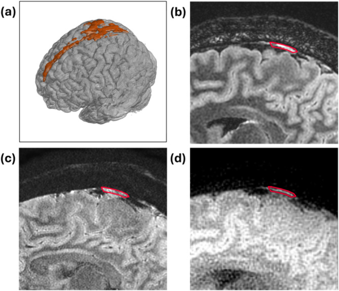
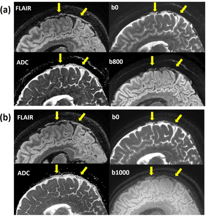
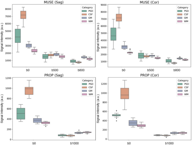
The parasagittal dura (PSD) is a thin channel along the sagittal sinus vein at the brain's upper convexities. Previous studies have shown that cerebrospinal fluid (CSF) flows directly into the PSD, with PSD dimensions and tracer clearance rates associated with aging and brain disorders. Since slow lymphatic drainage is sensitive to water diffusion, PSD circulation may be evaluated using diffusion-weighted imaging (DWI). However, traditional echo-planar DWI (EP-DWI) suffers from low resolution and image distortion, limiting its application to PSD assessment. This study employed high-resolution Multiplexed Sensitivity Encoding (MUSE) DWI and Periodically Rotated Overlapping Parallel Lines with Enhanced Reconstruction (PROPELLER) DWI to investigate PSD water diffusion. These advanced techniques reduce image distortion while enhancing spatial resolution. Our results demonstrated that PSD structures are clearly visible on high-resolution DWI and apparent diffusion coefficient (ADC) maps, correlating with PSD locations identified on T2 FLAIR imaging. In addition, mean ADC values of PSD (1843.1-2062.2 × 10- 6 mm2/sec) were higher than those of gray and white matter but lower than CSF. These findings highlight the potential of MUSE and PROPELLER DWI for assessing PSD diffusion, offering a promising non-invasive tool for studying PSD circulation and its role in neurological disorders.

Keywords: DWI; MUSE; Meningeal lymphatic vessels; PROPELLER; Parasagittal dura.


Conflict of interest statement

Declarations. Competing interests: The authors declare no competing interests.

Figures

Fig. 1
Fig. 1
(a) 3D FLAIR reconstruction showing the PSD structure in orange, (b) 2D T2 FLAIR image, (c) MUSE DWI, and (d) PROP DWI. The red contour outlines the PSD region, as identified on DWI, corresponding to the high signal in the PSD on FLAIR images. This overlay enhances geometric accuracy visualization and highlights the improved delineation of the PSD structure using MUSE and PROP techniques.
Fig. 2
Fig. 2
(a) The sagittal view displayed the PSD on T2 FLAIR image (up left), MUSE DWI b0 (up right), MUSE DWI b800 (down right), and MUSE ADC map (down left) as indicated by arrows. (b) The sagittal view displayed the PSD on T2 FLAIR image (up left), PROPELLER DWI b0 (up right), PROPELLER DWI b1000 (down right), and PROPELLER ADC map (down left) as indicated by arrows. The ADC of the PSD appeared darker than that of the CSF.
Fig. 3
Fig. 3
(a) The coronal view displayed the PSD on T2 FLAIR image (up left), MUSE DWI b0 (up right), MUSE DWI b800 (down right), and MUSE ADC map (down left) as indicated by arrows. (b) The coronal view displayed the PSD on T2 FLAIR image (up left), PROPELLER DWI b0 (up right), PROPELLER DWI b1000 (down right), and PROPELLER ADC map (down left) as indicated by arrows. The ADC of the PSD appeared darker than that of the CSF.
Fig. 4
Fig. 4
Box plots illustrating the DWI signal intensity of PSD, CSF, GM, and WM across b-values of 0, 500, 800, and 1000 in both sagittal and coronal views. (a) MUSE DWI in the sagittal plane, (b) MUSE DWI in the coronal plane, (c) PROPELLER DWI in the sagittal plane, and (d) PROPELLER DWI in the coronal plane.
Fig. 5
Fig. 5
The box plot illustrates the ADC values of PSD, CSF, GM, and WM based on b-values of 500, 800, and 1000, using the PWM and RBM methods in both sagittal (PWMsag, RBMsag) and coronal (PWMcor, RBMcor) views. Each label on the x-axis represents a specific condition: PSD500, PSD800, and PSD1000 represent PSD ADC values measured with b = 0 combined with b = 500 in the MUSE scan, b = 0 combined with b = 800 in the MUSE scan, and b = 0 combined with b = 1000 in the PROPELLER scan, respectively. Similarly, CSF500, CSF800, CSF1000 correspond to CSF measurements; GM500, GM800, GM1000 correspond to GM measurements; and WM500, WM800, WM1000 correspond to WM measurements under the same conditions.

References

    1. Secker, G. A. & Harvey, N. L. VEGFR signaling during lymphatic vascular development: from progenitor cells to functional vessels. Dev. Dyn.244, 323–331. 10.1002/dvdy.24227 (2015). - PubMed
    1. Iliff, J. J. et al. A paravascular pathway facilitates CSF flow through the brain parenchyma and the clearance of interstitial solutes, including amyloid beta. Sci. Transl Med.4, 147ra111. 10.1126/scitranslmed.3003748 (2012). - PMC - PubMed
    1. Louveau, A. et al. Structural and functional features of central nervous system lymphatic vessels. Nature523, 337–341. 10.1038/nature14432 (2015). - PMC - PubMed
    1. Ahn, J. H. et al. Meningeal lymphatic vessels at the skull base drain cerebrospinal fluid. Nature572, 62–66. 10.1038/s41586-019-1419-5 (2019). - PubMed
    1. Zhang, M. et al. Evaluation of glymphatic-meningeal lymphatic system with intravenous gadolinium-based contrast-enhancement in cerebral small-vessel disease. Eur. Radiol.33, 6096–6106. 10.1007/s00330-023-09796-6 (2023). - PubMed

LinkOut - more resources