

Morphological characteristics of microenvironment in the human thymus during fetal development
- PMID: 40033348
- PMCID: PMC11877800
- DOI: 10.1186/s13104-025-07109-2
Morphological characteristics of microenvironment in the human thymus during fetal development
Abstract
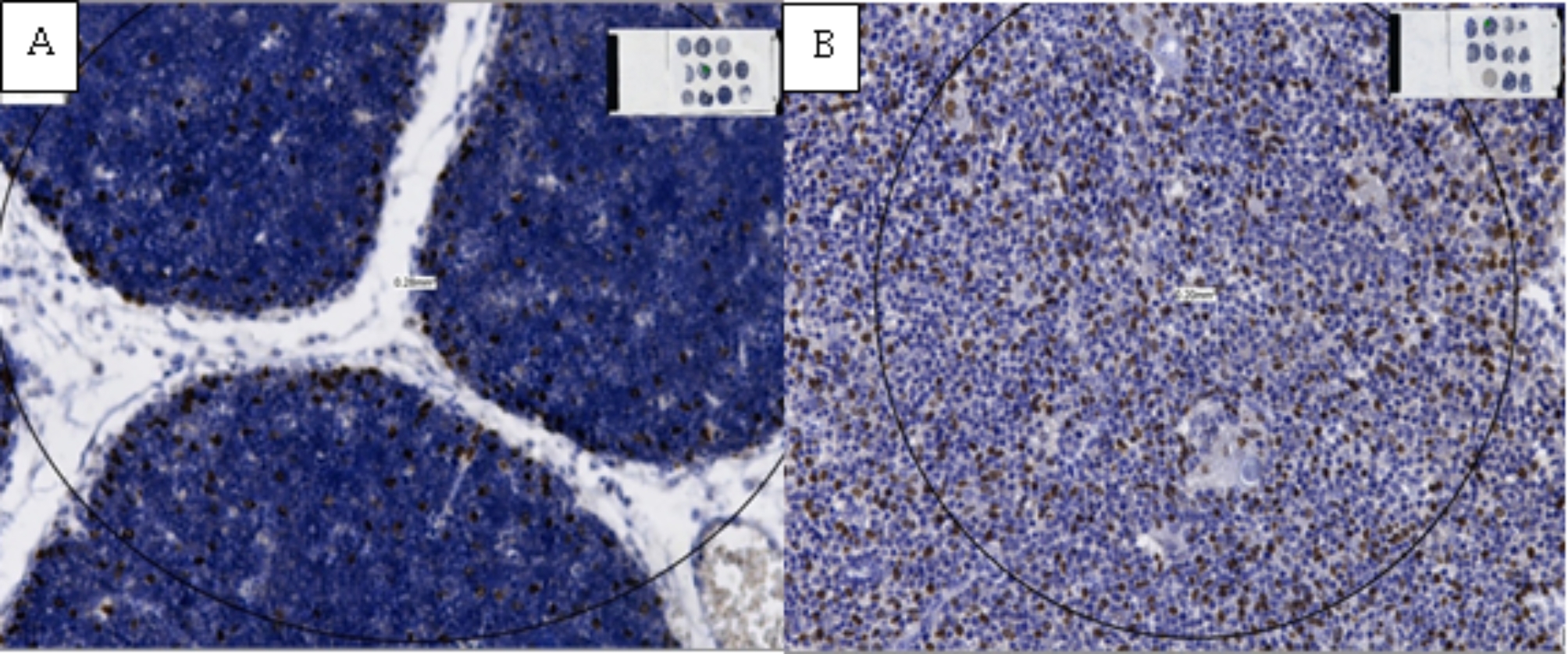
Background: The thymus is a key organ for the development of T cells. T cell precursors first migrate from the bone marrow to the thymus. During maturation, these precursors require interactions with various types of cells that form the thymic microenvironment, such as epithelial, mesenchymal, and other immune cells not belonging to the T lineage. The aim of this study was to examine the changes in the number and diameter of Hassall's corpuscles, as well as the density and distribution of epithelial cells (p63+) and macrophages (CD68+).
Methods: Twenty-five fetal thymus samples were examined, divided into five groups according to gestational age. The samples were processed using standard histological methods and immunohistochemical staining.
Results: The study showed that the number and diameter of Hassall's corpuscles gradually increased during fetal development, with a significant increase from the 14th to the 38th gestational week. The average diameter of Hassall's corpuscles was largest in the age group of 34-38 weeks. The density of p63 + epithelial cells decreased in correlation with gestational week, while the density of CD68 + macrophages significantly increased, particularly in the thymic medulla, towards the end of the fetal period.
Conclusions: An increase in the number and size of Hassall's corpuscles during fetal development was recorded, while the density of epithelial cells decreased and the density of macrophages increased.
Keywords: Epithelial cells; Hassall's corpuscles; Macrophages; Thymus.
© 2025. The Author(s).
Conflict of interest statement
Declarations. Ethics approval and consent to participate: All steps of the study were conducted in accordance with the Declaration of Helsinki [9]. The study protocol was reviewed and approved by the Ethics Committee of the University Clinical Center of Vojvodina, Novi Sad (decision from December 31, 2019, decision number: 00-1212). All samples were processed anonymously, respecting the privacy and confidentiality of information about the fetuses and their families. The material used in the study was collected exclusively from the archives of the Center for Pathology and Histology and from autopsies conducted in accordance with legal and ethical guidelines. All tissue analysis procedures were performed under laboratory conditions with strict adherence to ethical and professional standards. No aspect of the study compromised the physical or psychological well-being of the families, and all data were used solely for scientific purposes, without identifying personal information. Consent for publication: Not applicable. Competing interests: The authors declare no competing interests.
Figures


References
-
- Perez YE, Moran CA. The thymus: General concepts on embryology, anatomy, histology and immunohistochemistry. Semin Diagn Pathol. 2022;39(2):86–91. - PubMed
-
- Rebelatto MC. Chapter 24 - spleen, Lymph Nodes, and Thymus. In: Suttie AW, editor. Boorman’s Pathology of the Rat. 2nd ed. Academic; 2018. pp. 575–600.
-
- Jablonska-Mestanova V, Sisovsky V, Danisovic L, Polak S, Varga I. The normal human newborns thymus. Bratisl Lek Listy. 2013;114(7):402–8. - PubMed
MeSH terms
Substances
LinkOut - more resources
Full Text Sources
Medical
