Review
. 2025 Mar 3;23(1):161.
doi: 10.1186/s12951-025-03236-x.

Multidimensional applications of prussian blue-based nanoparticles in cancer immunotherapy

Affiliations
Review

Multidimensional applications of prussian blue-based nanoparticles in cancer immunotherapy

Jiayi Zhang et al. J Nanobiotechnology. .

Abstract

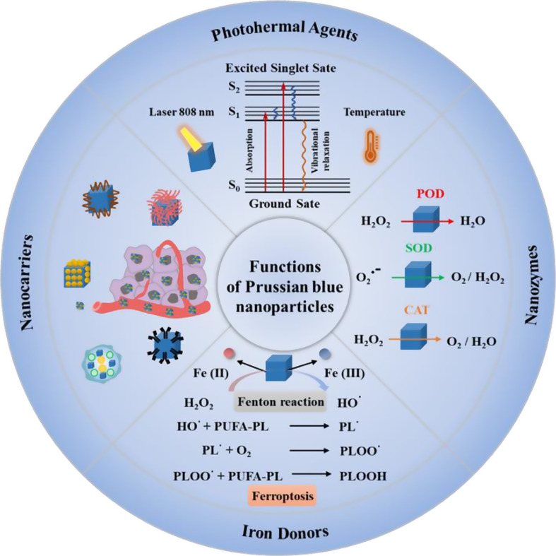
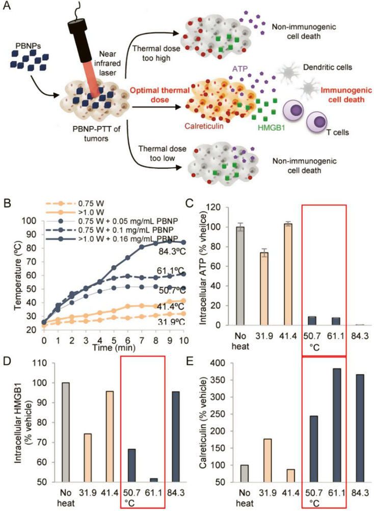
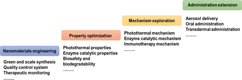
Immunotherapy holds notable progress in the treatment of cancer. However, the clinical therapeutic effect remains a significant challenge due to immune-related side effects, poor immunogenicity, and immunosuppressive microenvironment. Nanoparticles have emerged as a revolutionary tool to surmount these obstacles and amplify the potency of immunotherapeutic agents. Prussian blue nanoparticles (PBNPs) exhibit multi-dimensional immune function in cancer immunotherapy, including acting as a nanocarrier to deliver immunotherapeutic agents, as a photothermal agent to improve the efficacy of immunotherapy through photothermal therapy, as a nanozyme to regulate tumor microenvironment, and as an iron donor to induce immune events related to ferroptosis and tumor-associated macrophages polarization. This review focuses on the advances and applications of PBNPs in cancer immunotherapy. First, the biomedical functions of PBNPs are introduced. Then, based on the immune function of PBNPs, we systematically reviewed the multidimensional application of PBNPs in cancer immunotherapy. Finally, the challenges and future developments of PBNPs-based cancer immunotherapy are highlighted.

Keywords: Cancer immunotherapy; Ferroptosis; Nanoparticles; Nanozyme; Photothermal therapy; Prussian blue.


Conflict of interest statement

Declarations. Ethics approval and consent to participate: Not applicable. Consent for publication: Not applicable. Competing interests: The authors declare no competing interests.

Figures

Scheme 1
Scheme 1
Multidimensional application of Prussian blue-based nanoparticles in cancer immunotherapy. ICD, immunogenic cell death; TAA, tumor-associated antigen; CRT, calreticulin; HMGB1, high-mobility group protein box 1; ATP, adenosine triphosphate; iDCs, immature DCs; mDCs, mature DCs; PD1, programmed death receptor 1; PDL1, programmed death receptor ligand1; MHC, major histocompatibility complex; TCR, T cell receptor; TME, tumor micro-environment; TAMs: tumor-associated macrophage M2-TAMs, M2 type tumor-associated macrophages; M1-TAMs, M1 type tumor-associated macrophages; MDSC, myeloid-derived suppressor cells; Treg cell, regulatory T cells; Th cell, helper T cell; CTL, cytotoxic T lymphocyte; m6A, N6-methyladenosine; mRNA, messenger RNA; FPN, ferroportin
Fig. 1
Fig. 1
The main functions of Prussian blue nanoparticles. (1) the drug delivery function of PBNPs as nanocarriers; (2) the photothermal conversion function of PBNPs as photothermic agents; Reproduced with permission [–59]. Copyright 2021, Royal Society of Chemistry. (3) the TME regulatory function of PBNPs as POD, SOD, and CAT-like nanozymes for oxygen generation; (4) the ferroptosis induction function of PBNPs as iron-donors via Fenton reaction. S0, the ground state; S1, the first electron-excited singlet state; S2, the second electron-excited singlet state; POD, peroxidase; SOD, superoxide dismutase; CAT, catalase; PUFA, polyunsaturated fatty acid; PL, phospholipid
Fig. 2
Fig. 2
(A) Schematic illustration of Prussian blue nanoparticles-based photothermal therapy (PBNPs-PTT) generates a thermal window of immunogenic cell death. (B) Temperature–time profiles of samples containing ten million Neuro2a cells treated 0.75 W laser, > 1 W laser, 0.75 W laser + 0.05 mg·mL− 1 PBNPs, 0.75 W laser + 0.1 mg·mL− 1 PBNPs, and > 1 W laser + 0.16 mg·mL− 1 PBNPs. (C) Intracellular ATP, (D) Intracellular HMGB1 and (E) Surface calreticulin expression in the various treatment groups (as a % of the vehicle-treated group). Red boxes indicated the treatment temperature ranges for which all three markers of ICD are expressed/present (to varied degrees). Reproduced with permission [104]. Copyright 2018, Wiley-VCH-GmbH
Fig. 3
Fig. 3
(A) Schematic illustration of CpG oligodeoxynucleotide-coated Prussian blue nanoparticle (CpG-PBNPs)-mediated nano immunotherapy for neuroblastoma. (B) Induction of immunogenic cell death by CpG-PBNP-PTT in vitro. (C) Immunostimulatory properties of CpG-PBNPs, including activation of dendritic cells and percentage proliferation of CD8 + T cells. (D) Effect of CpG-PBNP-based on long-term survival and rejection of tumor rechallenge. Reproduced with permission [123]. Copyright 2019, Royal Society of Chemistry. (E) Schematic of the mechanism of action of the CpG-PBNPs-PTT-based nanoimmunotherapy in the TH-MYCN model of NB. The CpG-PBNPs-PTT-based nanoimmunotherapy mediates tumor cell priming along with ICD administered at a specific thermal dose, leading to T cell activation and generation of potent T cell memory, which can elicit long-term, tumor-free survival, and rejection of tumor rechallenge in a TH-MYCN model of NB. (F) CpG-PBNP-PTT generates a potent abscopal effect, which induces complete tumor regression on the treated flank and significantly slows tumor progression on the untreated flank, and improving animal survival in the TH-MYCN NB model [24]. Copyright 2021, Wiley-VCH GmbH. (G) illustration of the construction of pPBNPs-CpG@TD for photothermal-responsive ICD-driven in situ anti-tumor vaccine-like immunotherapy [124]. Copyright 2023, Elsevier Ltd
Fig. 4
Fig. 4
(A) Illustration of the construction of Mn-enriched MnPB-MnOx nanomedicines and the design principle of photothermal ablation synergizing with Mn2+-augmented cancer immunotherapy. (B) Proinflammatory cytokine type I interferons level in primary tumors from mice in each group on day 10 after various treatments. (C) DC maturation CD8, T cells, M1 (CD86 + macrophages in F4/80 + CD11b + CD45 + cells) and NK cells in primary. (D) Schematic illustration of the experiment design and time-dependent tumor volume curves for primary tumors (the former) and distant tumors (the latter) on mice after various treatments (group I: saline; group II: laser only; group III: MnPB + laser; group IV: MnPB-MnOx; group V: MnPB-MnOx + laser. The parameter of NIR-I laser was 808 nm wavelength, 1.5 W cm2, and 10 min exposure. Note: N.S. = not significant; **P < 0.01; ***P < 0.001. Reproduced with permission [138]. Copyright 2023, Elsevier Ltd. E) Schematic representation of the Mn(III)-doped nanoparticles for amplifying the cGAS-STING pathway, enhancing the antitumor immune response, and optimizing the efficiency of incomplete photothermal ablation therapy. F) The cGAS-STING activation in 4T1 tumor tissues by Western blot and the expression of TNF- α, IFN- β and IFN- γ in 4T1 tumor tissues by ELISA (n = 3). G) The production of cytotoxic T cells (n = 5) and H) dendritic cells (n = 6) in the tumor microenvironment. I) Tumor growth curves in the orthotopic breast tumor model (n = 5). J) Average tumor growth curves and representative photographic of the distant tumor. Data are shown as the mean values ± SD; Statistical significance was calculated by one-way ANOVA with Tukey’s test. ∗p < 0.05, ∗∗p < 0.01, ∗∗∗p < 0.001. n.s: no significance differences. Reproduced with permission [140]. Copyright 2023, Elsevier Ltd
Fig. 5
Fig. 5
(A) Schematic illustration of the incubation of S.oneidensis MR-1 and subsequently one-pot large-scale microbial synthesis of FDA-approved Prussian blue MOFs. (B) Schematic illustration of the biological precipitation of PB MOFs coated S. oneidensis MR-1 hybrid (S. oneidensis-MOFs). (C) Schematic illustration of the mitochondria-targeting MiBaMc system-induced ICD combined with aPDL1 for enhanced tumor immunotherapy. (D) Quantitative analysis of the matured DCs, the CD3 + CD8 + cytotoxic Tcells and CD3 + CD4 + helper Tcells as a percentage of CD3 + lymphocytes based on flow cytometric results. (n = 3mice). (E) 4T1 (the former) and MC38 (the latter) tumor volumes of different groups were measured every 2 days (n = 5 mice). Statistical analysis was conducted by one way ANOVA with Tukey’s tests. n.s. represents none of significance, *P < 0.05, **P < 0.01, ***P < 0.001, ****P < 0.0001. Reproduced with permission [157]. Copyright 2023, Nature. (F) Schematic diagram of the synthesis and preparation process of PB/PM/HRP/Apt biomimetic nanocomposite and the mechanism of collaborative therapy. (G) The relative content changes of IFN-γ, TNF-α, IL-6 and granzyme B in tumor tissues. (H) The tumor growth curves of 4T1 solid tumor-bearing mice monitored every 2 days after different treatments (n = 5), and representative digital photographs of dissected tumors. (I) H&E staining images of 4T1 solid tumor sections obtained after injection of PBS, PBS + Laser, PB/PM/HRP/Apt and PB/PM/HRP/Apt + Laser. Scale bar: 100 μm. Reproduced with permission [162]. Copyright 2023, Frontiers
Fig. 6
Fig. 6
(A) Schematic illustration of PBNPs-based photothermal therapy combined with checkpoint (CTLA-4) inhibition for photothermal immunotherapy of neuroblastoma. (B) Normalized tumor growth curves and Kaplan-Meier survival plots of neuroblastoma mice receiving photothermal immunotherapy and the other groups (log-rank test; p < 0.05); (n ≥ 5/group). X) Tumor growth curves and higher long-term survival in the rechallenged group compared to naïve mice (log-rank test, p < 0.05); (n ≥ 3/group). Copyright 2017 [163], Elsevier Ltd. D) αCD137-PBNPs synthesis scheme, treatment regimen and proposed mechanism of action of αCD137-PBNPs-PTT to trigger anti-tumor immunity in SM1 melanoma. E) Kaplan-Meier survival curve of all mice in this experiment. F) Serum AST and ALT activity reveals no increased hepatoxicity in αCD137-PBNP-PTT-treated versus control and untreated SM1 tumor-bearing mice. * p < 0.05; ** p < 0.01; **** p < 0.0001 compared with αCD137-PBNP-PTT group. Copyright 2024 [166], Taylor & Francis. G) The construction of MPB-3BP@CM NPs and their implementation in combined therapy for CRC. Copyright 2024 [171], Nature
Fig. 7
Fig. 7
(A) Schematic illustration of the PBNPs-PTT-mediated tumor-specific T cell expansion scheme. (B) T cells developed by co-culturing with DCs primed with PBNPs-PTT-treated U87 cells and (C) U87 cell lysates (donor 2 is excluded due to availability of PBMCs) and their expansion. (D) Phenotype of T cell populations measured post-PBNPs-PTT-mediated ex vivo expansion. (E) T cells developed to target U87 cells via PBNPs-PTT-mediated and (F) U87 cell lysate-mediated expansion were co-cultured with U87 cells at the listed E: T ratios generated using a fixed number of T cells and decreasing number of target cells, and IFN-ɣ release (n = 2/group). *P < 0.05 versus actin. (G) U87-specific T cells developed via PBNPs-PTT and lysis were co-cultured with U87 cells at the listed E: T ratios for 4 h. Cytotoxicity was measured by calcein release (n = 2 replicates/donor). (H) U87-specific T cells developed via PBNPs-PTT (donor 1 or donor 4) were co-cultured with NHAs (donor 1) or PBMCs from the corresponding healthy donors (donor 4) at the listed E: T ratios. Cytotoxicity was measured by calcein release. Values represent mean ± standard deviation (n = 2/group). Reproduced with permission [183]. Copyright 2023, Elsevier Ltd
Fig. 8
Fig. 8
(A) Schematic illustration of the preparation process and behavior of HMPB/BLZ945/anti-SIRPα@ATRA@fibrin for cancer immunotherapy in vivo. The immunotherapeutic gel at the surgical site released ATRA and HMPB/BLZ945/anti-SIRPα to induce CSC differentiation and TAMs reprogramming. Reproduced with permission [193]. Copyright 2023, Elsevier Ltd. (B) Schematic illustration of the synthetic of LMWHA-MPB nanoparticles and HMME-loading LMWHA-MPB nanoparticles inhibits the proliferation and metastasis of 4T1 tumor in vivo [194]. Copyright 2019, Europe PMC. (C) Schematic illustration of the preparation of G/APH-M and G/APH-M-based radiotherapy in combination with anti-PD-L1 to enhance glioblastoma radio-immunotherapy [195]. Copyright 2023, Wiley-VCH GmbH. (D) Schematic illustration of lactate consumption combined with siPD-L1 to synergistically improve tumor immunotherapy [199]. Copyright 2023, Elsevier Ltd. (E) Schematic illustration of the multifunctional nanoplatform (SP94-PB-SF-Cy5.5 NPs) for HCC-targeted multimodality imaging and combined PTT/SF treatment. Reproduced with permission [200]. Copyright 2019, American Chemical Society
Fig. 9
Fig. 9
(A) Schematic illustration showing the preparation of AuPB@LMHep. (B) the hepcidin-based nanocomposites for enhanced cancer immunotherapy by modulating iron export-mediated N6-methyladenosine RNA transcript. (C) Protein expression of ferroportin and LC3-I/II in Kasumi-1 cells treated with AuPB@LMHep or AuPB@LMT7 (0 to 50 µg mL− 1). (D) Intracellular Fe content in AuPB@LMHep or AuPB@LMT7 treated Kasumi-1 cells. (E) GSH/GSSH ratio and GPX4 activity in Kasumi-1 cells. (F) Dot blots showing m6A levels in the Kasumi-1 cells after AuPB@LMT7 treatment. (G) Quantification by flow cytometry of ratio of CD8 + T cell and CD4 + T cells populations, as well as Elisa assay of IFN-γ in the different treatment groups of leukemia-bearing mice. Reproduced with permission [211]. Copyright 2021, Wiley-VCH GmbH
Fig. 10
Fig. 10
(A) Schematic diagram of the preparation of Man HMPB/HCQ coated with TK-M hybrid membrane. (B) TK-M@Man-HMPB/HCQ potentiates cancer immunotherapy via mitigating hypoxia, reversing the TAMs phenotypes, and facilitating cytotoxic T lymphocyte infiltration. (C) Western blot and corresponding semi-quantitative analysis of M2 macrophages (RAW264.7 treated with IL-4 for 24 h) after different treatments. (D) In vivo antitumor efficacy investigation. (E) Relative quantification of M2-like macrophages (CD206+) and M1-like macrophages (CD86+) gating on F4/80 + cells (n = 5, mean ± SD). (F) Relative quantification of CD8 + and CD4 + T cells gating on CD3 + T cells (n = 5, mean ± SD). *p < 0.05, **p < 0.01, ***p < 0.001, by analysis of ANOVA with Tukey’s post-hoc test. Reproduced with permission [22]. Copyright 2022, Wiley-VCH GmbH
Fig. 11
Fig. 11
(A) Schematic illustration of the synthesis of PMo@CCM and the pathway of enhanced immunotherapy via the combined PTT/CDT treatment and improver presentation of tumor-associated antigens (TAAs) [217]. Copyright 2024, Wiley-VCH GmbH. (B) Schematic illustration of the CS-1@PB[HM] NPs for cancer comprehensive therapy by inducing pyroptosis [218]. Copyright 2023, Elsevier Ltd. (C) Schematic illustration showing the integration of NIR AIEgen with mesoporous PB nanocatalyzer to boost the theranostic performance for NIR-II fluorescence and PA imaging-guided robust cancer immunotherapy [219]. Copyright 2024 Wiley-VCH GmbH. (D) Schematic diagram of the homologous targeted tumor cocktail therapy based on M@P-PDR “Nano-targeted cells” [228]. Copyright 2021, BioMed Central
Fig. 12
Fig. 12
The current challenges and future development of PBNPs in tumor immunotherapy

References

    1. Mellman I, Coukos G, Dranoff G. Cancer immunotherapy comes of age. Nature. 2011;480(7378):480–9. - PMC - PubMed
    1. Hiam-Galvez KJ, Allen BM, Spitzer MH. Systemic immunity in cancer. Nat Rev Cancer. 2021;21(6):345–59. - PMC - PubMed
    1. Cogdill AP, Andrews MC, Wargo JA. Hallmarks of response to immune checkpoint blockade. Br J Cancer. 2017;117(1):1–7. - PMC - PubMed
    1. Morad G, Helmink BA, Sharma P, Wargo JA. Hallmarks of response, resistance, and toxicity to immune checkpoint blockade. Cell. 2021;184(21):5309–37. - PMC - PubMed
    1. Pardoll DM. Immunology beats cancer: a blueprint for successful translation. Nat Immunol. 2012;13(12):1129–32. - PMC - PubMed

LinkOut - more resources