. 2025 Mar 3;23(1):117.
doi: 10.1186/s12964-025-02108-z.

Lysine 2-hydroxyisobutyrylation of HXK1 alters energy metabolism and KATP channel function in the atrium from patients with atrial fibrillation

Affiliations

Lysine 2-hydroxyisobutyrylation of HXK1 alters energy metabolism and KATP channel function in the atrium from patients with atrial fibrillation

Hai-Tao Hou et al. Cell Commun Signal. .

Abstract

Background: Atrial fibrillation (AF) is the most common form of arrhythmia and is a growing clinical problem. Post-translational modifications (PTMs) constitute crucial epigenetic mechanisms but modification of lysine 2-hydroxyisobutyrylation (Khib) in AF is still unknown. This study aimed to investigate the role and mechanism of Khib in AF.

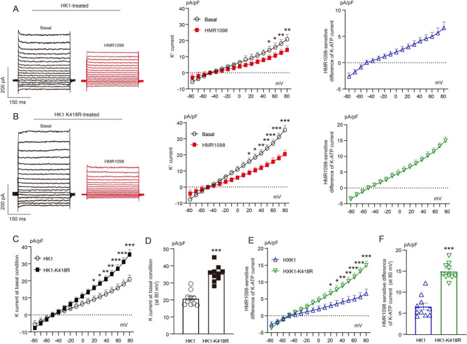
Methods: PTM proteomics was applied in the human atrial tissue from AF and sinus rhythm patients with heart valve disease during cardiac surgery to identify the Khib sites. The functional changes of differential modification sites were further validated at the cellular level. Cellular electrophysiology was performed to record the ion channel current and action potential duration (APD).

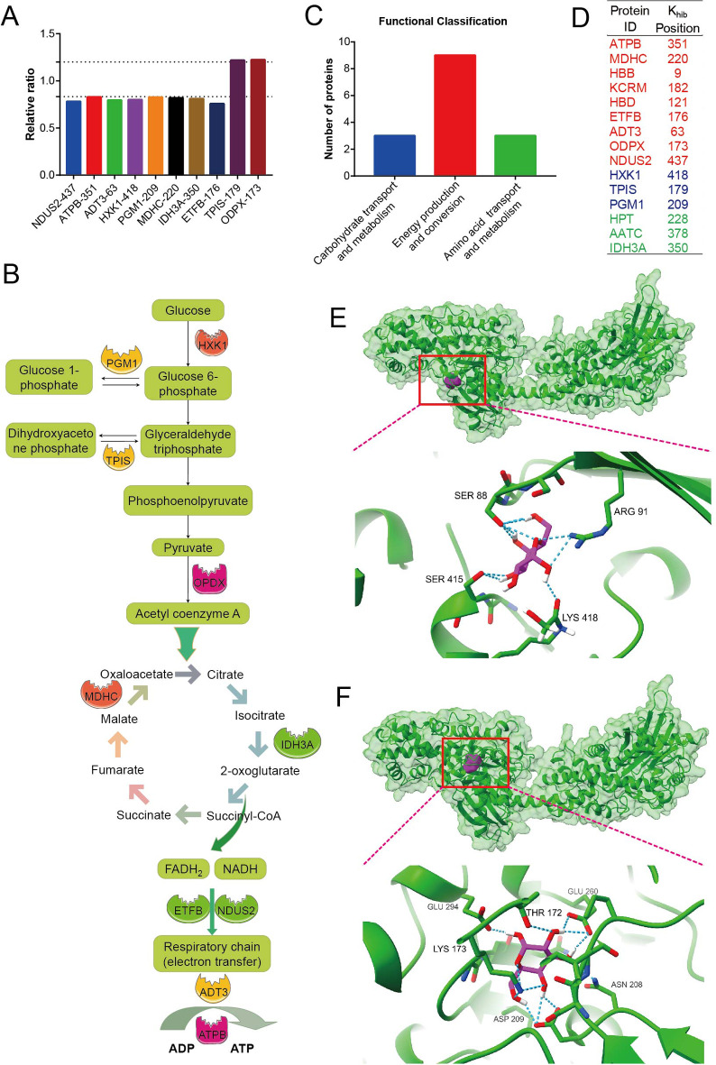
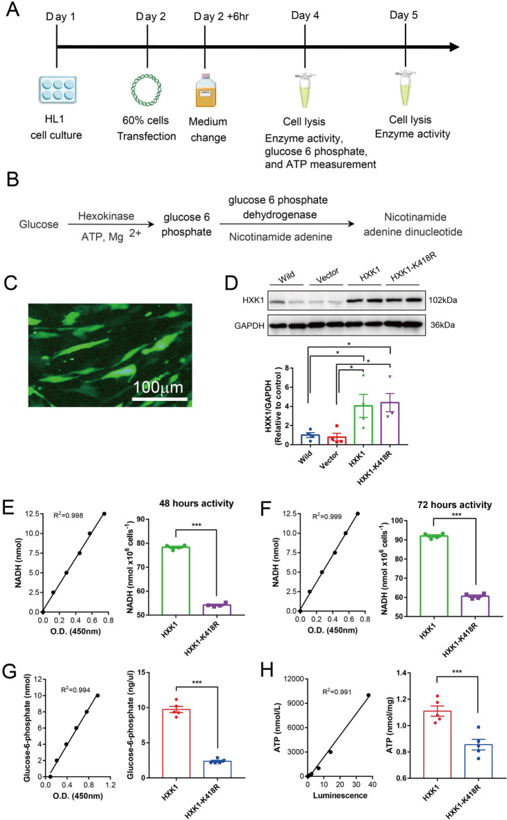
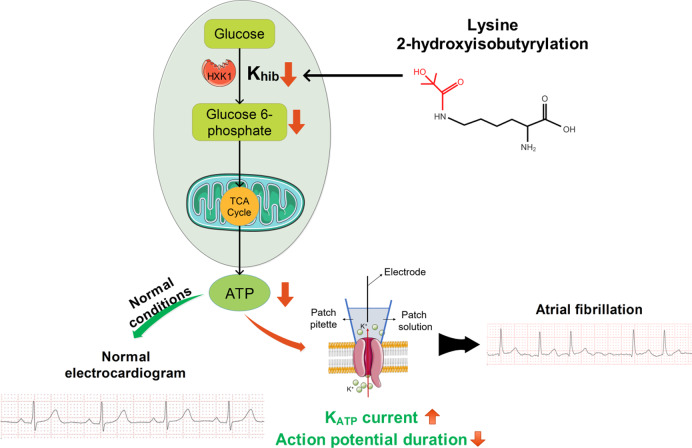
Results: The modification of 124 Khib sites in 35 proteins and 67 sites in 48 proteins exhibited significant increase or decrease in AF compared to sinus rhythm. Ten Khib sites were included in energy metabolism-related signaling pathways (HXK1, TPIS, PGM1, and ODPX in glycolysis; MDHC and IDH3A in tricarboxylic acid cycle; NDUS2, ETFB, ADT3, and ATPB in oxidative respiratory chain). Importantly, decreased HXK1 K418hib regulated by HDAC2 attenuated the original chemical binding domain between HXK1 and glucose, inhibited the binding ability between HXK1 and glucose, and reduced catalytic ability of the enzyme, resulting in low production of glucose-6-phosphate and ATP. Further, it also increased Kir6.2 protein and the current of KATP channel, and decreased APD.

Conclusions: This study demonstrates the importance of Khib to catalysis of HXK1 and reveals molecular mechanisms of HXK1 K418hib in AF, providing new insight into strategies of AF.

Keywords: 2-hydroxyisobutyrylation; Acylation; Atrial fibrillation; Catalysis; Post-translational modification.


Conflict of interest statement

Declarations. Competing interests: The authors declare no competing interests.

Figures

Fig. 1
Fig. 1
Model workflow for identifying 2-hydroxyisobutyrylated proteins between human AF and SR. (A) The schematic overview of the experimental approach. (B) The whole lysine 2-hydroxyisobutyrylation level between AF and SR. (C and D) Expression of P300 (n = 5) in AF was lower than that in SR. HDAC2 (n = 4) in AF was higher than that in SR. (E) Spectrums, peptides, proteins, and sites identified by mass spectrometry. (F) Peptide mass error. Mass error indicates the accuracy of identified peptides. All the mass errors were smaller than 3 ppm, demonstrating good accuracy of the MS data. (G) Relationship between 2-hydroxyisobutyrylation sites and proteins. (H) Peptide length distribution. Most of the identified peptides were from 7 to 22, consistent with the length of tryptic peptides. AF, atrial fibrillation; SR, sinus rhythm. *p < 0.05; **p < 0.01 by unpaired t test. Data are presented as mean ± SEM
Fig. 2
Fig. 2
The quantified differential 2-hydroxyisobutyrylated sites, typical motif, and regulation role of HDAC2 on HXK1. (A) Differentially expressed 2-hydroxyisobutyrylation sites and related proteins. (B) Subcellular distribution of above proteins. (C) Ten typical feature sequences of differentially expressed 2-hydroxyisobutyrylated sites. (D) The motif enrichment heatmap of upstream and downstream amino acids of all identified modification sites. Red color indicates that the amino acid is significantly enriched near the modification site, and green color indicates that the amino acid is significantly reduced near the modification site. (E) HXK1 peptides sequenced by mass spectrometry. b and y represent the fragment ions of the N and C termini in peptide backbone, respectively. m/z, mass-to-charge ratio. (F) HXK1 Khib verification (n = 4) by co-immunoprecipitation in human atrium. Tissue lysates were immunoprecipitated with anti-HXK1 antibody, followed by immunoblotting with a pan-2-hydroxyisobutyrylation antibody. (G) Expression of P300, HXK1, and HDAC2 in HL-1 cells (n = 6). (H and I) Verification of interaction of HDAC2 and HXK1 by co-immunoprecipitation in HL-1 cells. (J) Experimental scheme. Trichostatin A, a HDAC2 inhibitor, was added when the HL-1 cell grew to 90%. Cells were lysed when trichostatin A was added for 16 h. (K) The level of HXK1 Khib (n = 4) increased after adding trichostatin A. AF, atrial fibrillation; SR, sinus rhythm; GAPDH, glyceraldehyde-3-phosphate dehydrogenase; HDAC2, histone deacetylase 2; HXK1, hexokinase-1; TSA, Trichostatin A. *p < 0.05; ***p < 0.001 by unpaired t test. Data are presented as mean ± SEM
Fig. 3
Fig. 3
Lysine 2-hydroxyisobutyrylated enzymes involved in energy production. (A) Summarized bar graphs depicting 10 key glycolytic enzymes that were identified as differentially expressed 2-hydroxyisobutyrylated sites. (B) Above 10 enzymes were involved in glycolysis (HXK1, TPIS, PGM1, and ODPX), TCA circle (MDHC and IDH3A), and oxidative respiratory chain (NDUS2, ETFB, ADT3, and ATPB). (C) Three functional energy classification of proteins corresponding to differentially expressed Khib sites. (D) Specific proteins involved in above 3 energy functions. (E) In the presence of side chain modification (simulating high level of K418hib). Glucose formed a hydrogen bond with the modified Lys418. Glucose also interacted with other three amino acids (Ser88, Arg91, Ser415) and formed total 9 hydrogen bonds. The binding energy is -7.07 kcal/mol. (F) In the absence of side chain modification (simulating low level of K418hib). Glucose formed 12 hydrogen bonds with six amino acids (Thr172, Lys173, Asn208, Glu294, Glu260, Asp209). The binding energy is -3.74 kcal/mol
Fig. 4
Fig. 4
Lysine 2-hydroxyisobutyrylation reduced HXK1 activity and energy production. (A) Experimental design. (B) Reaction process of HXK1 and glucoses. (C) Representative fluorescence assay exhibiting a high transfection efficiency in HL-1 cells. Scale bar, 100 μm. (D) Expression of HXK1 (n = 4) in wild, empty vector PCDNA3.1, a vector encoding pcDNA3.1-HK1 (HXK1), and a vector encoding pcDNA3.1-HK1-K418R in HL-1 cells. (E and F) K418R group significantly reduced HXK1 activity at 48 (n = 4) and 72 h (n = 4) compared with HXK1 normal overexpression group. (G and H) HK1-K418R reduced the production of glucose-6-phosphate (n = 5) and adenosine 5’-triphosphate (ATP) (n = 5). HXK1, hexokinase 1; GAPDH, glyceraldehyde-3-phosphate dehydrogenase; NADH, nicotinamide adenine dinucleotide. *p < 0.05; ***p < 0.001 by unpaired t test. Data are presented as mean ± SEM
Fig. 5
Fig. 5
HK1-K418R increased ATP-sensitive potassium channel current (IK, ATP), reduced action potential duration (APD), and increased Kir6.2. (A and B) ATP-sensitive potassium current (IK, ATP) was significantly inhibited by glibenclamide in HL-1 atrial myocytes (n = 13) in both HK1 (22.26 ± 1.26 vs. 15.59 ± 1.14 pA/pF) and HK1-K418R (32.84 ± 1.95 vs. 19.98 ± 1.34 pA/pF) group. The left panel shows representative traces of whole-cell K+ currents. The mid panel shows the comparison of K+ currents with or without glibenclamide. The right panel shows the difference of the currents induced by glibenclamide (IK, ATP=IBasal-IGlibenclamide). (C and D) Comparisons of currents at basic levels (n = 13) between HK1 and HK1-K418R. The currents in HK1-K418R were significantly higher than that in HK from 50 to 80 mV. HK1-K418R increased whole-cell K + current from 22.26 ± 1.26 pA/pF to 32.84 ± 1.95 pA/pF. (E and F) Comparisons of glibenclamide-sensitive currents (IK, ATP, n = 13). The currents in HK1-K418R were significantly higher than that in HK from 40 to 80 mV. HK1-K418R increased IK, ATP current from 6.67 ± 0.37 to 12.89 ± 1.19 pA/pF. (G and H) HK1-K418R resulted in shortening of APD. (I, L, and M) HK1-K418R did not affect APD30, APA, and RMP. (J and K) Compared to HK1, APD50 decreased from 27.11 ± 2.09 to 18.26 ± 1.93 ms (n = 7) and APD90 decreased from 114.64 ± 8.67 to 73.21 ± 6.91 ms (n = 7). (N) HK1-K418R overexpression significantly increased the expression of Kir6.2 (n = 6) compared with HK1 normal overexpression group in HL-1 cell. (O) The expression of Kir6.2 (n = 8) in human atrial tissue with AF is significantly higher than that in SR atrial tissue. HK1, hexokinase-1; AF, atrial fibrillation; SR, sinus rhythm; GAPDH, glyceraldehyde-3-phosphate dehydrogenase; APA, action potential amplitude; RMP, resting membrane potential; NS, not significant. *p < 0.05; **p < 0.01; ***p < 0.001 by unpaired t test. Data are presented as mean ± SEM
Fig. 6
Fig. 6
HK1-K418R increased ATP-sensitive potassium channel current (IK, ATP). (A and B) ATP-sensitive potassium current was significantly inhibited by HMR1098 in HL-1 atrial myocytes (n = 10) in both HK1 and HK1-K418R. The left panel shows representative traces of whole-cell K+ currents. The mid panel shows the comparison of K+ currents with or without HMR1098. The right panel shows the difference of the currents induced by HMR1098 (IK, ATP=IBasal-IHMR1098). (C and D) Comparisons of currents at basic levels (n = 10) between HK1 and HK1-K418R. The currents in HK1-K418R were significantly higher than that in HK from 20 to 80 mV. HK1-K418R increased whole-cell K + current from 20.82 ± 1.32 pA/pF to 35.42 ± 1.46 pA/pF. (E and F) Comparisons of HMR1098-sensitive currents (IK, ATP, n = 10). The IK, ATP currents in HK1-K418R were significantly higher than that in HK from 20 to 80 mV. HK1-K418R increased IK, ATP currents from 6.62 ± 0.79 to 14.95 ± 0.62 pA/pF. *p < 0.05; **p < 0.01; ***p < 0.001 by unpaired t test. Data are presented as mean ± SEM
Fig. 7
Fig. 7
Proposed mechanism of Lysine 2-hydroxyisobutyrylation modification of HXK1 leading to atrial fibrillation. Low lysine 2-hydroxyisobutyrylation level decreased catalytic activity of HXK1 and production of glucose-6-phosphate and ATP. These metabolic changes resulted in the increasing of ATP-sensitive potassium channel (IK, ATP) currents and decreased action potential duration, triggering the onset of atrial fibrillation. HXK1, hexokinase 1; ATP, adenosine 5’-triphosphate

Similar articles

Cited by

References

    1. Nattel S, Atrial Fibrillation and Body Composition. Is it fat or lean that ultimately determines the risk?? J Am Coll Cardiol. 2017;69:2498–501. - PubMed
    1. Lazarte J, Laksman ZW, Wang J, Robinson JF, Dron JS, Leach E, Liew J, McIntyre AD, Skanes AC, Gula LJ, et al. Enrichment of loss-of-function and copy number variants in ventricular cardiomyopathy genes in ‘lone’ atrial fibrillation. Europace. 2021;23:844–50. - PMC - PubMed
    1. Kirchhof P, Benussi S, Kotecha D, Ahlsson A, Atar D, Casadei B, Castella M, Diener HC, Heidbuchel H, Hendriks J, et al. 2016 ESC guidelines for the management of atrial fibrillation developed in collaboration with EACTS. Eur Heart J. 2016;37:2893–962. - PubMed
    1. Thorolfsdottir RB, Sveinbjornsson G, Sulem P, Helgadottir A, Gretarsdottir S, Benonisdottir S, Magnusdottir A, Davidsson OB, Rajamani S, Roden DM, et al. A missense variant in PLEC increases risk of atrial fibrillation. J Am Coll Cardiol. 2017;70:2157–68. - PMC - PubMed
    1. Zhang D, Wu CT, Qi X, Meijering RA, Hoogstra-Berends F, Tadevosyan A, Cubukcuoglu Deniz G, Durdu S, Akar AR, Sibon OC, et al. Activation of histone deacetylase-6 induces contractile dysfunction through derailment of α-tubulin proteostasis in experimental and human atrial fibrillation. Circulation. 2014;129:346–58. - PubMed

LinkOut - more resources