Pelvic ring ratio: a novel indicator of comprehensive pelvic alignment assessment
- PMID: 40033729
- PMCID: PMC12242244
- DOI: 10.31616/asj.2024.0447
Pelvic ring ratio: a novel indicator of comprehensive pelvic alignment assessment
Abstract
Study design: A cross-sectional study.
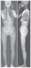
Purpose: To determine the effectiveness of the pelvic ring ratio as an indicator for assessing pelvic tilt (PT) from the frontal view and explore its correlation with various whole-body sagittal alignment (WBSA) parameters using EOS imaging technology.
Overview of literature: Traditional indicators of PT often rely on sagittal plane measurements, which can be challenging in cases of pelvic rotation or obesity. A new indicator such as the pelvic ring ratio could address these challenges and aid in the comprehensive assessment of pelvic alignment.
Methods: In total, 104 healthy participants (28 men, 76 women; mean age, 52.8±12.3 years) with no spinal disorders were recruited. Whole-body radiography using the EOS imaging system was performed to obtain sagittal and coronal parameters, including the pelvic ring ratio. Intra- and interobserver variability were assessed using intraclass correlation coefficients (ICCs) based on measurements by three spine surgery specialists. Correlation analyses among the pelvic ring ratio, age, body mass index, and WBSA parameters were conducted, and a multiple linear regression model was developed to predict PT.
Results: The mean pelvic ring ratio was 53.3%±11.5%. The intra- and interobserver ICCs were 0.89 and 0.87, respectively, indicating good reliability. The pelvic ring ratio was negatively correlated with age (r =-0.387, p <0.05) and PT (r =-0.598, p <0.05). The regression model revealed that the pelvic ring ratio and sex significantly predicted PT (p <0.05). Women had higher pelvic ring ratio (55.0%±11.3%) than men (48.6%±10.8%).
Conclusions: The pelvic ring ratio is a reliable and valuable indicator for PT assessment from the frontal view. It exhibits significant correlations with age and certain WBSA parameters, showing potential to improving the diagnostic accuracy and treatment planning for patients with spinal and hip disorders.
Keywords: Adult spinal deformity; Lower limb alignment; Pelvic ring ratio; Pelvic tilt; Sagittal alignment.
Conflict of interest statement
No potential conflict of interest relevant to this article was reported.
Figures


References
-
- Schwab F, Dubey A, Gamez L, et al. Adult scoliosis: prevalence, SF-36, and nutritional parameters in an elderly volunteer population. Spine (Phila Pa 1976) 2005;30:1082–5. - PubMed
-
- Diebo BG, Shah NV, Boachie-Adjei O, et al. Adult spinal deformity. Lancet. 2019;394:160–72. - PubMed
-
- Ames C, Gammal I, Matsumoto M, et al. Geographic and ethnic variations in radiographic disability thresholds: analysis of North American and Japanese operative adult spinal deformity populations. Neurosurgery. 2016;78:793–801. - PubMed
-
- Schwab FJ, Blondel B, Bess S, et al. Radiographical spinopelvic parameters and disability in the setting of adult spinal deformity: a prospective multicenter analysis. Spine (Phila Pa 1976) 2013;38:E803–12. - PubMed
-
- Nakashima H, Kawakami N, Ohara T, Saito T, Tauchi R, Imagama S. A new global spinal balance classification based on individual pelvic anatomical measurements in patients with adult spinal deformity. Spine (Phila Pa 1976) 2021;46:223–31. - PubMed
LinkOut - more resources
Full Text Sources
Research Materials

