. 2025 Mar 25;25(7):1787-1800.
doi: 10.1039/d4lc00848k.

Blood-perfused Vessels-on-Chips stimulated with patient plasma recapitulate endothelial activation and microthrombosis in COVID-19

Affiliations

Blood-perfused Vessels-on-Chips stimulated with patient plasma recapitulate endothelial activation and microthrombosis in COVID-19

Huub J Weener et al. Lab Chip. .

Abstract

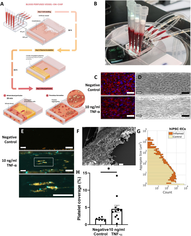
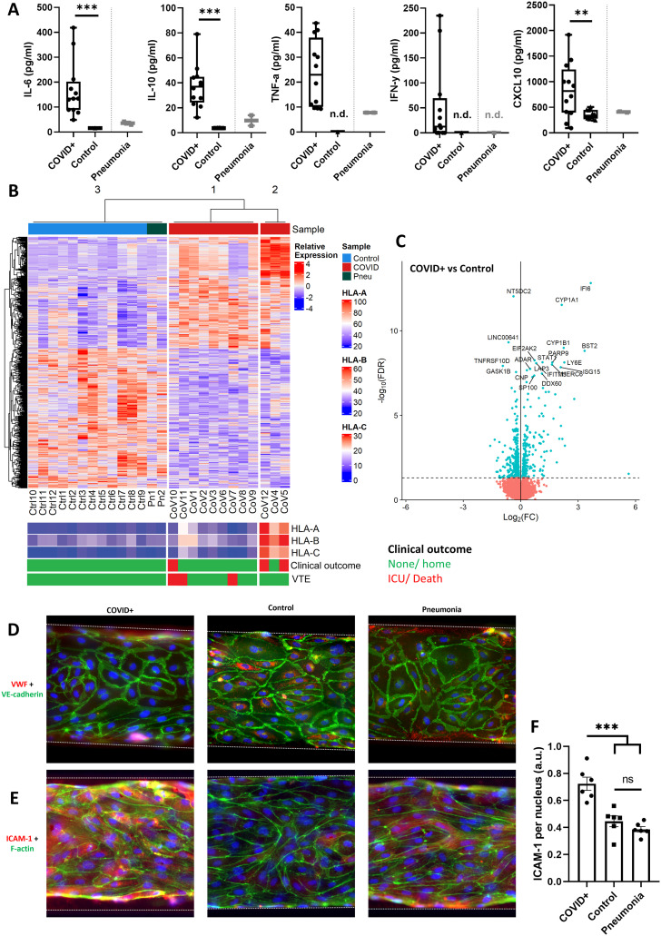
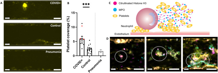
A subset of coronavirus disease 2019 (COVID-19) patients develops severe symptoms, characterized by acute lung injury, endothelial dysfunction and microthrombosis. Viral infection and immune cell activation contribute to this phenotype. It is known that systemic inflammation, evidenced by circulating inflammatory factors in patient plasma, is also likely to be involved in the pathophysiology of severe COVID-19. Here, we evaluate whether systemic inflammatory factors can induce endothelial dysfunction and subsequent thromboinflammation. We use a microfluidic Vessel-on-Chip model lined by human induced pluripotent stem cell-derived endothelial cells (hiPSC-ECs), stimulate it with plasma from hospitalized COVID-19 patients and perfuse it with human whole blood. COVID-19 plasma exhibited elevated levels of inflammatory cytokines compared to plasma from healthy controls. Incubation of hiPSC-ECs with COVID-19 plasma showed an activated endothelial phenotype, characterized by upregulation of inflammatory markers and transcriptomic patterns of host defense against viral infection. Treatment with COVID-19 plasma induced increased platelet aggregation in the Vessel-on-Chip, which was associated partially with formation of neutrophil extracellular traps (NETosis). Our study demonstrates that factors in the plasma play a causative role in thromboinflammation in the context of COVID-19. The presented Vessel-on-Chip can enable future studies on diagnosis, prevention and treatment of severe COVID-19.


Conflict of interest statement

Authors declare no competing interests.

Figures

Fig. 1
Fig. 1. Characterization of microthrombosis in the Vessel-on-Chip upon inflammation. A: Schematic of Vessel-on-Chip exposure to cytokines or patient plasma. B: Photograph of the Vessel-on-Chip perfused with human whole blood. C: Comparison of VE-cadherin morphology of hiPSC-ECs, in control conditions and upon activation with 10 ng mL−1 TNF-α. Scale bar: 100 μm. D: Brightfield microscopy images of hiPSC-EC monolayers in normal and activated state after perfusion with human whole blood, leading to adhered platelets. Scale bar: 100 μm. E: Fluorescence microscopy images of adherent blood platelets (orange) and fibrin (cyan) networks in control conditions and after activation with 10 ng ml−1 TNF-α. Scale bar: 100 μm. F: Scanning electron micrograph of a perfused channel with whole blood shows blood clots adhering to endothelial cells. Scale bar: 10 μm. G: Platelet aggregate size measured in normal and activated conditions. H: Platelet coverage in normal (n = 6) and activated (n = 12) conditions. Aggregate size and platelet coverage are determined using image analysis of fluorescence microscopy data. Error bars show standard error of mean. * p < 0.05.
Fig. 2
Fig. 2. Characterization of plasma samples from COVID-19 patients and their effects on endothelial cells in the Vessels-on-Chip. A: ELISA data shows elevated plasma concentrations of the cytokines IL-6, IL-10, TNF-α, IFN-γ, and CXCL10 in COVID-19 patients, ** p < 0.01; *** p < 0.001. B: Differentially expressed gene analysis of hiPSC-ECs treated with plasma samples from healthy controls (‘Ctrl’), patients with pneumonia (‘Pn’) and patients with COVID-19 (‘CoV’). The COVID-19 samples are divided into two sub-clusters, one with low expression, and one with elevated expression of class 1 HLA genes (‘HLA-A’, ‘HLA-B’, ‘HLA-C’). Individual plasma samples are also annotated with patient clinical outcome, with severe outcomes (admission to the intensive care unit, ICU, or death) labelled in red. C: Volcano plot of differentially expressed genes comparing hiPSC-ECs treated with plasma samples from COVID-19 patients and controls. Top 20 differentially expressed genes have been marked by name (blue: differentially expressed genes; red: genes with a −log10(FDR) value below 1). D: Representative immunofluorescence images of VWF, VE-cadherin and DNA in Vessels-on-Chip treated with plasma from COVID-19 patients, pneumonia patients or controls. Scale bar, 100 μm. E: Representative immunofluorescence microscopy images of ICAM-1, F-actin, and DNA in all aforementioned conditions. Scale bar, 100 μm. F: Quantification of ICAM-1 shows a significant increase in expression when hiPSC-ECs are stimulated with COVID+ plasma, *** p < 0.001, ns = non-significant.
Fig. 3
Fig. 3. Vessels-on-Chip treated with plasma from COVID-19 patients exhibit elevated platelet aggregation. A: Representative fluorescence microscopy images of three Vessels-on-Chip after perfusion with CD41-labelled human whole blood. All conditions showed platelet adhesion to the sides of the channel (yellow), but only in the Vessel-on-Chip treated with COVID-19 plasma (‘COVID+’) were dense clots formed in the middle of the channel. Scale bar, 100 μm. B: Mean platelet coverage in Vessels-on-Chip treated with plasma from COVID-19 patients (‘COVID+’), healthy controls (‘control’), and pneumonia patients (‘pneumonia’). Patients with VTE are indicated by red datapoints. Error bars: SEM, *** p < 0.001. C: Schematic representation of the process of NETosis, where neutrophils interacted with activated endothelium and associated platelet aggregates to release their nuclear contents. Their DNA strands are structured with citrullinated histone H3 (H3cit), and contain antiviral proteins like myeloperoxidase (MPO). The sticky DNA creates new areas to which platelets adhere, further increasing the size of aggregates. D: Representative images of different stages of NETosis as identified in Vessels-on-Chip treated with plasma from COVID-19 patients. Formed NETs show different morphologies of proteins CitH3 (magenta) and MPO (cyan), from minor (I) to more severe (II and III) patterns of secreted DNA and MPO, eventually forming web-like structures to which CD41-positive platelets (yellow) can bind. Scale bar, 100 μm.

Similar articles

References

    1. World Health Organization, 2023, data.who.int, WHO Coronavirus (COVID-19) Dashboard, https://data.who.int/dashboards/covid19, (accessed April 23, 2024)
    1. Tiwari N. R. Phatak S. Sharma V. R. Agarwal S. K. Thromb. Res. 2021;202:191–198. doi: 10.1016/j.thromres.2021.04.012. - DOI - PMC - PubMed
    1. Mitchell W. B. Paediatr. Respir. Rev. 2020;35:20–24. - PMC - PubMed
    1. Gu S. X. Tyagi T. Jain K. Gu V. W. Lee S. H. Hwa J. M. Kwan J. M. Krause D. S. Lee A. I. Halene S. Martin K. A. Chun H. J. Hwa J. Nat. Rev. Cardiol. 2021;18:194–209. doi: 10.1038/s41569-020-00469-1. - DOI - PMC - PubMed
    1. Middeldorp S. Coppens M. van Haaps T. F. Foppen M. Vlaar A. P. Müller M. C. A. Bouman C. C. S. Beenen L. F. M. Kootte R. S. Heijmans J. Smits L. P. Bonta P. I. van Es N. J. Thromb. Haemostasis. 2020;18:1995–2002. - PMC - PubMed