[Preprint]. 2025 Feb 27:2025.02.25.640204.
doi: 10.1101/2025.02.25.640204.

Cryo-EM structures of the Plant Augmin reveal its intertwined coiled-coil assembly, antiparallel dimerization and NEDD1 binding mechanisms

Affiliations

Cryo-EM structures of the Plant Augmin reveal its intertwined coiled-coil assembly, antiparallel dimerization and NEDD1 binding mechanisms

Md Ashaduzzaman et al. bioRxiv. .

Abstract

Microtubule (MT) branch nucleation is fundamental for building parallel MT networks in eukaryotic cells. In plants and metazoans, MT branch nucleation requires Augmin and NEDD1 proteins which bind along MTs and then recruit and activate the gamma-tubulin ring complex (γ-TuRC). Augmin is a fork-shaped assembly composed of eight coiled-coil subunits, while NEDD1 is a WD40 β-propellor protein that bridges across MTs, Augmin, and γ-TuRC during MT branch nucleation. Here, we reconstitute hetero-tetrameric and hetero-octameric Arabidopsis thaliana Augmin assemblies, resolve their subunit interactions using crosslinking mass spectrometry and determine 3.7 to 7.3-Å cryo-EM structures for the V-junction and extended regions of Augmin. These structures allowed us to generate a complete de novo plant Augmin model that reveals the long-range multi coiled-coil interfaces that stabilize its 40-nm hetero-octameric fork-shaped organization. We discovered the dual calponin homology (CH) domain forming its MT binding site at the end of its V-junction undertake open and closed conformations. We determined a 12-Å dimeric Augmin cryo-EM structure revealing Augmin undergoes anti-parallel dimerization through two conserved surfaces along Augmin's extended region. We reconstituted the NEDD1 WD40 β-propellor with Augmin revealing it directly binds on top its V-junction and enhances Augmin dimerization. Our studies suggest that cooperativity between the Augmin dual CH domains and NEDD1 WD40 binding site may regulate Augmin V-junction dual binding to MT lattices. This unique V-shaped dual binding and organization anchors Augmins along MTs generating a platform to recruit γ-TuRC and activate branched MT nucleation.

Keywords: AUG; Augmin; Cryo-EM; Haus; NEDD1; branch nucleation; gamma-tubulin ring complex; microtubule.


Figures
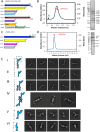
Figure 1:
Figure 1:. Biochemical and Structural characterization of A. thaliana Augmin assemblies
A) Schematic representation of plant Augmin hetero-octamer (AUG1,2,3,4,5,6,7,8) subunit organization, showing domain boundaries and purification tags. Note: AUG8 construct (residues 383–644) excludes the N-terminal MT binding domain (See Figure S1). B) Biochemical validation of hetero-octameric Augmin complex. Left: SEC-MALS analysis confirming monodisperse assembly with predicted octameric mass. Right: SDS-PAGE of purified complex showing all eight subunits (See Figure S1). C) Schematic of minimal Augmin hetero-tetramer (AUG1,3,4,5) subunit organization, including domain boundaries and purification tags. D) Biochemical validation of hetero-tetrameric complex. Left: SEC-MALS analysis demonstrating monodisperse assembly with tetrameric mass. Right: SDS-PAGE confirming presence of four subunits. E) Cryo-EM 2D-class averages revealing Augmin complex architecture. I: Monomeric AUG1,2,3,4,5,6,7,8: 40 nm tuning fork structure. II: Monomeric AUG1,2,3,4,5,6,7,8: V-junction focus with extended region. III: Dimeric AUG1,2,3,4,5,6,7,8: Two extended regions, single V-junction. IV: Dimeric AUG1,2,3,4,5,6,7,8: Two extended regions, two V-junctions. V: Focused V-junction from AUG1,2,3,4,5,6,7,8: 24 nm V-junction and stem regions. VI: Monomeric AUG1,3,4,5: 23 nm extended region.
Figure 2:
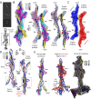
Figure 2:. Single particle Cryo-EM structures and models of Augmin assemblies.
A) Cryo-EM reconstructions of the AUG1,2,3,4,5,6,7,8 V-junction and stem. Left: 7.3-Å structure with segmented model of AUG1,2,3,4,5,6,7,8 (closed-state AUG6,7 CH-domain dimer). Right: 10 Å structure with open-state AUG6,7 CH-domain dimer (See Figures S2–S4). B) Model analysis. Left: AUG1,2,3,4,5,6,7,8 subunit organization in V-junction and stem. Right: Conformational comparison of closed (red) and open (blue) states. Inset: Vector representation of conformational transition. C) 3.7 Å reconstruction of AUG1,3,4,5, extended region with segmented model (See Figures S2–S5). D) Detailed subunit organization of AUG1,3,4,5 extended region model. Composite model of complete AUG1,2,3,4,5,6,7,8 complex derived from combined maps and models (See Figures S5A–C)
Figure 3:
Figure 3:. Architectural coiled-coil organization and conservation of the Augmin assembly.
A) Ribbon diagram highlighting individual subunit folds and organization. B) Subcomplex interaction analysis. Left: AUG3,5 heterodimer with interaction footprints. Right: AUG1,4 and AUG2,6,7,8 subcomplexes with corresponding interfaces. Insets: Topological organization of each subunit. C) Complete Augmin hetero-octamer assembly showing structural domains. D) Electrostatic surface potential map highlighting functional regions (Figure S9A–C). E) Surface conservation analysis across plant, animal, and insect (Figure S9F–H).
Figure 4:
Figure 4:. Cryo-EM structure and analysis of Augmin antiparallel dimer assembly.
A) Top: 2D class averages showing antiparallel organization of extended domains. Bottom: Low-resolution reconstruction with focused refinement of dimerization interface. B) Detailed structural analysis of extended region dimer. Left: Side view of segmented reconstruction with colored subunit organization. Middle: 90° rotated view showing subunit arrangement. Right: Isolated model highlighting subunit organization at dimer interface. C) Dimer interface analysis showing protomer organization. Red/blue: individual protomers. Yellow: Interface region between protomers. D) Conformational changes in dimer formation. Splayed view of protomers showing AUG1,3,4,5 organization. Interface zones highlighted in red at foot and belly regions. Arrows indicate domain movements during dimerization. E) Monomer-to-dimer transition analysis. Left: Single protomer structure. Middle: Overlay of monomeric and dimeric states. Right: Vector representation of conformational changes in tripod, belly, and leg regions.
Figure 5:
Figure 5:. XLMS validates features of Augmin hetero-octamer and Antiparallel Augmin dimer
A) XLMS analysis of hetero-tetrameric (AUG1,3,4,5) Augmin with mapped crosslinks on the cryo-EM generated model. This model of the flexible fold-back zone in AUG3,5, which is absent in the cryo-EM map, but is modeled from (AUG1,2,3,4,5,6,7,8) structure (See Figure S11). B) Comprehensive XLMS analysis of hetero-octameric (AUG1,2,3,4,5,6,7,8) Augmin model. Validated interactions throughout complex. Emphasis on joint and tripod zones. C) Crosslink mapping of AUG1,2,3,4,5,6,7,8 anti-parallel dimer assembly validating interface regions. For more details see Figure S11
Figure 6:
Figure 6:. Biochemical reconstitution and structure NEDD1 WD40 β-propeller-Augmin complex.
A) NEDD1 domain organization. N-terminal WD40 β-propeller domain. C-terminal helical domain (Note: Only WD40 β-propeller domain successfully purified). B) Complex formation analysis: AUG1,2,3,4,5,6,7,8 binds NEDD1-WD40 β-propellor and while AUG1,3,4,5 shows no binding (See Figure S12C–H). C) SEC analysis of complex formation. Red: AUG1,2,3,4,5,6,7,8 alone. Blue: AUG1,2,3,4,5,6,7,8 with NEDD1-WD40 β-propellor. Green: NEDD1-WD40 alone. D) Biochemical validation. SDS-PAGE showing co-migration of NEDD1-WD40 with AUG1,2,3,4,5,6,7,8 and comparison with AUG1,2,3,4,5,6,7,8 alone. E) Cryo-EM 2D-class average comparison NEDD1-WD40 β-propellor binding to Augmin. Top: AUG1,2,3,4,5,6,7,8 V-junction-stem with β-barrel density. Bottom: AUG1,2,3,4,5,6,7,8-NEDD1-WD40 β-propellor complex. F) Structural characterization of the complex. Left: 12 Å Cryo-EM segmented reconstruction with fitted models. Right: Ribbon representation of complex. G) 7.3-Å model showing β-barrel density bound to V-junction stem. Note the marked conformational change. H) Comparative analysis of V-junction. Top: Segmented maps of complex vs. AUG1,2,3,4,5,6,7,8 alone. Bottom: Atomic models of NEDD1-WD40 and β-barrel regions. I) NEDD1-WD40-β propellor interface analysis. Right: Surface views of Augmin interaction site. Left: Conservation analysis across species.
Figure 7:
Figure 7:. Mechanistic model of Augmin-NEDD1-MT interactions and MT branch nucleation.
A) MT lattice binding modes. Left: NEDD1-WD40 complex with closed AUG6,7 CH-domains. Center-left: β-barrel density impact on MT binding. Center-right: Open CH-domain configuration. Right: NDC80/Nuf2 CH-domain reference structure views shown in a side and a 90° rotated orientations. B) Proposed mechanism for Augmin function. Left: Transition from diffuse to NEDD1-WD40-bound state. Middle: Formation of anchored complex and dimerization. Right: γ-TuRC recruitment and MT branch nucleation.

Similar articles

References

    1. Akhmanova A. & Kapitein L. C. Mechanisms of microtubule organization in differentiated animal cells. Nat Rev Mol Cell Biol 23, 541–558 (2022). 10.1038/s41580-022-00473-y - DOI - PubMed
    1. Gudimchuk N. B. & McIntosh J. R. Regulation of microtubule dynamics, mechanics and function through the growing tip. Nat Rev Mol Cell Biol 22, 777–795 (2021). 10.1038/s41580-021-00399-x - DOI - PubMed
    1. Zupa E., Liu P., Wurtz M., Schiebel E. & Pfeffer S. The structure of the gamma-TuRC: a 25-years-old molecular puzzle. Curr Opin Struct Biol 66, 15–21 (2021). 10.1016/j.sbi.2020.08.008 - DOI - PubMed
    1. Liu P., Wurtz M., Zupa E., Pfeffer S. & Schiebel E. Microtubule nucleation: The waltz between gamma-tubulin ring complex and associated proteins. Curr Opin Cell Biol 68, 124–131 (2021). 10.1016/j.ceb.2020.10.004 - DOI - PubMed
    1. Yagi N., Fujita S. & Nakamura M. Plant microtubule nucleating apparatus and its potential signaling pathway. Curr Opin Plant Biol 82, 102624 (2024). 10.1016/j.pbi.2024.102624 - DOI - PubMed

Publication types

LinkOut - more resources