. 2024 Dec 31;28(3):111722.
doi: 10.1016/j.isci.2024.111722. eCollection 2025 Mar 21.

Design of a fragment crystallizable-engineered tetravalent bispecific antibody targeting programmed cell death-1 and vascular endothelial growth factor with cooperative biological effects

Affiliations

Design of a fragment crystallizable-engineered tetravalent bispecific antibody targeting programmed cell death-1 and vascular endothelial growth factor with cooperative biological effects

Tingting Zhong et al. iScience. .

Abstract

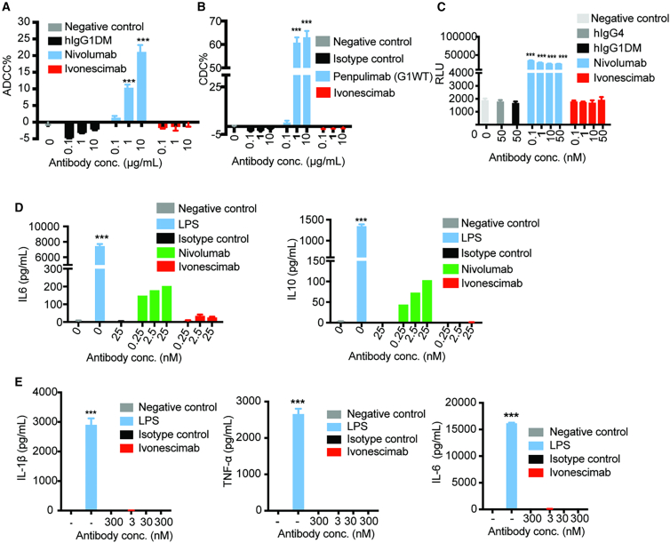
Clinical studies have shown that combination therapy of PD-1 and VEGF antibodies significantly improves clinical benefit over PD-1 antibody alone in certain settings. Ivonescimab, an on-market tetravalent anti-PD-1/VEGF bispecific antibody, was designed to improve efficacy and safety over combo therapy. In this study, the mechanism of action is investigated. In the presence of VEGF, ivonescimab forms soluble complexes with VEGF dimers, leading to the enhanced binding avidity of ivonescimab to PD-1 therefore promoting its increased potency on PD-1/PD-L1-signaling blockade. Likewise, PD-1 binding enhanced ivonescimab binding to VEGF, therefore enhancing VEGF-signaling blockade. Furthermore, ivonescimab treatment demonstrated statistically significant anti-tumor response in vivo. Moreover, ivonescimab contains Fc-silencing mutations abrogating FcγRI/IIIa binding and showed significantly reduced effector function in vitro which is consistent with the better safety profile of ivonescimab in monkeys and humans. Briefly, ivonescimab displays unique cooperative binding and promotes the increased in vitro functional bioactivities with a favorable safety profile.

Keywords: Cancer; Molecular biology; Therapeutics.


Conflict of interest statement

No potential conflicts of interest were disclosed by the authors.

Figures

None
Graphical abstract
Figure 1
Figure 1
Ivonescimab blocks PD-1/PD-L1 or VEGF/VEGFR signaling and inhibits VEGF-induced HUVECs proliferation (A and B) The binding kinetics of ivonescimab to PD-1 and VEGF simultaneously were determined by Fortebio Octet. Ivonescimab was fixed onto the AR2G sensor for 600 s and sequential binding to human PD-1 (PD-1-hFc, 300 s) and VEGF (VEGF-His, 300 s) or in reverse order was performed in two replicates and measured. Buffer only were used as controls. (C) Inhibition of human PD-L1-mFc binding to human PD1-hFc by ivonescimab and nivolumab were determined by ELISA. (D) Ivonescimab blocks PD-1/PD-L1 signaling. Luminescence signals in the co-culture of PD-L1 aAPC/CHO-K1 cells and PD-1 effector cells were detected by Steady-Glo Luciferase assay. RLU, relative light units. (E) Inhibition of human VEGFR2-mFc-Bio binding to human VEGF-His by ivonescimab and bevacizumab was determined by ELISA. (F) Ivonescimab blocks VEGF/VEGFR signaling in VEGF-mediated reporter assay. Luminescence signals in the 293T-KDR-NFAT-LUC cells treated with VEGF alone or VEGF with different concentrations of antibodies as indicated were assessed by Steady-Glo Luciferase assay. (G) Different concentrations of antibodies added with or without VEGF (20 nM) were co-cultured with the HUVEC cells for 3 days. The viable cells were quantified in CCK8 assay. Compared with the isotype control+20 nM VEGF, ∗∗∗p < 0.001.
Figure 2
Figure 2
Ivonescimab forms soluble complexes with VEGF (A–C) Ivonescimab alone (8 μM, A), VEGF alone (8 μM, B), or ivonescimab (8 μM) premixed with 2×VEGF (C) were analyzed on SEC-HPLC. Diagrams in the left panel represent ivonescimab, VEGF, and the proposed ivonescimab-VEGF complex structure. Also see Figure S1.
Figure 3
Figure 3
The presence of VEGF promotes binding avidity of ivonescimab to human PD-1 and facilitates better potency on the blockade of PD-1 signaling and T cell activation (A) Diagram representing the binding profile of ivonescimab to PD-1 with or without VEGF. (B) The binding kinetics of ivonescimab alone or ivonescimab-VEGF to immobilized PD-1-His-Biotin were determined by Fortebio Octet Molecular Interaction System. The binding kinetic parameters are shown above the sensorgram. Blue lines represent the real-time measurement of the binding affinity of ivonescimab with or without VEGF to PD-1, while red lines represent mathematical fitting curves based on blue lines. (C) Binding of ivonescimab and anti-PD-1 antibody penpulimab with or without VEGF on PD-1 transfected Jurkat cells via FACS. MFI, mean fluorescent intensity. (D) Cell surface PD-1 level on PD-1-expressing Jurkat cells, detected by FACS at different time points after ivonescimab incubation with or without VEGF. The reduction rates % were calculated from the decrease of surface PD-1 compared to its expression at 0 h. (E) Ivonescimab and penpulimab with or without VEGF blocked the interaction of PD-1 and PD-L1, leading to the enhancement of luminescence in the co-culture of PD-L1 aAPC/CHO-K1 cells and PD-1 effector cells. (F) IFN-γ and (G) IL-2 production in a mixed culture of hPBMCs and Raji-PD-L1 cells were analyzed by ELISA. Compared with the isotype control, ∗p < 0.05, ∗∗p < 0.01, ∗∗∗p < 0.001; compared with the isotype control+VEGF, ##p < 0.01, ###p < 0.001. Also see Figures S2 and S3.
Figure 4
Figure 4
The presence of PD-1 promotes the binding avidity of ivonescimab to human VEGF and facilitates enhanced VEGF signaling blockade (A) Diagram representing the binding profile of Ivonescimab to VEGF with or without PD-1. (B) Ivonescimab (7 nM) alone (left) or a mixture of ivonescimab (7 nM) with PD-1-human Fc (PD-1-hFc, 7 nM) (right) were immobilized on the AHC sensor. The binding kinetics of the serial dilution of human VEGF-His protein (1000–1.37 nM) to immobilized ivonescimab or ivonescimab-PD-1-hFc were determined by Fortebio Octet Molecular Interaction System. The binding kinetic parameters were shown above the sensorgrams. Blue lines represent the real-time measurement of the binding affinity of ivonescimab with or without PD-1 to VEGF, while red lines represent mathematical fitting curves based on blue lines. (C) Indicated antibodies were incubated with PD-1-His-Biotin-coated beads or control beads for 1 h in the presence of VEGF (5 nM). Then the supernatants without beads were measured for free VEGF by ELISA. Results of antibodies (PD-1+) were compared with the corresponding antibodies (PD-1-) group by t-test analysis, ∗p < 0.05, ∗∗p < 0.01, ∗∗∗p < 0.001, n.s. is not significant. (D) Luminescence signals in the 293T-KDR-NFAT-LUC cells treated with the same diluted supernatant in c as indicated were assessed by Steady-Glo Luciferase assay. Anti-HEL IgG1DM was used as an isotype control. Results of antibodies (PD-1+) were compared with the corresponding antibodies (PD-1-) group, ∗p < 0.05, ∗∗p < 0.01, ∗∗∗p < 0.001 and n.s. represents no significance.
Figure 5
Figure 5
Ivonescimab demonstrates superior anti-tumor efficacy in both single/combination treatment in mice (A–C) Each mouse was inoculated subcutaneously at the right hind flank with HCC827 cells, PBMCs, and ivonescimab, bevacizumab, or isotype control anti-HEL mixture on day 0. Different doses of antibodies were then continuously intravenously injected on day 7, 14, 21, 28, and 35. Tumor volume (A) and body weight (C) were measured (n = 7–8 for each group). (B) Spaghetti plots of all mice in each of the treatment groups are shown. (D) MDA-MB-231 cells were subcutaneously inoculated in the SCID/Beige mice. Mice were grouped when tumor volume reached 120 mm3 and intraperitoneally injected with activated hPBMCs. AK117 (anti-CD47 mAb) was then continuously intravenously injected twice per week for eight times and ivonescimab was administrated weekly for four times. Tumor volume was then monitored. (E) MC38-hPD-L1/hCD73 cells were subcutaneously inoculated in the C57BL/6-hPD-1/hPD-L1/hCD73 transgenic mice. Mice were grouped when tumor volume reached 80–120 mm3 and ivonescimab, AK119 (anti-CD73 mAb) or ivonescimab with AK119 were then intraperitoneally injected biweekly. Tumor volume was then monitored. ∗p < 0.05, ∗∗p < 0.01, ∗∗∗p < 0.001. Comparisons of tumor growth among groups were analyzed using two-way ANOVA followed by Bonferroni’s multiple comparison test. Also see Figure S4.
Figure 6
Figure 6
Favorable immunosafety profile of ivonescimab compared to nivolumab (A) ADCC of ivonescimab was determined by measuring lactase dehydrogenase (LDH) release in the mixed culture of CHO-K1-PD-1 cells and human PBMCs. (B) CDC of ivonescimab was determined by measuring LDH release from CHO-K1-PD-1 cells. The normal human serum complement was used as the source of complement. (C) ADCP activities of ivonescimab were measured by reporter assay. Jurkat-NFAT-CD64CD32H cells and CHO-K1-PD-1 cells were cocultured for 5 h in the presence of indicated antibodies. (D) IL-6 and IL-10 release by HPMMs cocultured with CHO-K1-PD-1 cells were examined by ELISA. (E) The release of inflammatory cytokines IL-1β, TNF-α, and IL-6 from human PBMCs when treated with ivonescimab were assessed by ELISA. LPS was used as a positive control. Data are expressed as mean ± SEM and analyzed using one-way ANOVA. Compared with the negative control, ∗∗∗p < 0.001. Also see Figure S5.
Figure 7
Figure 7
Model diagram of ivonescimab cooperative mechanisms of action Conceptually, ivonescimab can bind with VEGF dimers and PD-1 simultaneously. When all 4 binding sites of ivonescimab are occupied by VEGF and PD-1 (top row), this “cluster” complex becomes the most stable structure in which the binding of VEGF to ivonescimab would strengthen its binding to PD-1, and vice versa. This propensity and interdependency type of interaction requiring the engagement of the tetravalent antibody and its two antigens is defined as cooperative binding. This complex structure is very stable and relevant to a mono-specific anti-VEGF antibody (such as bevacizumab, middle row) or an anti-PD-1 antibody (such as penpulimab, bottom row).

References

    1. Carmeliet P. VEGF as a key mediator of angiogenesis in cancer. Oncology. 2005;69:4–10. doi: 10.1159/000088478. - DOI - PubMed
    1. Garcia J., Hurwitz H.I., Sandler A.B., Miles D., Coleman R.L., Deurloo R., Chinot O.L. Bevacizumab (Avastin®) in cancer treatment: A review of 15 years of clinical experience and future outlook. Cancer Treat Rev. 2020;86 doi: 10.1016/j.ctrv.2020.102017. - DOI - PubMed
    1. Villaruz L.C., Socinski M.A. The Role of Anti-angiogenesis in Non-small-cell Lung Cancer: an Update. Curr. Oncol. Rep. 2015;17:26. doi: 10.1007/s11912-015-0448-y. - DOI - PMC - PubMed
    1. Zhou C., Ren S., Luo Y., Wang L., Xiong A., Su C., Zhang Z., Li W., Zhou J., Yu X., et al. A phase Ib/II study of AK112, a PD-1/VEGF bispecific antibody, as first- or second-line therapy for advanced non–small cell lung cancer (NSCLC) J. Clin. Orthod. 2022;40:9040. doi: 10.1200/JCO.2022.40.16_suppl.9040. - DOI
    1. Zhao Y., Chen G., Chen J., Zhuang L., Du Y., Yu Q., Zhuang W., Zhao Y., Zhou M., Zhang W., et al. AK112, a novel PD-1/VEGF bispecific antibody, in combination with chemotherapy in patients with advanced non-small cell lung cancer (NSCLC): an open-label, multicenter, phase II trial. eClinicalMedicine. 2023;62 doi: 10.1016/j.eclinm.2023.102106. - DOI - PMC - PubMed

LinkOut - more resources