

Repair capacity of Taenia solium extraparenchymal cysts: radiological and in vitro evidence
- PMID: 40035503
- PMCID: PMC11980455
- DOI: 10.1080/17460913.2025.2472594
Repair capacity of Taenia solium extraparenchymal cysts: radiological and in vitro evidence
Abstract
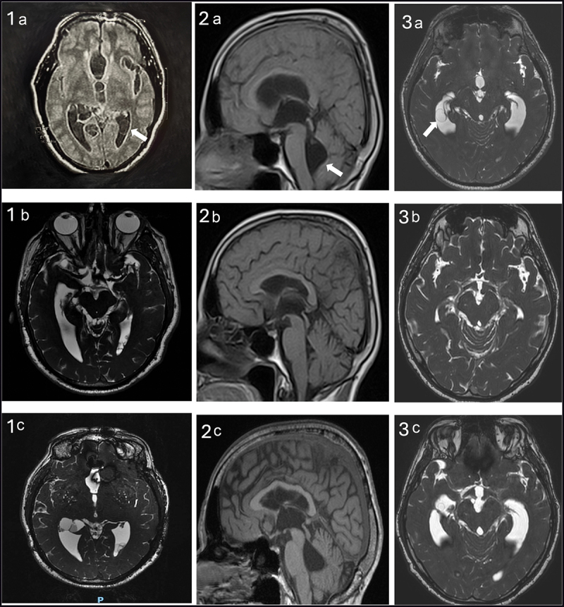
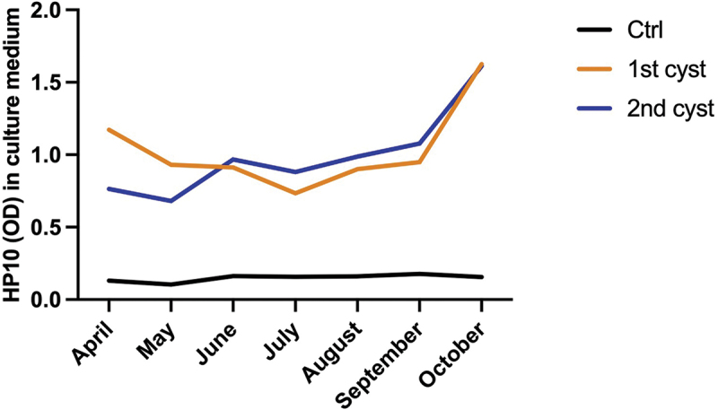
Extraparenchymal neurocysticercosis (EP-NC) responds poorly to anthelmintic treatment. Several factors are involved in this low responsiveness, including the host's heterogeneous immune response and the ability of the parasite to evade it. In this study, we present radiological and in vitro findings that demonstrate that Taenia solium cysts have the capacity to repair from injuries. Six patients (three with cases of subarachnoid, two with cases of intraventricular, and one with a case of mixed subarachnoid and intraventricular cysts) presented with neurological complaints and underwent either medical or surgical treatment. Follow-up magnetic resonance imaging (MRI) showed apparent resolution of the cysts. However, months later (10-56) new MRI scans revealed cysts at the same sites observed before treatment. Cysts surgically removed were maintained in RPMI-1640 medium supplemented with 10% fetal bovine serum. Monthly assessments demonstrated the growth of the parasites and the release of HP-10. Our findings demonstrate the ability of T. solium extraparenchymal cysts to grow and repair themselves. This capacity is likely another factor involved in the disease's poor treatment response.
Keywords: Neurocysticercosis; Taenia solium; extraparenchymal neurocysticercosis; in vitro tests; magnetic resonance imaging.
Conflict of interest statement
The authors have no relevant affiliations or financial involvement with any organization or entity with a financial interest in or financial conflict with the subject matter or materials discussed in the manuscript. This includes employment, consultancies, honoraria, stock ownership or options, expert testimony, grants or patents received or pending, or royalties.
No writing assistance was utilized in the production of this manuscript.
Figures


Similar articles
-
[Giant racemose subarachnoid and intraventricular neurocysticercosis: A case report].Rev Argent Microbiol. 2015 Jul-Sep;47(3):201-5. doi: 10.1016/j.ram.2015.07.001. Epub 2015 Aug 28. Rev Argent Microbiol. 2015. PMID: 26321177 Review. Spanish.
-
Taenia solium: Development of an Experimental Model of Porcine Neurocysticercosis.PLoS Negl Trop Dis. 2015 Aug 7;9(8):e0003980. doi: 10.1371/journal.pntd.0003980. eCollection 2015. PLoS Negl Trop Dis. 2015. PMID: 26252878 Free PMC article.
-
Relevance of 3D magnetic resonance imaging sequences in diagnosing basal subarachnoid neurocysticercosis.Acta Trop. 2015 Dec;152:60-65. doi: 10.1016/j.actatropica.2015.08.017. Epub 2015 Aug 29. Acta Trop. 2015. PMID: 26327445
-
Intraventricular Neurocysticercosis: An atypical presentation of a little-known and poorly understood disease - A case report.Parasitol Int. 2025 Dec;109:103108. doi: 10.1016/j.parint.2025.103108. Epub 2025 Jun 13. Parasitol Int. 2025. PMID: 40517948
-
Subarachnoid neurocysticercosis: emerging concepts and treatment.Curr Opin Infect Dis. 2020 Oct;33(5):339-346. doi: 10.1097/QCO.0000000000000669. Curr Opin Infect Dis. 2020. PMID: 32868512 Free PMC article. Review.
References
-
- Rodríguez-Rivas R, Flisser A, Norcia LF, et al. Neurocysticercosis in Latin America: current epidemiological situation based on official statistics from four countries. PLOS Negl Trop Dis. 2022;16(8):e0010652. doi: 10.1371/journal.pntd.0010652 - DOI - PMC - PubMed
-
• Interesting work showing the current epidemiological trend of NC in Latin America.
MeSH terms
Substances
LinkOut - more resources
Full Text Sources
Research Materials
Miscellaneous