. 2025 Oct 1;166(10):2285-2299.
doi: 10.1097/j.pain.0000000000003560. Epub 2025 Mar 4.

Evidence for peripheral neuroinflammation after acute whiplash

Affiliations

Evidence for peripheral neuroinflammation after acute whiplash

Colette Ridehalgh et al. Pain. .

Abstract

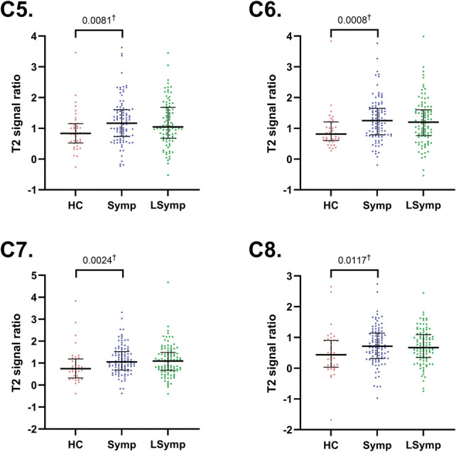
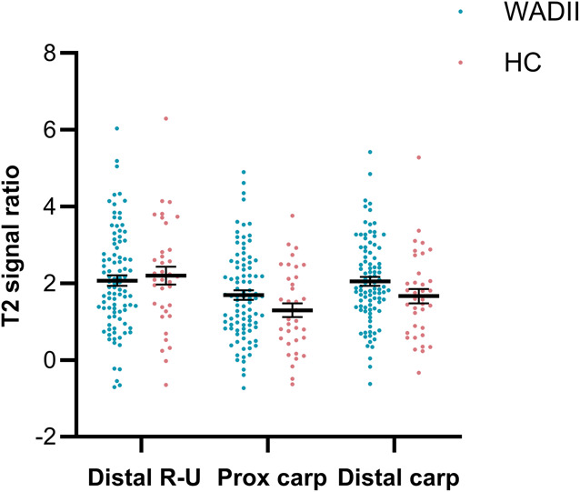
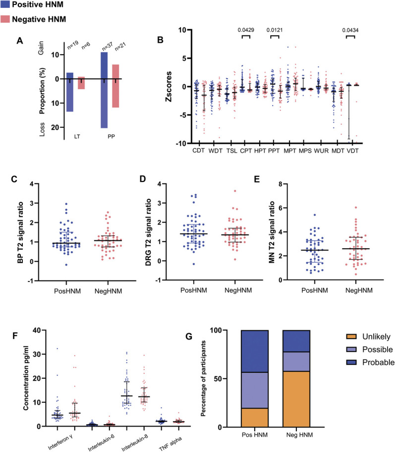
Whiplash injury is associated with high socioeconomic costs and poor prognosis. Most people are classified as having whiplash-associated disorder grade II (WADII), with neck complaints and musculoskeletal signs, in the absence of frank neurological signs. However, evidence suggests that there is a subgroup with underlying nerve involvement in WADII, such as peripheral neuroinflammation. This study aimed to investigate the presence of neuroinflammation in acute WADII using T2-weighted magnetic resonance imaging of the brachial plexus, dorsal root ganglia and median nerve, and clinical surrogates of neuroinflammation: heightened nerve mechanosensitivity (HNM), raised serum inflammatory mediators, and somatosensory hyperalgesia. One hundred twenty-two WADII participants within 4 weeks of whiplash and 43 healthy controls (HCs) were recruited. Magnetic resonance imaging T2 signal ratio was increased in the C5 root of the brachial plexus and the C5-C8 dorsal root ganglia in WADII participants compared with HCs but not in the distal median nerve trunk. Fifty-five percent of WADII participants had signs of HNM. Inflammatory mediators were also raised compared with HCs, and 47% of WADII participants had somatosensory changes on quantitative sensory testing. In those WADII individuals with HNM, there was hyperalgesia to cold and pressure and an increased proportion of neuropathic pain. Many people with WADII had multiple indicators of neuroinflammation. Overall, our results present a complex phenotypic profile for acute WADII and provide evidence suggestive of peripheral neuroinflammation in a subgroup of individuals. The results suggest that there is a need to reconsider the management of people with WADII.

Trial registration: ClinicalTrials.gov NCT04940923.

Keywords: Heightened nerve mechanosensitivity; Magnetic resonance imaging; Neuritis; Neuroinflammation; Neuropathic pain; Quantitative sensory testing; Whiplash; Whiplash associated disorder.


Conflict of interest statement

The authors have no conflicts of interest to declare.

Sponsorships or competing interests that may be relevant to content are disclosed at the end of this article.

Figures

Figure 1.
Figure 1.
Image analysis of T2 weighted images of the brachial plexus, dorsal root ganglia (DRG), and median nerve at the wrist. (A) Example T2 maximum intensity projection image formed from extracted selected slices. A bounding box is shown positioned around the right C6 DRG (green square). (B) Freehand mask drawn around the roots of the plexus on the maximum intensity projection image. (C) Segmented regions overlaid to produce a complete segmented image of the roots of the brachial plexus. (D) Regions of interest overlaid on C5 to C8 roots. (E) Bounding boxes positioned around the C5-C8 DRG. (F) C5 to T1 vertebral bodies showing the 4 control ROIs (10 × 10 voxels). (G–I) Example axial slices of the median nerve at the wrist, at the (G) distal radioulnar joint, (H) proximal carpal row, and (I) distal carpal row. Upper left of (G–I): Enlarged image of the median nerve. Lower left of (G–I): Freehand mask drawn around nerve and segmented median nerve (yellow). Right of (G–I): Complete axial slice across wrist showing the segmented median nerve (yellow) and the control region of interest in the underlying bone (yellow circle). The T2-weighted scans were from WADII participants. Note the brighter C5 and C6 roots of the brachial plexus on the left side (*), which corresponded to the most symptomatic side in this participant. ROI, regions of interest; WADII, whiplash-associated disorder grade II.
Figure 2.
Figure 2.
Symptom distribution in WADII participants. Percent of participants reporting symptoms is indicated in blue, with darker colours indicating higher percentages. WADII, whiplash-associated disorder grade II.
Figure 3.
Figure 3.
T2-signal ratios for the roots of the brachial plexus. Pooled data from the healthy control (HC) group is compared with symptomatic side of WADII participants (†Mann–Whitney tests), Symptomatic (Symp) and less symptomatic (LSymp) sides are also compared (#Wilcoxon signed-rank test). The median values are shown (horizontal bars). Upper and lower horizontal bars represent IQRs. α = 0.0125. IQR, interquartile range; WADII, whiplash-associated disorder grade II.
Figure 4.
Figure 4.
T2 signal ratios for the C5-C8 dorsal root ganglia. Pooled data from the healthy control (HC) group is compared with symptomatic side of WADII participants (†Mann–Whitney tests), Symptomatic (Symp) and less symptomatic (LSymp) sides are also compared. The median values are shown (horizontal bars). Upper and lower horizontal bars represent IQRs. α = 0.0125. IQR, interquartile range; WADII, whiplash-associated disorder grade II.
Figure 5.
Figure 5.
T2 signal ratios for the median nerve at the wrist of the most symptomatic side at 3 levels: Distal R-U, distal radioulnar joint; Prox carp, proximal carpal row; Distal carp, distal carpal row. HC, healthy control.
Figure 6.
Figure 6.
Heightened nerve mechanosensitivity measures. (A) Elbow extension range of motion during the ULNT1 (median nerve bias). (B) Digital palpation of the brachial plexus. The proportion of participants with no pain, discomfort, and local and referred pain is shown. (C) Pressure pain thresholds over the median nerve at the carpal tunnel. (D) Pressure pain thresholds over the ulnar nerve at the cubital tunnel. In (A, C, and D), pooled data from both sides are compared (whiplash and HC groups), as well as data from the symptomatic and less symptomatic sides; the medians are shown (horizontal bars). Upper and lower error bars represent the IQRs. HC, healthy control; IQR, interquartile range; LSymp, less symptomatic side; ROM: range of motion; Symp, symptomatic side; ULNT1, upper limb neurodynamic test-1.
Figure 7.
Figure 7.
Blood serum concentrations of inflammatory mediators. Values for interferon-γ (IFN-γ) and IL-10 have been cropped to 100 and 2 pg/mL for display (missing values IFN-γ n = 2, 108.6, 138.89; IL-10 n = 1, 5.74, α = 0.01). HC, healthy control; IL, interleukin; WADII, whiplash-associated disorder grade II.
Figure 8.
Figure 8.
(A) Proportion of participants with sensory changes upon bedside neurological examination to light touch and pin prick in the upper limb dermatomes on the symptomatic side (C5-T1; n = 122). (B) Quantitative sensory testing profiles over the index finger in WADII participants and HCs. Z-scores cropped to −10 for display (missing values CDT n = 6, range −10.438 to −26.723, VDT n = 21, range −14.667 to −39.796). (C) Proportion of WADII participants with a gain or loss of sensory function based on QST measures (individuals with Z-scores ±1.96 SD of HCs). CDT, cold detection threshold; CPT, cold pain threshold; HC, healthy control; HPT, heat pain threshold; LT, light touch; MDT, mechanical detection threshold; MPT, mechanical pain threshold; MPS, mechanical pain sensitivity; PP, pin prick; PPT, pressure pain threshold; QST, quantitative sensory testing; TSL, thermal sensory limen; VDT, vibration detection threshold; WADII, whiplash-associated disorder grade II; WDT, warm detection threshold; WUR, windup ratio.
Figure 9.
Figure 9.
Comparison of clinical measures in WADII participants with and without heightened nerve mechanosensitivity. (A) Proportion of participants with sensory changes to light touch (LT) or pin prick (PP) in at least one upper limb dermatome (n = 118; participants with both a gain and loss of function in one of the dermatomes; LT n = 0, PP n = 7). (B) Quantitative sensory testing z-scores. Z-scores for VDT cropped to −10 for display (missing values n = 21, range −14.667 to −39.796). (C–E) MRI T2 signal ratios for (C) brachial plexus, (D) DRG, and (E) median nerve. Individual values represent the highest T2 value for the levels of the roots of the brachial plexus, DRG, and median nerve location. (F) Serum inflammatory mediator concentrations. (G) Proportion of participants with unlikely, possible and probable neuropathic pain according to the presence of heightened nerve mechanosensitivity (P = 0.0002; n with positive HNM = 65, n with negative HNM = 55). In (A–F), positive HNM = blue, negative HNM = red. CDT, cold detection threshold; CPT, cold pain threshold; DRG, dorsal root ganglia; HNM, heightened nerve mechanosensitivity; HPT, heat pain threshold; LT, light touch; MDT, mechanical detection threshold; MRI, magnetic resonance imaging; MPS, mechanical pain sensitivity; MPT, mechanical pain threshold; PP, pin prick; PPT, pressure pain threshold; TSL, thermal sensory limen; VDT, vibration detection threshold; WDT, warm detection threshold; WADII, whiplash-associated disorder grade II; WUR, windup ratio.

References

    1. Andersen TE, Hansen M, Ravn SL, Seehuus R, Nielsen M, Vaegter HB. Validation of the PTSD-8 scale in chronic pain patients. Pain Med 2018;19:1365–72. - PubMed
    1. Baumer P, Dombert T, Staub F, Kaestel T, Bartsch AJ, Heiland S, Bendszus M, Pham M. Ulnar neuropathy at the elbow: MR neurography—nerve T2 signal increase and caliber. Radiology 2011;260:199–206. - PubMed
    1. Beaudoin FL, Gaither R, DeLomba WC, McLean SA. Tolerability and efficacy of duloxetine for the prevention of persistent musculoskeletal pain after trauma and injury: a pilot three-group randomized controlled trial. PAIN 2023;164:855–63. - PMC - PubMed
    1. Blankenburg M, Boekens H, Hechler T, Maier C, Krumova E, Scherens A, Magerl W, Aksu F, Zernikow B. Reference values for quantitative sensory testing in children and adolescents: developmental and gender differences of somatosensory perception. PAIN 2010;149:76–88. - PubMed
    1. Boretius S, Gadjanski I, Demmer I, Bahr M, Diem R, Michaelis T, Frahm J. MRI of optic neuritis in a rat model. Neuroimage 2008;41:323–34. - PubMed

Associated data