

Exosomes containing miR-148a-3p derived from mesenchymal stem cells suppress epithelial-mesenchymal transition in lens epithelial cells
- PMID: 40036306
- PMCID: PMC11878568
- DOI: 10.1093/stcltm/szae091
Exosomes containing miR-148a-3p derived from mesenchymal stem cells suppress epithelial-mesenchymal transition in lens epithelial cells
Abstract
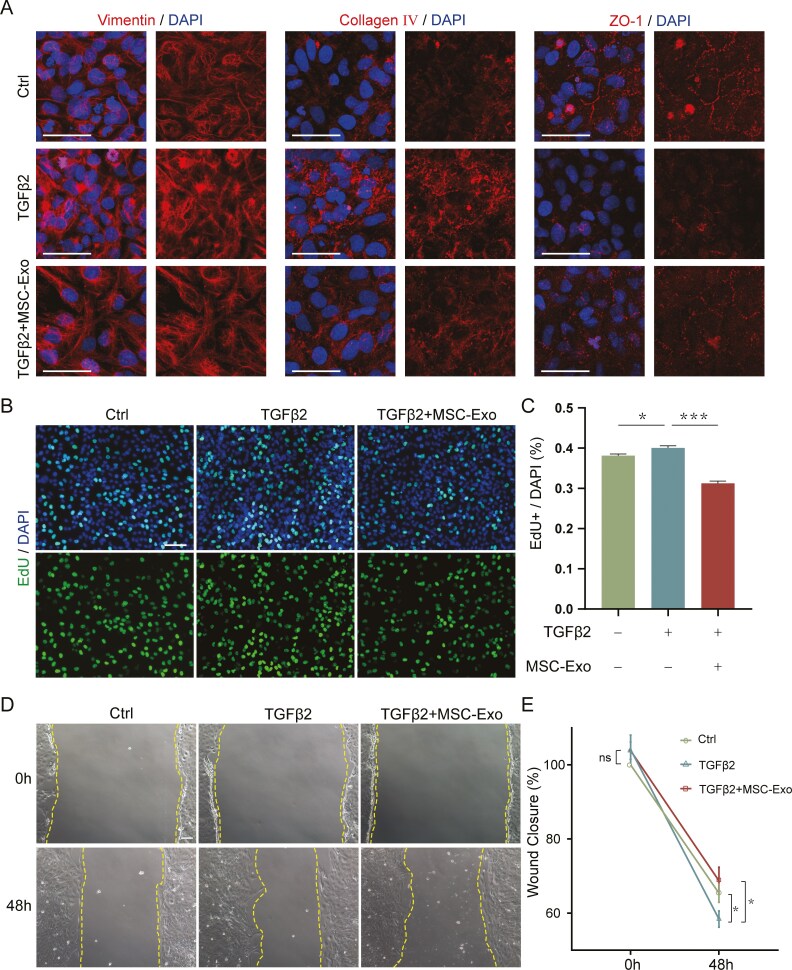
Epithelial-mesenchymal transition (EMT) of lens epithelial cells (LECs) is responsible for the development of fibrotic cataracts, which contribute to severe visual impairment. Recent evidence has shown that mesenchymal stem cell-derived exosomes (MSC-Exo) can attenuate EMT in several tissues. However, the effect of MSC-Exo on EMT in LECs (LECs-EMT) has not been determined. In this study, we isolated exosomes from human umbilical cord MSCs (hucMSC-Exo) and evaluated their effect on LECs-EMT both in vitro and in vivo. HucMSC-Exo application significantly suppressed the expression of mesenchymal cell-associated genes while increasing the expression of epithelial cell-associated genes. Cell proliferation and migration of LECs undergoing EMT were inhibited after hucMSC-Exo treatment. The volume of EMT plaques in mice with injury-induced anterior subcapsular cataract (ASC) was significantly reduced in the hucMSC-Exo-treated group. Furthermore, miR-148a-3p was abundant in hucMSC-Exo. After transfection with miR-148a-3p inhibitor, the anti-fibrotic effect of hucMSC-Exo was attenuated in LECs-EMT. A dual-luciferase reporter assay identified PRNP as a direct target gene of miR-148a-3p. Furthermore, we verified that hucMSC-Exo inhibited LECs-EMT through the miR-148a-3p/PRNP axis and the potential downstream ERK signaling pathway. Taken together, our work reveals the inhibitory effect of hucMSC-Exo on LECs-EMT and the underlying mechanism involved, which may provide potential therapeutic options for fibrotic cataracts.
Keywords: EMT; LECs; PRNP; hucMSC-derived exosomes; miR-148a-3p.
© The Author(s) 2025. Published by Oxford University Press.
Conflict of interest statement
The authors declared no potential conflicts of interest.
Figures


Similar articles
-
Human umbilical cord mesenchymal stem cell-derived exosomal miR-27b attenuates subretinal fibrosis via suppressing epithelial-mesenchymal transition by targeting HOXC6.Stem Cell Res Ther. 2021 Jan 7;12(1):24. doi: 10.1186/s13287-020-02064-0. Stem Cell Res Ther. 2021. PMID: 33413548 Free PMC article.
-
MicroRNA-23b-3p promotes the proliferation, migration, and epithelial-mesenchymal transition of lens epithelial cells by targeting Sprouty2.Acta Histochem. 2019 Aug;121(6):704-711. doi: 10.1016/j.acthis.2019.05.007. Epub 2019 Jun 22. Acta Histochem. 2019. PMID: 31235073
-
MiR-148a-3p Loaded Human Umbilical Cord Mesenchymal Stem Cell-Derived Extracellular Vesicles Alleviates Silica-Induced Pulmonary Fibrosis by Inhibiting β-Catenin Signaling.Int J Nanomedicine. 2025 Apr 9;20:4319-4336. doi: 10.2147/IJN.S506542. eCollection 2025. Int J Nanomedicine. 2025. PMID: 40230541 Free PMC article.
-
Human umbilical cord mesenchymal stem cell-derived exosomal microRNA-148a-3p inhibits neointimal hyperplasia by targeting Serpine1.Arch Biochem Biophys. 2022 Apr 15;719:109155. doi: 10.1016/j.abb.2022.109155. Epub 2022 Feb 24. Arch Biochem Biophys. 2022. PMID: 35218720
-
Let-7c-3p suppresses lens epithelial-mesenchymal transition by inhibiting cadherin-11 expression in fibrotic cataract.Mol Cell Biochem. 2024 Apr;479(4):743-759. doi: 10.1007/s11010-023-04758-4. Epub 2023 May 12. Mol Cell Biochem. 2024. PMID: 37171723
References
-
- Wormstone IM, Wormstone YM, Smith AJO, Eldred JA.. Posterior capsule opacification: What’s in the bag? Prog Retin Eye Res. 2021;82:100905. https://doi.org/10.1016/j.preteyeres.2020.100905 - DOI - PubMed
-
- Blindness GBD, Vision Impairment C, Vision Loss Expert Group of the Global Burden of Disease S. Causes of blindness and vision impairment in 2020 and trends over 30 years, and prevalence of avoidable blindness in relation to VISION 2020: the Right to Sight: an analysis for the Global Burden of Disease Study. Lancet Glob Health. 2021;9:e144-ee60. - PMC - PubMed
-
- Apple DJ, Peng Q, Visessook N, et al.Eradication of posterior capsule opacification: documentation of a marked decrease in Nd:YAG laser posterior capsulotomy rates noted in an analysis of 5416 pseudophakic human eyes obtained postmortem. Ophthalmology. 2020;127:S29-S42. https://doi.org/10.1016/j.ophtha.2020.01.026 - DOI - PubMed
-
- Bremond-Gignac D, Daruich A, Robert MP, Valleix S.. Recent developments in the management of congenital cataract. Ann Transl Med. 2020;8:1545. https://doi.org/10.21037/atm-20-3033 - DOI - PMC - PubMed
-
- Elgohary MA, Beckingsale AB.. Effect of posterior capsular opacification on visual function in patients with monofocal and multifocal intraocular lenses. Eye (Lond). 2008;22:613-619. https://doi.org/10.1038/sj.eye.6702661 - DOI - PubMed
MeSH terms
Substances
Grants and funding
LinkOut - more resources
Full Text Sources
Miscellaneous
