. 2025 Apr 17;34(9):821-834.
doi: 10.1093/hmg/ddaf029.

A novel recurrent ARL3 variant c.209G > A p.(Gly70Glu) causes variable non-syndromic dominant retinal dystrophy with defective lipidated protein transport in human retinal stem cell models

Affiliations

A novel recurrent ARL3 variant c.209G > A p.(Gly70Glu) causes variable non-syndromic dominant retinal dystrophy with defective lipidated protein transport in human retinal stem cell models

Julio C Corral-Serrano et al. Hum Mol Genet. .

Abstract

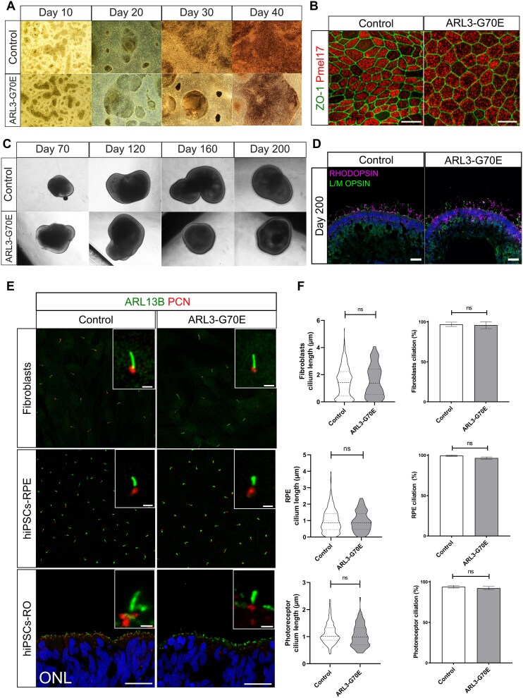
Inherited retinal dystrophies (IRDs) are characterized by their high clinical and genetic heterogeneity. Despite significant advances in the identification of genes associated with IRDs, many individuals and families still have not received a definite molecular diagnosis. Here, we performed clinical examinations and conducted genetic testing in five families with IRD. Whole exome sequencing in the five index cases revealed a heterozygous missense variant, c.209G > A, p.(Gly70Glu) in the ARL3 gene (NM_004311.4). A de novo occurrence was demonstrated in one affected individual and autosomal dominant inheritance in nine affected individuals from four families. Their phenotypes displayed variable expressivity, and ranged from rod-cone to cone-rod dystrophy with photophobia. Human induced pluripotent stem cells (hiPSCs) were generated from dermal fibroblasts from the individual with the de novo ARL3 variant and were differentiated to retinal pigment epithelium cells (RPE) and retinal organoids. Immunofluorescence analyses in these models showed decreased INPP5E localization within the cilia of RPE and connecting cilia of retinal organoids, as well as reduced PDE6⍺ in the organoid outer segments, suggesting that the p.(Gly70Glu) variant causes IRD by defective lipidated protein transport in photoreceptors and/or RPE. This is the first study of ARL3 dysfunction in human retinal cells, highlighting its importance for retinal homeostasis, as well as a variability in the clinical presentation of ARL3-associated IRD.

Keywords: ARL3; cilia; inherited retinal disease; retinal dystrophy; retinal organoids.


Figures

Figure 1
Figure 1
Genetic examination of five families with the ARL3 c.209G > A variant causing non-syndromic autosomal dominant retinal dystrophy. (A) Pedigrees of five unrelated families showing the affected individuals with the heterozygous change c.209G > A, p.(Gly70Glu) in ARL3. (B) Sanger sequencing confirmation of the variant in selected probands and family members. (C) Multispecies protein sequence alignment with conservation of the Gly (G) residue across evolution. Residues that are fully conserved are depicted in bold. M1: ARL3 c.209G > A, p.(Gly70Glu); WT: Wild-type. (D) AlphaFold prediction of ARL3 wild-type protein (ARL3-WT) structure bound to GTP (pink, top) or GDP (green, bottom), and (E) ARL3-G70E bound to GTP (pink, top) or GDP (green, bottom).
Figure 2
Figure 2
Clinical investigation of all families with the ARL3 c.209G > A variant causing non-syndromic autosomal dominant retinal dystrophy (A) Fundoscopy of family 1, individual II.2 showing a largely preserved macular area with fine spicular intraretinal pigmentation in the periphery and normal vascular calibers. OCT of the macula showing well-preserved retinal architecture and thickness. Blue-light autofluorescence imaging showing largely preserved macular autofluorescence surrounded by granular hyperautofluorescence. (B) Family 2, individual IV.1. Fundus color imaging shows areas of atrophy, nasal to the optic disc, with an absence of pigmentary changes. OCT shows reduced ONL outside the fovea. AF images show a hypoautofluorescent ring around the fovea and hypoautofluorescent areas nasal to the optic nerve. (C) Family 3 individual II.4. AF images show a hyperautofluorescent ring in the right eye, with a lamellar pseudohole on OCT. the left eye has areas of hypoautofluorescence around the fovea on AF imaging and reduced ONL outside the fovea. (D) Family 4, individual II.2. Fundus color imaging shows areas of atrophy with an absence of bone spicules. OCT of the right eye shows a scar from previous choroidal neovascular membrane (CNV) and left eye a lamellar pseudohole. AF images show hypoautofluorescent areas nasal to the disc, as well as hypoautofluorescence at the fovea. (E) Family 5, individual II.3. Fundus imaging reveals sparse bone spicules. OCT shows reduced ONL outside the fovea. A hyperautofluorescent ring can be seen on AF imaging. AF: Autofluorescence; OCT: Optical coherence tomography; ONL: Outer nuclear layer; RE: Right eye. LE: Left eye.
Figure 3
Figure 3
The ARL3-G70E variant does not have a major effect on ciliation or cilium length. (A and B) hiPSCs-RPE differentiation. (A) Bright-field images of control and ARL3-G70E hiPSCs-derived RPE cells (hiPSCs-RPE) during the differentiation process at days 10, 20, 30 and 40. (B) Immunofluorescence analysis of hiPSCs-RPE cells stained with the cell junction marker ZO-1 (green) and the melanosome marker PMEL17 (red). Scale bars: 20 μm. (C and D) hiPSCs differentiation to retinal organoids (hiPSCs-ROs). (C) Bright-field images of control and ARL3-G70E hiPSCs-ROs during the differentiation process at days 70, 120, 160, and 200. (D) Immunofluorescence analysis of ROs at day 200 stained with the rod photoreceptor marker RHODOPSIN (magenta), and the cone photoreceptor marker L/M OPSIN (green). DAPI marks the nuclei (blue). Scale bars: 20 μm. (E) Cilia staining in fibroblasts, hiPSC-RPE, and photoreceptors in hiPSC-ROs, as indicated, with the markers ARL13B (green) and PCN (red). DAPI (blue) marks the photoreceptor nuclei from ROs. Scale bars are 1 μm for insets, and 20 μm for the photoreceptor ONL images. (F) Cilia length was analyzed using CiliaQ as a measure of the ARL13B length and PCN as ciliary base. For fibroblasts, n = 373 control cilia and n = 148 ARL3-G70E cilia; for RPE, n = 285 control cilia and n = 112 ARL3-G70E cilia; for ROs, n = 93 control cilia and n = 77 ARL3-G70E cilia. P-value: Ns, not significant, as assessed by Mann–Whitney test.
Figure 4
Figure 4
ARL3-G70E hiPSCs-derived RPE (hiPSCs-RPE) and retinal organoids (hiPSCs-ROs) show a reduction of ciliary INPP5E and PDE6⍺. (A) Upper panels: Cilia were stained with INPP5E (green) and acetylated tubulin (magenta) in fibroblasts, hiPSCs-RPE, and photoreceptors of hiPSCs-ROs. hiPSCs-ROs were differentiated for 120 days to examine photoreceptor cilia. Scale bars: 2 μm (fibroblasts), 1 μm (hiPSCs-RPE and hiPSCs-RO). Lower panels: The INPP5E/acetylated tubulin fluorescence intensity ratio was measured. For fibroblasts, n = 86 control cilia and n = 41 ARL3-G70E cilia; for RPE, n = 88 control cilia and n = 82 ARL-G70E cilia, for ROs, n = 23 control cilia and n = 18 ARL3-G70E cilia. Ns, not significant. *P-value < 0.05, ***P-value < 0.001 as assessed by Mann–Whitney test. (B) hiPSCs-ROs were differentiated for 200 days and stained with PDE6⍺ (green), rhodopsin (magenta) and DAPI (blue). Scale bars: 100 μm for RO1 and RO2 images; 25 μm for RO3 image, and 5 μm for the RO3 insets. (C) The PDE6α to rhodopsin (RHO) colocalization coefficient in the OS region was analysed using JACoP on ImageJ. For controls, 8 images from n = 4 ROs were measured. For ARL3-G70E, 9 images from n = 5 ROs were measured. *P-value < 0.05 as assessed by Mann–Whitney test. RO: Retinal organoid. OS: Outer segments.

References

    1. Hanany M, Shalom S, Ben-Yosef T. et al. Comparison of worldwide disease prevalence and genetic prevalence of inherited retinal diseases and variant interpretation considerations. Cold Spring Harb Perspect Med 2024;14:a041277. - PMC - PubMed
    1. Ben-Yosef T. Inherited retinal diseases. Int J Mol Sci 2022;23:13467. - PMC - PubMed
    1. Strom SP, Clark MJ, Martinez A. et al. De novo occurrence of a variant in ARL3 and apparent autosomal dominant transmission of retinitis Pigmentosa. PLoS One 2016;11:e0150944. - PMC - PubMed
    1. Holtan JP, Teigen K, Aukrust I. et al. Dominant ARL3-related retinitis pigmentosa. Ophthalmic Genet 2019;40:124–128. - PubMed
    1. Ratnapriya R, Jacobson SG, Cideciyan AV. et al. A novel ARL3 gene mutation associated with autosomal dominant retinal degeneration. Front Cell Dev Biol 2021;9. - PMC - PubMed

MeSH terms

Substances