. 2025 Mar;15(3):240242.
doi: 10.1098/rsob.240242. Epub 2025 Mar 5.

Pigment-dispersing factor neuropeptides act as multifunctional hormones and modulators in tardigrades

Affiliations

Pigment-dispersing factor neuropeptides act as multifunctional hormones and modulators in tardigrades

Soumi Dutta et al. Open Biol. 2025 Mar.

Abstract

Pigment-dispersing factors (PDFs) are neuropeptides that play key roles in controlling the circadian rhythms in various insects, whereas their function remains elusive in other protostomes including tardigrades (water bears). Here we show that the three PDFs of the tardigrade Hypsibius exemplaris are co-localized in two pairs of inner lobe cells in the brain, whereas only one PDF occurs in four additional cerebral and two extracerebral cells. The axons of the inner lobe cells pass through the contralateral brain hemisphere, descend to the ventral nerve cord and terminate in two pairs of potential release sites in the posteriormost trunk ganglion. Using in vitro assays, we demonstrate that all three PDFs and their deorphanized receptor (PDFR) are functional. Widespread localization of PDFR suggests that tardigrade PDFs may act as multifunctional hormones and neuromodulators that control major functions including light detection, neural processing, locomotion, feeding, digestion, osmoregulation, growth, embryonic development and oogenesis/reproduction.

Keywords: G protein-coupled receptor; Hypsibius exemplaris; Tardigrada; gene expression; nervous system; neuromodulator.


Conflict of interest statement

We declare we have no competing interests.

Figures
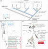
Evolutionary history of PDF/PDH peptides in ecdysozoans and phylogeny of PDF peptides and PDF receptor proteins.
Figure 1.
Evolutionary history of PDF/PDH peptides in ecdysozoans and phylogeny of PDF peptides and PDF receptor proteins. (a) Scenario on evolution of PDF peptides in ecdysozoans. Note the presence of two PDF peptides in the last common ancestors of Panarthropoda and Ecdysozoa. Modified from Mayer et al. [21]. (b) Maximum likelihood tree of PDFs across ecdysozoans. Note the position of monophyletic eutardigrade PDFs nested within the arthropod PDF/PDH clade and the occurrence of two distinct clades of parachelan PDFs (PDF-1 and PDF-2/3). PDF peptides of H. exemplaris are highlighted in red. (c) Maximum likelihood tree of approximately 1000 class B G protein-coupled receptor proteins (GPCRs). The dataset was obtained previously [20] from cluster analysis of approximately 18 000 bilaterian GPCRs. Both PDFR splice variants (He-PDFR-A and He-PDFR-B) of H. exemplaris fall into the monophyletic group of PDF receptors of protostomes (highlighted in red).
Genomic structure of pdf and pdfr genes of eutardigrades, and results of in vitro functional analyses of PDFs and PDFR isoforms in H. exemplaris.
Figure 2.
Genomic structure of pdf and pdfr genes of eutardigrades, and results of in vitro functional analyses of PDFs and PDFR isoforms in H. exemplaris. (a) Diagrams of genomic scaffolds illustrating the location and orientation of pdf genes in H. exemplaris and Ramazzottius varieornatus. Exons are depicted as coloured rectangles. Dashed lines indicate the position of introns. Note the proximity of He-pdf-2 and He-pdf-3 on a single scaffold (MTYJ01000058.1). (b) Comparison of amino acid sequences of mature tardigrade PDF peptides to selected arthropod PDF/PDHs. GenBank accession numbers are given for each peptide. Black and grey backgrounds indicate either identical or physicochemically similar amino acids to those of β-PDH from the crustacean Uca pugilator, respectively. Note the relatively low identities of tardigrade PDFs to β-PDH (33−44%). (c) Diagram of genomic structure of pdfr gene of H. exemplaris. Exons are depicted as rectangles. Dashed lines indicate the position of introns. Red rectangle (exon 13 of He-pdfr-B) indicates an alternative 3' splice junction (acceptor site) within the last exon of He-pdfr in H. exemplaris. (d,e) BRET ratio changes of HEK293T cells transfected with long (He-pdfr-A) and short variants (He-pdfr-B) of the PDF receptor from H. exemplaris after activation with synthetic PDFs (He-PDF-1, He-PDF-2 and He-PDF-3) in a dose-dependent manner. EC50 values are given for each peptide. Note decreasing BRET ratios (i.e. increasing intracellular cAMP levels) at higher concentrations of peptide indicating a stronger response to stimuli. Box plots of eight biological replicates (n = 8 for each dosage). Boxes correspond to interquartile ranges. The circle inside each box represents the arithmetic mean. Negative controls were performed without pdfr transfection (n = 4; −He-pdfr-A; −He-pdfr-B) and PDF stimuli (n = 8; −PDF). Positive controls were conducted with Forskolin/IBMX (at least n = 3; +Forsk./IBMX), which causes high intracellular concentrations of cAMP.
Localization of He-PDF−1 and He-pdf−1 transcripts in the brain of the tardigrade H. exemplaris.
Figure 3.
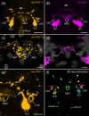
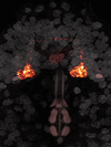
Localization of He-PDF-1 and He-pdf-1 transcripts in the brain of the tardigrade H. exemplaris. Antibody (a,c,e) and mRNA labelling (b,d). 3D reconstruction (f) and projections of confocal substacks (a–e) in dorsal view. Anterior is up in all images. Pharyngeal placoids are autofluorescent in (a). DNA staining is illustrated in grey in (c) and (d). (a) Localization of the He-PDF1 peptide in inner lobe cells, lateral cells and median cells. (b) Localization of He-pdf-1 mRNA. Note correspondence to immunolabelling in (a). (c) Detail of immunolabelled median cells with their nuclei (NU1 and NU2). Asterisks indicate varicosities of neurites labelled with He-PDF-1 antibody. (d) Detail of He-pdf-1 mRNA expression in median cells. Note correspondence to immunolabelling in (c), except that varicosities have not been labelled. (e) Detail of right brain hemisphere from specimen in (a) showing individual axons of inner lobe cells (open arrowheads) and neurites of lateral cell (arrows). (f) 3D reconstruction of He-PDF-1 immunoreactivity illustrating number and position of somata of individual neurons with nuclei (coloured). Note immunoreactivity in two pairs of inner lobe cells (yellow and cyan), two median cells (green) and two lateral cells (red) within the brain. Arrowheads indicate varicosities of axonal projections of left (filled arrowheads) and right pairs of inner lobe cells (open arrowheads) that cross over to the other brain hemisphere. Abbreviations: AX-LH axonal projections from left brain hemisphere, AX-RH axonal projections from right brain hemisphere, BR brain, ILC inner lobe PDF-immunoreactive cells, LAC lateral PDF-immunoreactive cells, MEC median PDF-immunoreactive cells, NU1 nucleus of median cell 1, NU2 nucleus of median cell 2, PLA pharyngeal placoids. Scale bars: 10 µm (a), 5 µm (b,e) and 2 µm (c,d).
Localization of each of the three PDFs in the nervous system of H. exemplaris.
Figure 4.
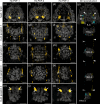
Localization of each of the three PDFs in the nervous system of H. exemplaris. Antibody labelling (orange in a–o), and 3D reconstruction (yellow and cyan in p). DNA staining is illustrated in grey. Projections of confocal substacks of the head region in dorsal view (a,f,k) and trunk ganglia in ventral view (b–e,g–j,l–o). Anterior is up in all images. (a–o) Note prominent expression of all three peptides in two pairs of somata in inner lobes of brain. Varicose axonal projections pass through first three trunk ganglia and terminate in anterior region of fourth trunk ganglion (arrows in e,j,o). (p) 3D reconstruction of left (yellow) and right pairs (cyan) of inner lobe cells and trajectories of their contralateral fibres (filled and open arrowheads) based on He-PDF-1 immunolabelling. Dorsal perspective. Note position of potential terminal buttons in anterior region of fourth trunk ganglion. Abbreviations: ILC inner lobe PDF-immunoreactive cells, TRG-1TRG-4 first to fourth trunk ganglia. Scale bars: 10 µm (a,f,k) and 5 µm (b–e,g–j,l–o).
Localization of He-PDF−1 and He-pdf−1 transcripts in the ventral extracerebral PDF-ir cells of H. exemplaris.
Figure 5.
Localization of He-PDF-1 and He-pdf-1 transcripts in the ventral extracerebral PDF-ir cells of H. exemplaris. Specimens are presented in ventral perspective. Anterior is up. Antibody labelling is illustrated in orange in (a), mRNA labelling in magenta in (b) and DNA staining in grey in (a–c). (a,b) Projections of confocal substacks. (a) Antibody labelling of He-PDF-1 peptide. Arrowheads indicate immunoreactive fibres of inner lobe cells in inner connectives. (b) Localization of He-pdf-1 mRNA transcripts, which corresponds to distribution of He-PDF-1 peptide in (a). (c) 3D reconstruction of immunolabelled anterior body region. Note the position of somata of both ventral extracerebral PDF-ir cells outside the brain, anterior to first trunk ganglion. (d) Diagram of anterior body region illustrating position of ventral extracerebral PDF-ir cells that are widely separated from other PDF-ir cells across the z-axis. Abbreviations: BR brain, BR-IL inner lobe of brain, BR-OL outer lobe of brain, CN-IN inner connectives, CN-OU outer connectives, EY eyes, ILC inner lobe PDF-immunoreactive cells, LAC lateral PDF-immunoreactive cells, LE-1 first leg, TRG-1 first trunk ganglion, VEC ventral extracerebral PDF-immunoreactive cells. Scale bars: 5 µm (b,c).
Differences in expression levels of the three pdf genes and MALDI-TOF mass spectra of the three PDFs in H. exemplaris.
Figure 6.
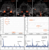
Differences in expression levels of the three pdf genes and MALDI-TOF mass spectra of the three PDFs in H. exemplaris. (a–c) Detection of He-pdf-1, He-pdf-2 and He-pdf-3 mRNA using same settings of confocal microscope. Brains in dorsal view. Anterior is up. DNA staining is shown in grey. Buccal tube, stylets and pharyngeal placoids are autofluorescent. Note that He-pdf-1 shows highest and He-pdf-3 lowest signal in inner lobe cells. (d,e) Relative expression levels of He-pdf-1, He-pdf-2 and He-pdf-3 based on comparative analysis of raw sequence reads from four independent RNA-seq experiments using short read mapping approach, with housekeeping gene GAPDH as reference. Boxes correspond to interquartile ranges. Circle inside each box represents arithmetic mean. (d) Linear chart. (e) Logarithmic chart using same dataset as in (d). (f,g) Representative MALDI-TOF mass spectra obtained from single dissected brain by direct tissue profiling (f) and extract of 100 brains (g) from sample no. 2 (see §4; electronic supplementary material, figure S10). Ion signals are marked and represent single charged peptides [M+H]+. Note that using direct tissue profiling only He-PDF-1 and He-PDF-2 could be detected, whereas ion masses corresponding to all three predicted He-PDFs were obtained from brain extracts. Abbreviations: BTU buccal tube, ILC inner lobe PDF-immunoreactive cells, MEC median PDF-immunoreactive cells, PLA pharyngeal placoids, STY stylet. Scale bars: 5 µm (a–c).
Localization of PDF receptor mRNA in various tissues and cells of H. exemplaris.
Figure 7.
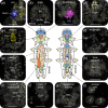
Localization of PDF receptor mRNA in various tissues and cells of H. exemplaris. Labelling using He-pdfr probes is illustrated in yellow in (a–l) (see electronic supplementary material, figure S15 for positive controls). Projections of confocal substacks (a–l) and diagrams of tardigrade anatomy in dorsal and ventral views (centre). Dotted white lines indicate contours of organs and tissues. Anterior is up in all images. Grey colour shows DNA staining in (a–h), (j) and (k), and autofluorescence in (i) and (l). Pharyngeal placoids are autofluorescent in (d) and (e), and claws in (j). (a) Expression of He-pdfr in the rhabdomeric cell of the eye labelled with r-opsin-v mRNA probe (blue). (b) Expression of He-pdfr in the brain. (c) Expression of He-pdfr in inner lobe cells double labelled with He-pdf-1 probe (magenta). (d–l) Expression of He-pdfr in various other tissues and cells. Asterisks indicate storage cells that occur within body cavity. Abbreviations: BR brain, BR-IL inner lobe of brain, BR-ML median lobe of brain, BR-OL outer lobe of brain, CLG claw gland, CL claw (visualized by cuticular autofluorescence), ESO esophagus, ILC inner lobe PDF-immunoreactive cells, MAT Malpighian tubules, MIG midgut, OOC autofluorescent yolk granules of an oocyte, PHA pharynx, PLA pharyngeal placoids, RHC rhabdomeric cell, TRG-3 third trunk ganglion, TRC trophocytes. Scale bars: 10 µm (b,d–h), 5 µm (a,c,i–k) and 2 µm (l).
Summary diagrams of localization of PDFs in the head of H. exemplaris.
Figure 8.
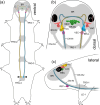
Summary diagrams of localization of PDFs in the head of H. exemplaris. Brain, trunk ganglia and connectives are indicated in grey. (a) Overview illustrating the position of PDF-ir neurons (coloured), ventral extracerebral PDF-ir cells and trajectories of inner lobe cells (yellow and blue). Arrowheads point to button-like terminals on the antero-ventral surface of the fourth trunk ganglion. (b) Diagram illustrating the number, position, morphology and connectivity of PDF-ir cells in the head in dorsal view. Note eight cerebral neurons exhibiting different morphologies and two ventral extracerebral cells associated with inner connectives. Note also that lateral PDF-ir neurons send their projections to the eyes and central part of the brain. (c) Same as in (b), but in lateral view. Abbreviations: BR brain, BR-IL inner lobe of brain, BR-ML median lobe of brain, BR-OL outer lobe of brain, CN-IN inner connectives, CN-OU outer connectives, EY eye, ILC inner lobe PDF-immunoreactive cells, LAC lateral PDF-immunoreactive cells, LE1 first leg, MEC median PDF-immunoreactive cells, TRG-1TRG−4 first to fourth trunk ganglia, VEC ventral extracerebral PDF-immunoreactive cells.
None

References

    1. Kleinholz LH. 1975. Purified hormones from the crustacean eyestalk and their physiological specificity. Nature 258, 256–257. (10.1038/258256a0) - DOI - PubMed
    1. Fernlund P. 1976. Structure of a light-adapting hormone from the shrimp, Pandalus borealis. Biochim. Biophys. Acta 439, 17–25. (10.1016/0005-2795(76)90155-0) - DOI - PubMed
    1. Rao KR, Riehm JP. 1993. Pigment-dispersing hormones. Ann. NY Acad. Sci. 680, 78–88. (10.1111/j.1749-6632.1993.tb19676.x) - DOI - PubMed
    1. Homberg U, Würden S, Dircksen H, Rao KR. 1991. Comparative anatomy of pigment-dispersing hormone-immunoreactive neurons in the brain of orthopteroid insects. Cell Tissue Res. 266, 343–357. (10.1007/BF00318190) - DOI
    1. Stengl M, Homberg U. 1994. Pigment-dispersing hormone-immunoreactive neurons in the cockroach Leucophaea maderae share properties with circadian pacemaker neurons. J. Comp. Physiol. 175, 203–213. (10.1007/bf00215116) - DOI - PubMed

LinkOut - more resources