Review
. 2025 Jul 30;27(6):1419-1433.
doi: 10.1093/neuonc/noaf058.

Applications of artificial intelligence and advanced imaging in pediatric diffuse midline glioma

Affiliations
Review

Applications of artificial intelligence and advanced imaging in pediatric diffuse midline glioma

Atlas Haddadi Avval et al. Neuro Oncol. .

Abstract

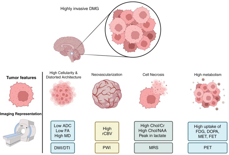
Diffuse midline glioma (DMG) is a rare, aggressive, and fatal tumor that largely occurs in the pediatric population. To improve outcomes, it is important to characterize DMGs, which can be performed via magnetic resonance imaging (MRI) assessment. Recently, artificial intelligence (AI) and advanced imaging have demonstrated their potential to improve the evaluation of various brain tumors, gleaning more information from imaging data than is possible without these methods. This narrative review compiles the existing literature on the intersection of MRI-based AI use and DMG tumors. The applications of AI in DMG revolve around classification and diagnosis, segmentation, radiogenomics, and prognosis/survival prediction. Currently published articles have utilized a wide spectrum of AI algorithms, from traditional machine learning and radiomics to neural networks. Challenges include the lack of cohorts of DMG patients with publicly available, multi-institutional, multimodal imaging and genomics datasets as well as the overall rarity of the disease. As an adjunct to AI, advanced MRI techniques, including diffusion-weighted imaging, perfusion-weighted imaging, and Magnetic Resonance Spectroscopy (MRS), as well as positron emission tomography (PET), provide additional insights into DMGs. Establishing AI models in conjunction with advanced imaging modalities has the potential to push clinical practice toward precision medicine.

Keywords: artificial intelligence; deep learning; diffuse intrinsic pontine glioma; diffuse midline glioma; radiomics.


Conflict of interest statement

A.R.: Co-founder and consultant for MRIMatch, Inc., and consultant for Arterys, Inc.

Other authors declare no conflicts of interest.

Figures

Figure 1.
Figure 1.
A case of DMG tumor in axial T1w (A), T1 post-contrast (B and F), T2w (C), T2 FLAIR (D and H), and mid-sagittal T2 FLAIR (G) sequences. The bottom row illustrates automatically generated segmentation masks (F showing the contrast enhanced region, G and H showing the whole tumor mask) along with some key quantitative parameters (E) extracted by our in-house Automated Lesion and Feature Extraction (pyALFE) program (github.com/reghbali/pyalfe). Abbreviations: FO, First Order radiomics features.
Figure 2.
Figure 2.
An example of a radiomics workflow. In the first block, medical images are gathered from multimodal magnetic resonance imaging. These images are then split into training, validation, and testing sets on a patient level. The second block shows image pre-processing steps such as image registration with anatomic atlases, resampling to isotropic volumes, bias field correction, and normalization of the intensity values. In the next block, the tumor mask is generated via automatic or manual segmentation. Radiomic features are extracted including first-order histogram, second- and higher-order texture, and filtered image feature subclasses. Features are then harmonized to improve their reliability. In the fourth block, feature selection algorithms pick a limited number of radiomics features and feed them into classifiers. In the final stage, the combination of feature selectors and classifiers is tested on the testing set; Results and charts are generated to represent which model yields the best performance.
Figure 3.
Figure 3.
The DMG tumor volume in the brainstem (the arrow, please refer to Figure 1H for the segmentation mask) is represented as mixed hyperintensity and hypointensity in axial DWI (A) and in the corresponding ADC map (B). The colored Fractional Anisotropy map and the Mean Diffusivity maps are represented in C and D, respectively. The colored Fractional Anisotropy map (C) codes voxels based on the predominant direction of the diffusion of water molecules: left-right, superior-inferior, and anterior-posterior. White matter fiber tractography (E) and orientation distribution functions (ODFs) derived at each voxel from diffusion imaging data (F, G), all color-coded by the direction of diffusion, demonstrate distorted white matter tracts by the pontine mass. Diffusion imaging provides quantitative values that can be measured within the tumor and its surrounding structures, including the distorted white matter fibers.
Figure 4.
Figure 4.
A case of DMG tumor in a 5-year-old patient experiencing clinical deterioration after the completion of radiotherapy. The contrast-enhanced area of the tumor (the arrow) is visible in T1c (A). Multi-voxel MR spectroscopy of the tumor and its surrounding area is shown in (B). In each of the voxel boxes, metabolite concentration peaks are represented on the X-axis from right to left (C). The bottom row visualizes the perfusion-weighted Arterial Spin Labeling (ASL) magnetic resonance imaging quantification (in mL/100 g/min) (D) and 18FET-PET (E) images of the same patient, with tumor-to-brain ratios shown in the colormap (F). Higher values indicate metabolically active tumor areas. In this case, while FET PET (Figure 4E and 4F) can distinctly visualize the active tumor area, the tumor region in ASL (Figure 4D) demonstrates low perfusion values, demonstrating discrepancies between methods and opportunities for further research.
Figure 5.
Figure 5.
Schematic overview of possible advanced MR imaging findings in Diffuse Midline Glioma. Advanced imaging modalities aid in its characterization: diffusion-weighted imaging (DWI) and diffusion tensor imaging (DTI) show low apparent diffusion coefficient (ADC), low fractional anisotropy (FA), and high mean diffusivity (MD) indicative of high cellularity. Magnetic resonance perfusion (MRP) highlights elevated relative cerebral blood volume (rCBV) due to neovascularization. Magnetic resonance spectroscopy (MRS) reveals metabolic changes, such as increased choline-to-creatine (Chol/Cr) and choline-to-N-acetylaspartate (Chol/NAA) ratios and lactate peaks. Positron emission tomography (PET) demonstrates high uptake of tracers like FDG; DOPA, MET, and FET, reflecting tumor metabolism. This figure was created in BioRender, https://BioRender.com/r88t111
Figure 6.
Figure 6.
A typical step-by-step workflow for future artificial intelligence research on DMG tumors. Step 1 involves identifying the research question, such as focusing on the diagnosis, classification, prognosis, or treatment response. Step 2 ensures the inclusion of diverse patient populations by gender, ethnicity, and race. Step 3 emphasizes multimodal data collection, including genomics, clinical, laboratory, and radiological data. Step 4 applies AI analyses through radiomics, machine learning, and deep learning pipelines. Step 5 refines algorithms with human–computer interaction to enhance the performance. Step 6 concludes with reporting results using comprehensive charts and graphs, summarizing key metrics. This workflow facilitates AI-driven insights into DMG characterization and treatment strategies.”

Similar articles

References

    1. Miguel Llordes G, Medina Pérez VM, Curto Simón B, et al. Epidemiology, diagnostic strategies, and therapeutic advances in diffuse midline glioma. J Clin Med. 2023;12(16):5261. - PMC - PubMed
    1. Vuong HG, Le HT, Jea A, McNall-Knapp R, Dunn IF.. Risk stratification of H3 K27M-mutant diffuse midline gliomas based on anatomical locations: An integrated systematic review of individual participant data. J Neurosurg Pediatr. 2022;30(1):99–106. - PMC - PubMed
    1. Vallero SG, Bertero L, Morana G, et al. Pediatric diffuse midline glioma H3K27- altered: A complex clinical and biological landscape behind a neatly defined tumor type. Front Oncol. 2023;12:1082062. - PMC - PubMed
    1. Pachocki CJ, Hol EM.. Current perspectives on diffuse midline glioma and a different role for the immune microenvironment compared to glioblastoma. J Neuroinflammation. 2022;19(1):276. - PMC - PubMed
    1. Argersinger DP, Rivas SR, Shah AH, Jackson S, Heiss JD.. New developments in the pathogenesis, therapeutic targeting, and treatment of H3K27M-mutant diffuse midline glioma. Cancers (Basel). 2021;13(21):5280. - PMC - PubMed

Grants and funding