. 2025 Feb 27;53(5):gkaf123.
doi: 10.1093/nar/gkaf123.

Mug20-Rec25-Rec27 binds DNA and enhances meiotic DNA break formation via phase-separated condensates

Affiliations

Mug20-Rec25-Rec27 binds DNA and enhances meiotic DNA break formation via phase-separated condensates

Max F Wang et al. Nucleic Acids Res. .

Abstract

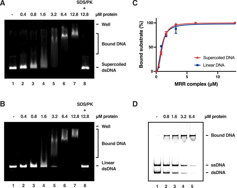
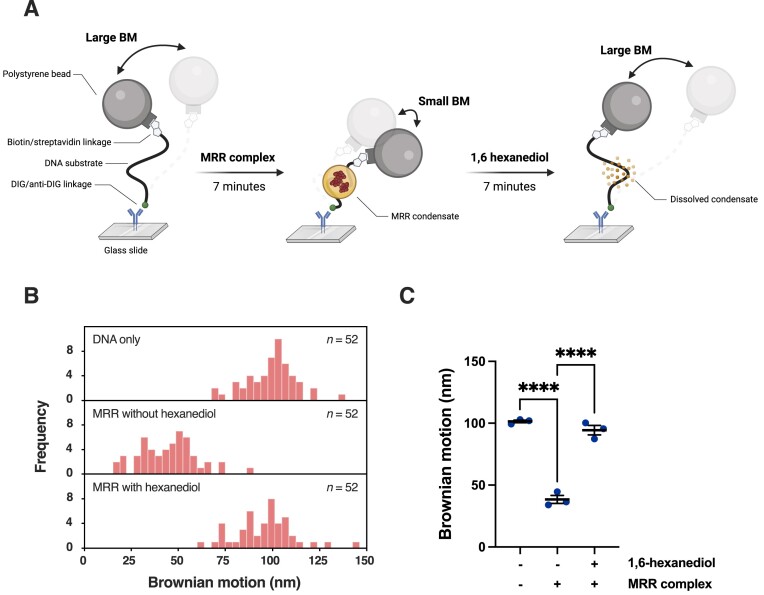
During meiosis, programmed DNA double-strand breaks (DSBs) are formed at hotspots to initiate homologous recombination, which is vital for reassorting genetic material. In fission yeast, the linear element (LinE) proteins Mug20, Rec25, and Rec27 interdependently bind chromosomal hotspots with high specificity and are necessary for high-level DSB formation. However, their mechanistic role in regulating the meiotic DSB machinery remains unknown. Here, using purified Mug20-Rec25-Rec27 (MRR) complex and functional intracellular analyses, we reveal that the MRR-DNA nucleoprotein complex assembles phase-separated condensates that compact the DNA. Notably, MRR complex formation is a prerequisite for DNA binding and condensate assembly, with Rec27 playing a pivotal role in directly binding DNA. Consistent with this finding, failure to form MRR-DNA condensates results in defective intracellular meiotic DSB formation and recombination. Our results provide mechanistic insights into how LinEs enhance meiotic DSB formation and provide a paradigm for studies in other species.


Conflict of interest statement

The authors declare no competing interests.

Figures

Graphical Abstract
Graphical Abstract
Figure 1.
Figure 1.
Expression and purification of S. pombe MRR complex. (A) Plasmid maps of Rec25, Mug20, and His6-SUMO-tagged Rec27. T7P and T7T represent the T7 promoter and terminator, respectively. (B) Coomassie brilliant blue-stained SDS-PAGE and immunoblotting (hexahistidine-tag monoclonal antibody) of the whole cell extracts harboring the His6-SUMO-Rec27 and Rec25–Mug20 expression vectors incubated with or without IPTG. The hexahistidine tag monoclonal antibody was used in the immunoblotting analysis. (C) SDS-PAGE of the purified untagged MRR complex (3 μg was loaded). (D) The identities of Mug20, Rec25, and Rec27 were determined by LC–MS/MS. The amino acids identified in the fragments are underlined.
Figure 2.
Figure 2.
DNA binding properties of the MRR complex. (AB) The indicated concentration of MRR complex was incubated with (A) supercoiled or (B) linear plasmid DNA substrates (∼3 kb). The reaction mixture in lane eight was treated with SDS and proteinase K (PK) to deproteinize the nucleoprotein complex as a control. (C) Quantification of the gel mobility shift assay with supercoiled and linear plasmid DNA substrates. Data are shown as mean ± SD from at least two independent experiments and fitted to the Hill equation. The fitted dissociation constants (Kd) are 0.98 μM (supercoiled) and 0.86 μM (linear) with coefficients of determination (R2) of 0.997 and 0.982, respectively. (D) The indicated concentration of MRR complex was co-incubated with a mixture of the 80-bp dsDNA and its constituent ssDNA. DNA was used at a concentration of 5 nM molecules.
Figure 3.
Figure 3.
DNA-driven MRR condensate assembly in vitro. (A) Prediction of Mug20, Rec25, and Rec27 droplet-promoting regions using the FuzDrop server [56]. The amino acid position is shown in the x-axis, and the y-axis indicates the droplet-promoting probability (pDP), where pDP = 0.6 (dotted line) is a cutoff value above which residues are considered capable of promoting phase separation. The bars indicate the predicted droplet-promoting regions. (B) Representative micrograph of 5 μM MRR–DNA nucleoprotein condensates. The boxes indicate two zoomed-in regions of interest, expanded on the right. (C) (i) Representative micrograph of 5 μM MRR–DNA nucleoprotein condensates assembled after 5 and 30 minutes. (ii) Normalized foci number and average foci area at different time points. Numbers and average foci area were normalized to the mean of the sample assembled after 5 and 30 minutes, respectively. Data are shown as mean ± SEM from three independent experiments. (D) The impact of KCl and protein concentrations on MRR–DNA nucleoprotein condensate formation. The relative fluorescence intensity of condensation is represented as the average foci area multiplied by mean fluorescence intensity (A × M). (E) The normalized absorbance at 595 nm of the clarified supernatant of the MRR complex. The vertical dashed line intersects the abscissa at ∼10 μM, the inferred saturation concentration (csat) for the MRR–DNA nucleoprotein condensates. BSA was included as a control, as shown by the gray dashed line. (F) (i) Representative micrographs of pre-assembled MRR–DNA nucleoprotein condensates challenged with 8.3% 1,6-hexanediol or 1 M KCl. Condensates were assembled for 30 minutes before being challenged. (ii) Quantification data were normalized to the mean of the control group. Data are shown as mean ± SEM from three independent experiments. Statistical significance was determined using one-way ANOVA with Dunnett's post hoc test. ***p < 0.001, ****p < 0.0001.
Figure 4.
Figure 4.
MRR compacts duplex DNA. (A) Schematic of the single-molecule TPM experiment. BM and DIG represent Brownian motion and digoxigenin, respectively. (B) Representative distribution of median BM of the DNA-tethered beads under different conditions. n is the number of DNA molecules measured. Median BM values were taken from 500 frames. (C) Mean of the median BM of the DNA-tethered beads under different conditions. Data are shown as mean ± SEM from three independent experiments. Statistical significance was determined using one-way ANOVA with Šidák's post hoc test. ****p < 0.0001.
Figure 5.
Figure 5.
Basic residues in Rec27 account for DNA binding activity and condensate assembly. (A) Amino acid sequence alignment of the 50 N-terminal residues of Rec27 from different Schizosaccharomyces spp. was performed using Clustal Omega [77]. Identical amino acids are shaded. Five conserved lysine (K) or arginine (R) residues were mutated to glutamic acid (E), which are indicated by asterisks. (B) SDS-PAGE of the purified MRR-5E complex (3 μg was loaded). (C) The indicated concentrations of the WT MRR complex and its 5E mutant variant were incubated with 3-kb supercoiled plasmid DNA substrates. Binding was analyzed by an electrophoretic mobility shift assay. (D) (i) Representative micrographs of WT MRR complex and its 5E mutant variant with different concentrations in the presence or absence of supercoiled plasmid DNA. (ii) Normalized foci number and average foci area from different protein concentrations (μM). Quantification data were normalized to the mean of the WT MRR complex. Data are shown as mean ± SEM from three independent replicates. The scale bar is 10 μm. DNA was used at a concentration of 5 nM molecules.
Figure 6.
Figure 6.
The rec27-283 (5E) mutant exhibits reduced meiotic recombination, DNA DSB frequency, and abnormal linear structures. (AB) Frequencies of meiotic (A) gene conversion to ade6+ at the ade6 locus and (B) cross-over in the (i) ade6arg1 and (ii) ade6ura4 intervals in rec27 mutants. Data are from Supplementary Table S3. Statistical significance was determined using the unpaired t-test. ***p < 0.001. (C) (i) DSBs at the ade6 locus on chromosome III. The black triangle shows breakage at the ade6-3049 hotspot, and the asterisk shows a meiosis-independent DSB site. DSBs in the (ii) NotI-D and (iii) NotI-J regions on chromosome I. DSB frequencies of five hotspots in (ii) NotI-D [1–5] and two hotspots in (iii) NotI-J (mbs1 and mbs2) are quantified in Supplementary Table S4. Meiotically induced cells of WT, rec27-283 (5E), and rec27-184 (rec27Δ) were collected at the indicated time points. DNA was prepared, digested with (i) PmeI or (ii and iii) NotI, and analyzed by gel electrophoresis and Southern blot hybridization with the indicated DNA probes previously described [47]. (D) Rec25–GFP protein was observed in rec27-283 (5E) cells of h90 (homothallic) strains using live-cell super-resolution fluorescence microscopy at 25°C. Hoechst 33342 stains nuclear DNA. Each image is representative of at least 20 cells examined. Images show the maximal projection of the entire Z-stack of image sections. Dotted lines outline the cells. The scale bar is 2 μm.
Figure 7.
Figure 7.
Individual Mug20, Rec25, and Rec27 proteins neither bind DNA nor assemble condensates. (A) The indicated concentrations (μM) of the WT MRR complex, Rec25, Rec27, and Mug20, were incubated with 3-kb supercoiled plasmid DNA substrates. (B) The indicated concentrations (μM) of Rec27 and Rec27-5E proteins were incubated with 10 μM Rec25 and Mug20 and supercoiled plasmid DNA. (C) (i) Representative micrographs of the WT MRR complex and the Rec27 protein with different concentrations in the presence or absence of 3-kb supercoiled DNA. (ii) Normalized foci number and average foci area from different protein concentrations. Quantification data were normalized to the mean of the WT MRR complex. Data are shown as mean ± SEM from three independent replicates. DNA was used at a concentration of 5 nM molecules.

References

    1. Zickler D, Kleckner N Meiotic chromosomes: integrating structure and function. Mol Syst Biol. 1999; 33:10952–7.10.1146/annurev.genet.33.1.603. - DOI - PubMed
    1. Marston AL, Amon A Meiosis: cell-cycle controls shuffle and deal. Nat Rev Mol Cell Biol. 2004; 5:983–97.10.1038/nrm1526. - DOI - PubMed
    1. Keeney S, Giroux CN, Kleckner N Meiosis-specific DNA double-strand breaks are catalyzed by Spo11, a member of a widely conserved protein family. Cell. 1997; 88:375–84.10.1016/S0092-8674(00)81876-0. - DOI - PubMed
    1. Baudat F, Manova K, Yuen JP et al. . Chromosome synapsis defects and sexually dimorphic meiotic progression in mice lacking Spo11. Mol Cell. 2000; 6:989–98.10.1016/S1097-2765(00)00098-8. - DOI - PubMed
    1. Cervantes MD, Farah JA, Smith GR Meiotic DNA breaks associated with recombination in S. pombe. Mol Cell. 2000; 5:883–8.10.1016/S1097-2765(00)80328-7. - DOI - PubMed

MeSH terms

LinkOut - more resources