. 2025 Feb 27;53(5):gkaf133.
doi: 10.1093/nar/gkaf133.

Tuning the tropism and infectivity of SARS-CoV-2 virus-like particles for mRNA delivery

Affiliations

Tuning the tropism and infectivity of SARS-CoV-2 virus-like particles for mRNA delivery

Qi Yang et al. Nucleic Acids Res. .

Abstract

Severe acute respiratory syndrome coronavirus 2 (SARS-CoV-2) virus-like particles (VLPs) are ∼100-nm-sized bioinspired mimetics of the authentic virus. We undertook molecular engineering to optimize the VLP platform for messenger RNA (mRNA) delivery. Cloning the nucleocapsid protein upstream of M-IRES-E resulted in a three-plasmid (3P) VLP system that displayed ∼7-fold higher viral entry efficiency compared with VLPs formed by co-transfection with four plasmids. More than 90% of human ACE2-expressing cells could be transduced using these 3P VLPs. Viral tropism could be programmed by switching glycoproteins from other viral strains, including other betacoronaviruses and the vesicular stomatitis virus G protein. An infectious two-plasmid VLP system was also advanced where one vector carried the viral surface glycoprotein and the second carried the remaining SARS-CoV-2 structural proteins and reporter gene. SARS-CoV-2 VLPs could be engineered to carry up to four transgenes, including functional Cas9 mRNA for genome editing. Gene editing of specific target cell types was feasible by modifying VLP tropism. Successful mRNA delivery to mouse lungs suggests that the SARS-CoV-2 VLPs can overcome natural biological barriers to enable pulmonary gene delivery. Overall, the study describes the advancement of the SARS-CoV-2 VLP platform for robust mRNA delivery both in vitro and in vivo.


Conflict of interest statement

A US provisional patent application No. 63/754,897 related to this paper has been filed by the University at Buffalo on behalf of Q.Y. and S.N. All other authors declare no conflicts of interest.

Figures

Graphical Abstract
Graphical Abstract
Figure 1.
Figure 1.
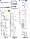
3P SARS2 VLPs are superior to 4P SARS2 VLPs. (A) 4P and 3P SARS2 Luc-PS9 VLPs were produced by co-transfection of either four or three plasmids into 293T producer cells. These plasmids encode for the viral structural proteins, along with a luciferase reporter coupled to PS9 packaging signal (Luc-PS9). (B) Luciferase assay showed ∼7-fold higher luminescence intensity in 293T-hACE2 cells upon using 3P versus 4P VLPs. (C) VLP concentrate (10 μl for M and E, 2 μl for S2 and N) was loaded in each lane. Western blot analysis suggests incorporation of all SARS2 structural components in VLPs, with 3P SARS2 Luc-PS9 VLPs displaying more intense protein bands compared with 4P VLPs. (D) Higher Luc-PS9 transcript levels were observed using RT-PCR in the case of 3P SARS2 Luc-PS9 VLPs. (E) Cryo-TEM images of 3P SARS2 Luc-PS9 VLPs showed spherical ∼100-nm-sized VLPs with double-layered membrane structures. (F) 3P SARS2 EGFP-PS9 VLPs produced with EGFP reporter efficiently infected 293T-hACE2 and A549-hACE2-TMPRSS2 cells. Fluorescence images were acquired 24 h post-infection. (G) Flow cytometry VLP entry time-course showed peak fluorescence for 3P SARS2 EGFP-PS9 VLPs at 24 h followed by decrease at larger times. This was observed both for 293T-hACE2 and A549-hACE2-TMPRSS2 cells. Abbreviations: aa, amino acid; nt, nucleotide. Data are mean ± STD. *P < .05, **P < .01, ***P < .001, ****P < .0001, NS: not significant.
Figure 2.
Figure 2.
Tuning VLP viral tropism by altering viral glycoprotein. (A) Different types of VLPs were produced using the 3P system by varying the viral glycoprotein (VSV-G, SARS2 spike, SARS spike, or MERS spike) and reporter genes (luciferase or EGFP). (B) One microgram of the viral glycoprotein was used to create various 3P Luc-PS9 VLPs and these were used to infect five cell types: WT 293T (293T), 293T-hACE2, A549-hACE2-TMPRSS2, Calu-3, and 293T-DPP4. SARS2 and SARS spike VLPs displayed similar tropism and entered only hACE2-expressing cells (293T-hACE2, A549-hACE2-TMPRSS2, and Calu-3), with SARS exhibiting higher luminescence intensity compared with SARS2. MERS VLPs infected Calu-3 cell at low level and efficiently entered 293T-DPP4 cells. VSV-G VLPs entered all cell types. (C) 3P EGFP-PS9 VLPs were produced with 4 μg VSV-G, 1 μg SARS2 spike, or 1 μg MERS spike plasmid. VSV-G VLPs entered all cell types. SARS2 VLPs only entered ACE2 cells. MERS VLPs only entered DPP4 cells. Methods provide detailed steps for VLP production. Data are mean ± STD. *P < .05, **P < .01, ***P < .001, ****P < .0001, NS: not significant.
Figure 3.
Figure 3.
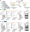
Streamlining VLP technology using a 2P system. (A) Four constructs were developed with two independent promoters driving expression of reporter gene and SARS2 structural proteins. The promoters were separated by insulator and terminator sequences to minimize promoter interference: synthetic polyA (spa); a G-rich sequence from β-actin (Tactb); chicken hypersensitive site 4 (cHS4); and a synthetic MAR sequence 8 (sMAR8) at the end of the E protein. (B, C) Each of these constructs was transfected into 293T cells along with spike plasmid to produce four different 2P SARS2 Luc-PS9 VLPs. 2P.2 VLPs displayed highest luminescence intensity (B). Its signal was comparable to 4P VLP but lower than 3P VLP (C). (D) Western blots of SARS2 structural proteins showed different patterns of protein expression for different 2P plasmids. 2P.2 SARS2 Luc-PS9 VLPs displayed more intense protein bands compared with 2P.3 and 2P.4, but this was lower than 2P.1. (E) 2P.2.EGFP VLPs were created by replacing the luciferase reporter with EGFP. VLP entry of 2P.2.EGFP SARS2 VLPs into A549-hACE2-TMPRSS2 and 293T-hACE2 cells was measured using flow cytometry. Data are mean ± STD. ***P < .001, ****P < .0001, NS: not significant.
Figure 4.
Figure 4.
Delivery of four transgenes using 3P SARS2 VLPs. (A) Schematic of six PS9 constructs with successive addition of four different reporter genes: EGFP, dTomato, TagBFP, and/or luciferase. These constructs were employed to develop 3P VLPs for transgene delivery. (B) Fluorescence and luminescence signal for multiple biological replicates using different 3P constructs (same amount of VLP in each case). The relation between payload size and viral entry was measured based on reporter intensity (upper panels) and % fluorescent cells (lower panels). While percentage of infected cells decreased gradually upon increasing mRNA package size, the decrease in fluorescence intensity was more dramatic. Data are mean ± STD. **P < .01, ***P < .001, ****P < .0001, NS: not significant.
Figure 5.
Figure 5.
Mechanisms regulating VLP function and efficacy. (A) VLPs with either EGFP reporter (downstream of CMV promoter) or luciferase reporter (downstream of CMV or IRES) were produced using the 3P system. Various parameters were measured as illustrated. (B, C) The individual panels in these plots from left to right present: (i) reporter signal intensity in producer cells; (ii) N protein ELISA levels for individual VLP preparations; (iii) ddPCR quantitation of PS9 mRNA levels in VLPs; (iv) reporter signal in target cells following VLP infection; and (v) western blot of VLP structural proteins. EGFP fluorescence data are presented in panel (B) and luciferase reporter data in panel (C). The data showed that VLPs with similar structural compositions and mRNA copy numbers were produced for all payloads. However, increasing payload size progressively decreased reporter signal in target cells. Data are mean ± STD. ***P < .001, ****P < .0001, NS: not significant.
Figure 6.
Figure 6.
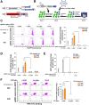
VLPs deliver functional Cas9 mRNA into target cells to achieve gene editing. (A) 3P Cas9-P2A-dTo-T20 VLPs (dTo: dTomato) were used for gene editing studies, with surface glycoprotein encoding for either VSV-G or SARS2 spike. (B) General workflow of gene editing study performed in panels (C)–(E). sgRNAs were transfected into cells on day −1 using plasmids carrying BFP reporter. VLP-carrying spCas9 mRNA was introduced into cells on day 0. Tropism of the VLP depends on surface glycoprotein. Gene editing efficiency was quantified on day 6. Editing efficiency quantified percentage of BFP(+) population that either turned EGFP(−) (panels C and E) or lost ACE2 expression based on anti-hACE2 binding (panel D). (C) sgRNAs targeting EGFP were introduced into 293T-hACE2-EGFP and spCas9 mRNA was delivered using 3P SARS2 Cas9-P2A-dTo-T20 VLPs. (D) sgRNAs against hACE2 were introduced to knockout the receptor in 293T-hACE2 cells using 3P SARS2 Cas9-P2A-dTo-T20 VLPs. (E) sgRNAs targeting EGFP were introduced in 293T-EGFP cells, with genome editing being performed using 3P Cas9-P2A-dTo-T20 VLPs bearing either VSV-G or SARS2 spike. In all panels, the target gene (EGFP or hACE2) was knocked out in 20%–35% of cells expressing sgRNA. Higher VLP amount resulted in greater editing. (F) 293T-hACE2 stably expressed sgRNAs against SLC35A1 were infected with 3P SARS2 Cas9-P2A-dTo-T20 VLPs (1.885 μg/μl N protein equivalent) or without SARS2 spike (1.385 μg/μl N protein equivalent). Gene editing efficiency was evaluated based on increase in fluorescent peanut agglutinin lectin (PNA) binding to cells. More than 70% gene editing was observed upon using 3P VLPs to edit endogenous genes. Volume of VLP used in each assay is specified in individual panels. Data are mean ± STD. ***P < .001, ****P < .0001, NS: not significant.
Figure 7.
Figure 7.
In vivo pulmonary gene delivery using VLPs. (A) VLPs bearing either VSV-G or maSARS2 were instilled into mice at time = 0. Twenty-four hours post-instillation, luciferase activity was measured in tissue extracts from left lung, right lung, or trachea. Protein concentration in lysate was used to normalize luminescence signal, with untreated/mock values being set to 1.0 for all in vivo studies. (B) VSV-G VLPs were instilled via either o.p.a. (100 μl) or i.n. (50 μl) routes. o.p.a. resulted in VLP administration to mouse lung. (C) Q493K and N501Y mutations were introduced into SARS2 spike to generate maSARS2 spike. 3P VLPs with VSV-G, SARS2, maSARS2, and no spike were produced with N protein equivalents of 1.859, 1.149, 1.334, and 2.024 μg/μl, respectively. A total of 50 μl of VLP was used to infect three target cell types: 293T, 293T-hACE2, and 293T-mACE2. VSV-G VLPs infected all three cell types, SARS2 spike only infected 293T-hACE2 (hACE2), whereas maSARS2 spike was permissive to both 293T-hACE2 and 293T-mACE2. VSV-G luminescence was set to 1.0 in this panel. (D) Hundred microliters of VSV-G VLPs or maSARS2 VLPs, both 0.820 μg/μl N protein equivalent, were instilled via o.p.a. into mice. Whole lung tissue was harvested. Mice without VLPs served as negative control. Both VSV-G and maSARS2 VLPs enabled luciferase signal in mouse lung with VSV-G being more efficient. Data are mean ± STD. N = 5–6 for each mouse treatment group. *P < .05, **P < .01, ***P < .001, ****P < .0001, NS: not significant.

References

    1. Jackson CB, Farzan M, Chen B et al. Mechanisms of SARS-CoV-2 entry into cells. Nat Rev Mol Cell Biol. 2022; 23:3–20. 10.1038/s41580-021-00418-x. - DOI - PMC - PubMed
    1. Hoffmann MAG, Yang Z, Huey-Tubman KE et al. ESCRT recruitment to SARS-CoV-2 spike induces virus-like particles that improve mRNA vaccines. Cell. 2023; 186:2380–91. 10.1016/j.cell.2023.04.024. - DOI - PMC - PubMed
    1. Yilmaz IC, Ipekoglu EM, Bulbul A et al. Development and preclinical evaluation of virus-like particle vaccine against COVID-19 infection. Allergy. 2022; 77:258–70. 10.1111/all.15091. - DOI - PMC - PubMed
    1. Gao X, Xia Y, Liu X et al. A perspective on SARS-CoV-2 virus-like particles vaccines. Int Immunopharmacol. 2023; 115:109650. 10.1016/j.intimp.2022.109650. - DOI - PMC - PubMed
    1. Sharifzadeh M, Mottaghi-Dastjerdi N, Soltany Rezae Raad M A review of virus-like particle-based SARS-CoV-2 vaccines in clinical trial phases. Iran J Pharm Res. 2022; 21:e127042. 10.5812/ijpr-127042. - DOI - PMC - PubMed

Substances