

CeDaD-a novel assay for simultaneous tracking of cell death and division in a single population
- PMID: 40038265
- PMCID: PMC11880512
- DOI: 10.1038/s41420-025-02370-7
CeDaD-a novel assay for simultaneous tracking of cell death and division in a single population
Abstract
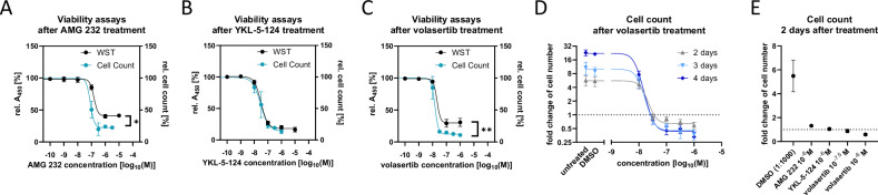
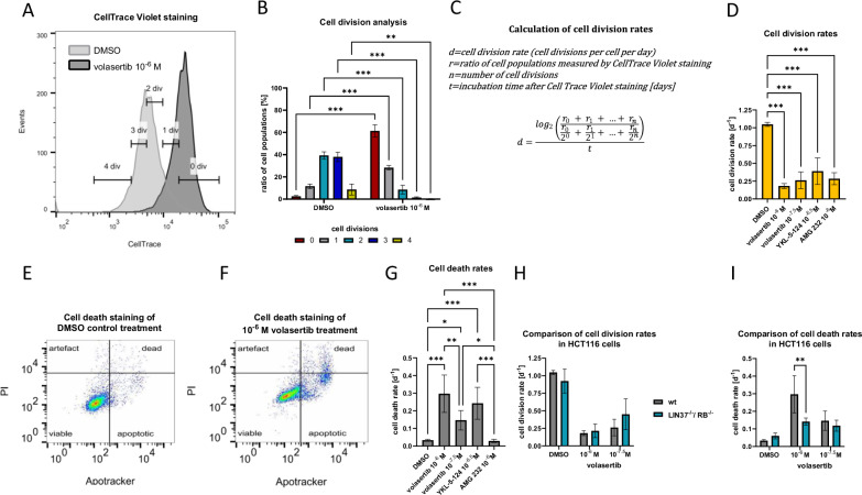
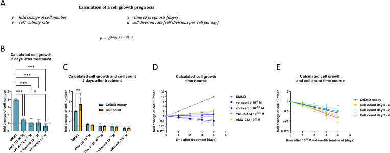
The cell division cycle and the various forms of programmed cell death are interconnected. A prominent example is the tumor suppressor p53, which not only induces apoptosis but also plays an important role in the arrest of the cell cycle. Consequently, simultaneous analysis of cell division and cell death is frequently of significant interest in cell biology research. Traditionally, these processes require distinct assays, making concurrent analysis challenging. To address this, we present a novel combined assay, called CeDaD assay-Cell Death and Division assay-which allows for the simultaneous quantification of cell division and cell death within a single-cell population. This assay utilizes a straightforward flow cytometric approach, combining a staining based on carboxyfluorescein succinimidyl ester (CFSE) to monitor cell division with an annexin V-derived staining to assess the extent of cell death.
© 2025. The Author(s).
Conflict of interest statement
Competing interests: The authors declare no competing interests. Ethics approval: All methods were performed in accordance with the relevant guidelines and regulations. The experiments did not require ethical approval, as no studies on animals or human participants were carried out.
Figures
Similar articles
-
Mathematical models for CFSE labelled lymphocyte dynamics: asymmetry and time-lag in division.J Math Biol. 2014 Dec;69(6-7):1547-83. doi: 10.1007/s00285-013-0741-z. Epub 2013 Dec 13. J Math Biol. 2014. PMID: 24337680
-
Asymmetry of Cell Division in CFSE-Based Lymphocyte Proliferation Analysis.Front Immunol. 2013 Sep 2;4:264. doi: 10.3389/fimmu.2013.00264. Front Immunol. 2013. PMID: 24032033 Free PMC article. Review.
-
Identification and isolation of slow-dividing cells in human glioblastoma using carboxy fluorescein succinimidyl ester (CFSE).J Vis Exp. 2012 Apr 29;(62):3918. doi: 10.3791/3918. J Vis Exp. 2012. PMID: 22565048 Free PMC article.
-
Carboxyfluorescein succinimidyl ester-based proliferative assays for assessment of T cell function in the diagnostic laboratory.Immunol Cell Biol. 1999 Dec;77(6):559-64. doi: 10.1046/j.1440-1711.1999.00870.x. Immunol Cell Biol. 1999. PMID: 10571678
-
Flow cytometric analysis of cell division by dilution of CFSE and related dyes.Curr Protoc Cytom. 2013;Chapter 9:9.11.1-9.11.12. doi: 10.1002/0471142956.cy0911s64. Curr Protoc Cytom. 2013. PMID: 23546777 Review.
Cited by
-
BRCA1 and BRCA2 gene expression: p53- and cell cycle-dependent repression requires RB and DREAM.Cell Death Differ. 2025 Aug 22. doi: 10.1038/s41418-025-01566-9. Online ahead of print. Cell Death Differ. 2025. PMID: 40841483
References
-
- Evan GI, Vousden KH. Proliferation, cell cycle and apoptosis in cancer. Nature. 2001;411:342–8. 10.1038/35077213. - PubMed
-
- Heim A, Rymarczyk B, Mayer TU. Regulation of cell division. Adv Exp Med Biol. 2017;953:83–116. 10.1007/978-3-319-46095-6_3. - PubMed
-
- Hatakeyama M, Weinberg RA. The role of RB in cell cycle control. Prog Cell Cycle Res. 1995;1:9–19. 10.1007/978-1-4615-1809-9_2. - PubMed
-
- Sherr CJ. Cancer cell cycles. Science. 1996;274:1672–7. 10.1126/science.274.5293.1672. - PubMed
-
- Hochegger H, Takeda S, Hunt T. Cyclin-dependent kinases and cell-cycle transitions. Does one fit all? Nat Rev Mol Cell Biol. 2008;9:910–6. 10.1038/nrm2510. - PubMed
Grants and funding
LinkOut - more resources
Full Text Sources
Research Materials
Miscellaneous
