. 2025 Mar 4;16(1):2196.
doi: 10.1038/s41467-025-57481-7.

AAV9-mediated MYBPC3 gene therapy with optimized expression cassette enhances cardiac function and survival in MYBPC3 cardiomyopathy models

Affiliations

AAV9-mediated MYBPC3 gene therapy with optimized expression cassette enhances cardiac function and survival in MYBPC3 cardiomyopathy models

Amara Greer-Short et al. Nat Commun. .

Abstract

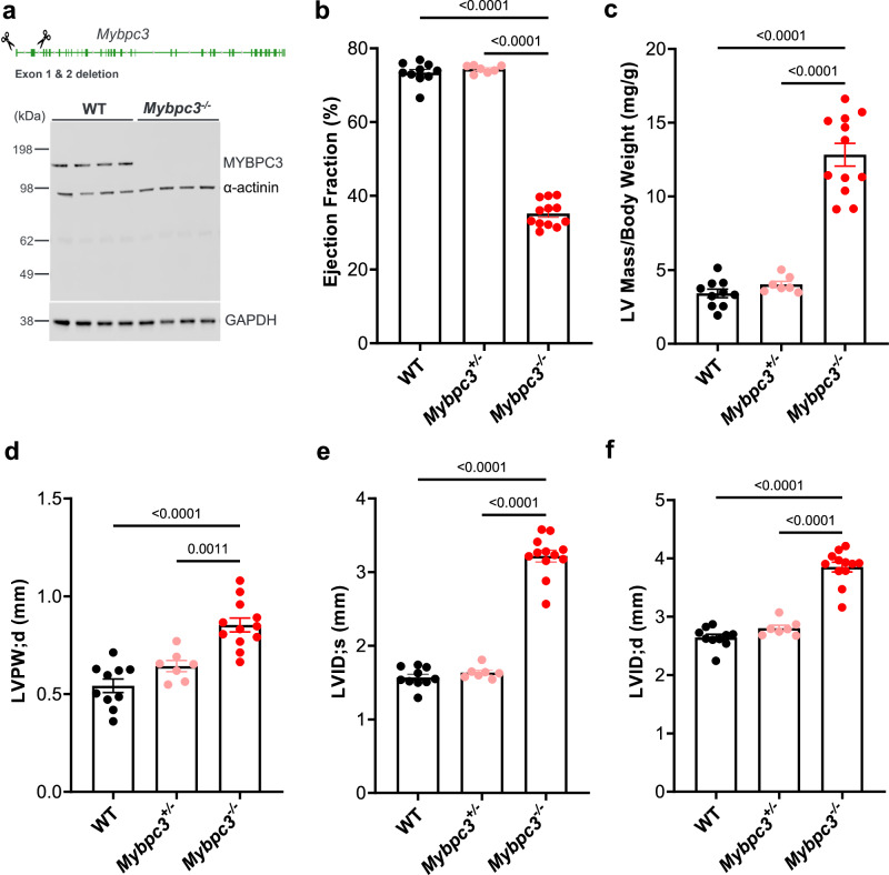
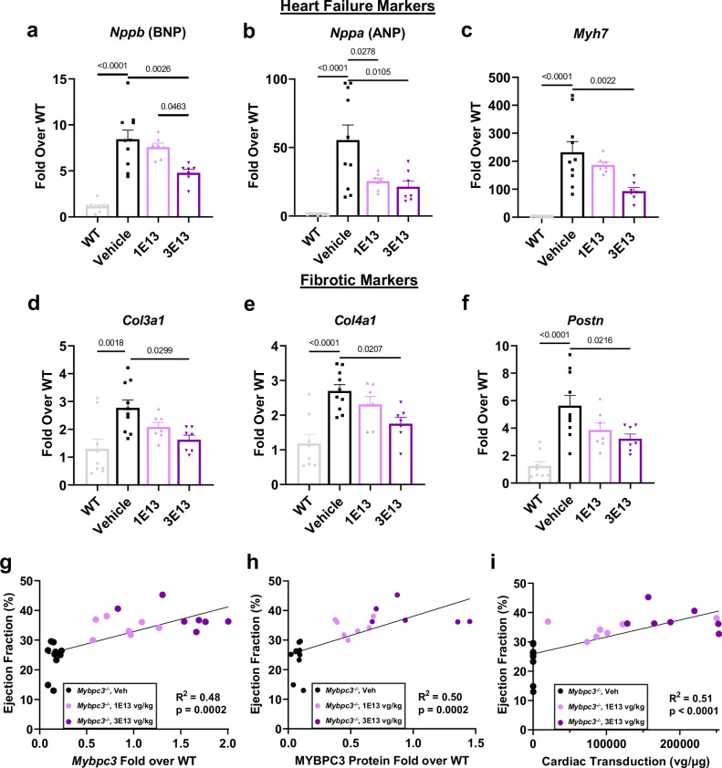
Hypertrophic cardiomyopathy (HCM) affects approximately 600,000 people in the United States. Loss-of-function mutations in Myosin Binding Protein C3, MYBPC3, are the most common genetic cause of HCM, with the majority of mutations resulting in haploinsufficiency. To restore cardiac MYBPC3, we use an adeno-associated virus (AAV9) vector and engineer an optimized expression cassette with a minimal promoter and cis-regulatory elements (TN-201) to enhance packaging efficiency and cardiomyocyte expression. Rather than simply preventing cardiac dysfunction preclinically, we demonstrate in a symptomatic MYBPC3-deficient murine model the ability of AAV gene therapy to reverse cardiac hypertrophy and systolic dysfunction, improve diastolic dysfunction, and prolong survival. Dose-ranging efficacy studies exhibit restoration of wild-type MYBPC3 protein levels and saturation of cardiac improvement at the clinically relevant dose of 3E13 vg/kg, outperforming a previously published construct. These findings suggest that TN-201 may offer therapeutic benefits in MYBPC3-associated cardiomyopathy, pending further validation in clinical settings.


Conflict of interest statement

Competing interests: The authors of this publication declare the following competing interests: All authors of this publication were employed by Tenaya Therapeutics at the time of the study and hold or held equity in Tenaya Therapeutics. L.M.L. is an inventor on published U.S. Patent No.: US 2023/0372541 A1 held by Tenaya Therapeutics that covers gene therapy cassettes for treating heart disease.

Figures

Fig. 1
Fig. 1. AAV genomic cassette and promoter engineering to drive potent and specific expression.
a In order to validate the assay system, we confirmed transgene expression resulted in proper localization in human MYBPC3-/- iPSC-CMs transduced with AAV:MYBPC3. Immunofluorescence analysis was performed seven days post-infection (scale bars, 25 µm). b Cassette schematic indicating the genomic size of a standard cassette and the alterations tested. c Human MYBPC3-/- iPSC-CMs were transduced with AAV6-packaged constructs encoding human MYBPC3 driven by various promoter versions of human cardiac troponin T (TNNT2) (pCard0, pCard1, and pCard2) and harvested 5 days post-infection (n = 1–2/condition). d A head-to-head comparison of the independently packaged versions of the pCard0 and pCard1 constructs. MYBPC3-/- iPSC-CMs were harvested five days post-infection with CR9-01-packaged constructs (n = 2/condition). e To determine if the optimized promoter also increased expression in vivo and whether selectivity was maintained, adult female mice were retro-orbitally injected with 4E13 vg/kg AAV9 encoding the pCard0 (n = 3) and pCard1 (n = 4) constructs. Heart, skeletal muscle (tibialis anterior), liver, and whole brain samples were harvested two weeks post-injection. P-value per two-sided Student’s t-test with 1.0% False Discovery Rate with Two-stage step-up (Benjamini, Krieger, and Yekutieli). Matched transduction (f), RNA (g), and protein (h) analysis in MYBPC3-/- iPSC-CMs demonstrated near-WT levels of protein expression at 1 vg/dg compared to parental wild-type control iPSC-CMs one-week post-infection. WT No Inf (n = 3), MYBPC3-/- No Inf (n = 2), MYBPC3-/- 1 K (n = 3), MYBPC3-/- 3 K (n = 3), WT GFP 100 K (n = 3), and WT GFP 100 K (n = 3). 1 K = multiplicity of infection of 1000, or 1000 vector genomes/cell. Data are shown as means ± SEM. Source data are provided as a Source Data file.
Fig. 2
Fig. 2. Murine Mybpc3-/- model for treatment of HCM.
a A null mutant allele of Mybpc3 was generated by excision of exons one and two using CRISPR-Cas9 and confirmed by demonstrating MYBPC3 depletion in an immunoblot from cardiac tissue (n = 4/condition). Echocardiography analysis of homozygous mutants, heterozygous mutants, and WT littermates demonstrated a significant decrease in cardiac systolic function in homozygous animals based on b ejection fraction (%) at two weeks of age. c Homozygous mice exhibited marked LV hypertrophy at two weeks of age as evidenced by their LV mass normalized by body weight and d increased left ventricular posterior wall thickness during diastole (LVPW;d). Left ventricular internal diameters during e systole (LVID;s) and f diastole (LVID;d) were also significantly increased in homozygotes at two weeks of age. WT (n = 10; 4 M/6 F), Mybpc3+/- (n = 7; 4 M/3 F) and Mybpc3-/- (n = 12; 6 M/6 F). Heterozygous mice were not significantly different from WT in any parameter. P-value per one-way ANOVA with Tukey’s multiple comparisons test. Data are shown as means ± SEM. Source data are provided as a Source Data file.
Fig. 3
Fig. 3. AAV9:mMybpc3 and TN-201 improved hypertrophy, cardiac dysfunction, and premature lethality of Mybpc3-/- mice.
a LV mass normalized to body weight progression over time and b at 31 weeks post-delivery. c EF progression and d EF at 31 weeks post-delivery. e Left ventricular posterior wall thickness in diastole (LVPW;d) and f QT interval at 31 weeks post-delivery. g Kaplan–Meier survival curve with animals followed out until 20 months of age. Median survival for Mybpc3-/- vehicle animals was 14 months, and all animals died by 16 months of age. For TN-201-treated animals, median survival was 16 months (i.e. 2 months lifespan extension from vehicle), and all animals died by 19 months of age. AAV9:mMybpc3 treatment extended lifespan, with only one animal euthanized due to skin lesions not related to heart failure by 20 months. WT (n = 12; 5 M/7 F), Mybpc3-/- Veh (n = 11; 5 M/6 F; 10 at 31 weeks), Mybpc3-/- AAV9:mMybpc3 (n = 12; 6 M/6 F) and Mybpc3-/- TN-201 (n = 9; 4 M/5 F). Data are shown as means ± SEM. WT mice were significantly different from all groups for all parameters, with the exception of AAV9:mMybpc3-treated animals for QT interval. P-value per one-way ANOVA with Tukey’s multiple comparisons test. Mantel–Cox test used for Kaplan–Meier survival curve analysis. Source data are provided as a Source Data file.
Fig. 4
Fig. 4. AAV9:mMybpc3 improves cardiac function at a dose as low as 1E13 vg/kg.
Dose-dependent improvement compared to vehicle-treated Mybpc3-/- mice in a LV mass normalized to body weight, b EF, and c QT interval at 31 weeks post-delivery. d No group differences in body weight at 31 weeks post-delivery. Data are shown as means ± SEM. WT mice were significantly different from all groups for all echocardiographic parameters, with the exception of 3E13 vg/kg and 1E14 vg/kg AAV9:mMybpc3-treated animals for QT interval. WT (n = 7; 4 M/3 F), Mybpc3-/- Veh (n = 7; 4 M/3 F), Mybpc3-/- 1E13 vg/kg (n = 4; 3 M/1 F), Mybpc3-/- 3E13 vg/kg (n = 6; 4 M/2 F), and Mybpc3-/- 1E14 vg/kg (n = 6; 4 M/2 F). P-value per one-way ANOVA with Tukey’s multiple comparisons test. Source data are provided as a Source Data file.
Fig. 5
Fig. 5. Cardiac restoration of wild-type MYBPC3 protein levels.
Homozygous mice were dosed retro-orbitally IV with vehicle or the indicated doses (vg/kg) of AAV9:mMybpc3, and cardiac protein analyzed two weeks and six weeks post-injection. Restoration of WT levels of cardiac MYBPC3 protein upon dosing Mybpc3-/- mice at two weeks of age with 3E13 and 1E14 vg/kg based upon a immunoblot and b ELISA. c Mybpc3 transgene RNA two weeks and six weeks post-injection. WT (n = 3; 1 M/2 F), Mybpc3-/- Veh (n = 2; 2 F), Mybpc3-/- 3E13 vg/kg (n = 3; 1 M/2 F) and Mybpc3-/- 1E14 vg/kg (n = 3; 1 M/2 F). d Immunohistochemistry for MYBPC3 4 weeks post-injection. Homozygous mice were dosed retro-orbitally IV with vehicle or the indicated doses (vg/kg) of AAV9:mMybpc3 at two weeks of age. WT littermates were harvested, and samples processed simultaneously (n = 2/condition; 10× scale bars: 2 mm; 40× scale bars: 25 µm). e Confocal images for assessment of sarcomere structure in animals six weeks following viral dosing (n = 4/condition, 90× scale bars: 25 µm). f Adult WT male mice were dosed retro-orbitally IV with vehicle or the indicated doses (vg/kg) of AAV9:mMybpc3 and cardiac transgene expression was analyzed 10 weeks post-injection by qPCR for Mybpc3 RNA (normalized to Gapdh). Protein assessment in WT mice was performed by ELISA with equivalent total protein for each sample, as well as g immunoblot; Vehicle, Veh (n = 6), 3E13 vg/kg (n = 5) and 3E14 vg/kg (n = 6). P-value per one-way ANOVA with Tukey’s multiple comparisons test. Data are shown as means ± SEM. Source data are provided as a Source Data file.
Fig. 6
Fig. 6. AAV9:mMybpc3 improved diastolic dysfunction in Mybpc3-/- mice.
Homozygous mice were dosed retro-orbitally IV with vehicle or 3E13 vg/kg of AAV9:mMybpc3 at two weeks of age and assessed for diastolic dysfunction 12 weeks post-injection. a Representative tissue (top) and pulsed-wave transmitral (bottom) Doppler tracings. Mitral annular e’ velocity, maximal mitral E-wave velocity (E), and maximal mitral A-wave velocity (A) shown. b Quantitation of mitral E-wave velocity (MV E), c mitral annular e’ velocity, and d isovolumic relaxation time (IVRT) demonstrated dysfunction in Mybpc3-/- mice that was ameliorated by treatment, n = 9 (5 M/4 F)/group. P-value per one-way ANOVA with Tukey’s multiple comparisons test. Data are shown as means ± SEM. Source data are provided as a Source Data file.
Fig. 7
Fig. 7. Dose-dependent inhibition of expression of genes associated with heart failure and fibrosis.
Transcriptional analysis of cardiac tissue from homozygous mice dosed retro-orbitally IV with vehicle or the indicated doses (vg/kg) of AAV9:mMybpc3 at two weeks of age and WT littermates for heart failure markers a Nppb, b Nppa, and c Myh7. Dose-dependent decreases in fibrotic marker expression in treated animals as assessed for d Col3a1, e Col4a1, and f Postn. Significant correlation between ejection fraction and MYBPC3 restoration as analyzed by transgene g RNA expression, h protein expression, and i cardiac transduction. Values are based on a simple linear regression model in GraphPad Prism. Analysis was performed 14 weeks post-injection. WT (n = 9; 5 M/4 F), Mybpc3-/- Veh (n = 10; 5 M/5 F), Mybpc3-/- 1E13 vg/kg (n = 7; 4 M/3 F), and Mybpc3-/- 3E13 vg/kg (n = 7; 3 M/4 F); preserved protein lysate unavailable for one 3E13 vg/kg animal. Data are shown as means ± SEM. P-value per one-way ANOVA with Tukey’s multiple comparisons test. Source data are provided as a Source Data file.
Fig. 8
Fig. 8. Optimized construct outperforms published construct in late-stage Mybpc3-/- mice.
a Homozygous mice with advanced disease (2.5 months of age) were injected retro-orbitally with 3E13 vg/kg or 1E14 vg/kg of AAV9 vector encoding Mybpc3 in the context of the published 5.4 kb cassette (denoted “Published”) or the TN-201 mouse surrogate (AAV9:mMybpc3, denoted mTN-201), or injected with vehicle control. Both cassettes utilize sequence from the human cardiac troponin T (TNNT2) promoter. b EF was measured to represent contractile ability at 27 weeks post-delivery. c LV mass was measured to represent hypertrophy and was normalized to body weight at 27 weeks post-delivery. WT (n = 8; 4 M/4 F), Mybpc3-/- Veh (n = 7; 3 M/4 F), Mybpc3-/- mTN-201 3E13 (n = 6; 2 M/4 F), Mybpc3-/- Published 3E13 (n = 7; 4 M/3 F), Mybpc3-/- mTN-201 1E14 (n = 7; 4 M/3 F), and Mybpc3-/- Published 1E14 (n = 6; 4 M/2 F). d Sustained cardiac transduction 20-months post-injection drove e sustained cardiac Mybpc3 RNA at ~4× levels of endogenous transcript and sustained restoration of wild-type protein levels in dosed homozygotes 20-months post-injection. MYBPC3 protein expression was assessed by ELISA with equivalent total protein for each sample, as well as immunoblot analysis of equivalent total protein, with MYBPC3 intensities normalized to WT (f, representative blot). n = 11 mice/group; 4 M/7 F for WT and 6 M/5 F for mMybpc3. P-value per one-way ANOVA with Tukey’s multiple comparisons test. Error bar: mean ± Standard Error of the Mean (SEM). Source data are provided as a Source Data file.
Fig. 9
Fig. 9. Normalization of calcium handling abnormalities and relaxation defects in heterozygous iPSC-CMs.
MYBPC3+/- iPSC-CMs and their WT isogenic control iPSC-CMs were compared side-by-side for a Fluorescence Transient Amplitude (∆F/Fmin) and b Fluorescence Rise Rate ((∆F/Fmin)/s) using Fluo-4 AM dye on the Nautilai (CuriBio) fluorescence imager (n = 8/group). iPSC-CMs were stimulated at 10 volts, 3 ms, 1 Hz. As above, but with transduced iPSC-CMs one-week post-transduction 10k MOI of “Empty” (CR9-01: cassette without a transgene) or TN-201* for c Fluorescence Transient Amplitude (∆F/Fmin) and d Fluorescence Rise Rate ((∆F/Fmin)/s). WT Empty (n = 15), MYBPC3+/- Empty (n = 16), and MYBPC3+/- TN-201* (n = 16). MYBPC3 protein analysis of transduced iPSC-CMs one-week post-transduction by e immunoblotting [WT Empty (n = 10), MYBPC3+/- Empty (n = 8), and MYBPC3+/- TN-201* (n = 8)] and f ELISA (n = 10/group). Impaired time to 50% relaxation (RT50) (g) and impaired time to 90% relaxation (RT90) (h) of MYBPC3+/- engineered heart tissues was corrected with transduction; 10k MOI of “Empty” (CR9-01: cassette without a transgene) or TN-201*; WT Empty (n = 8), MYBPC3+/- Empty (n = 7), and MYBPC3+/- TN-201* (n = 7). P-value per one-way ANOVA with Tukey’s multiple comparisons test. Error bar: mean ± Standard Error of the Mean (SEM). Source data are provided as a Source Data file.

References

    1. Ho, C. Y. et al. Genotype and lifetime burden of disease in hypertrophic cardiomyopathy: insights from the Sarcomeric Human Cardiomyopathy Registry (SHaRe). Circulation138, 1387–1398 (2018). - PMC - PubMed
    1. Norrish, G. & Kaski, J. P. The risk of sudden death in children with hypertrophic cardiomyopathy. Heart Fail Clin.18, 9–18 (2022). - PubMed
    1. Miron, A. et al. A validated model for sudden cardiac death risk prediction in pediatric hypertrophic cardiomyopathy. Circulation142, 217–229 (2020). - PMC - PubMed
    1. Maron, B. J. & Maron, M. S. Hypertrophic cardiomyopathy. Lancet381, 242–255 (2013). - PubMed
    1. Maron, B. J., Maron, M. S. & Semsarian, C. Genetics of hypertrophic cardiomyopathy after 20 years: clinical perspectives. J. Am. Coll. Cardiol.60, 705–715 (2012). - PubMed