. 2025 Mar 4;23(1):119.
doi: 10.1186/s12964-025-02115-0.

Emerging role of IGF1R and IR expression and localisation in adrenocortical carcinomas

Affiliations

Emerging role of IGF1R and IR expression and localisation in adrenocortical carcinomas

Rosa Catalano et al. Cell Commun Signal. .

Abstract

Background: The insulin-like growth factor 2 (IGF2) is overexpressed in 90% of adrenocortical carcinomas (ACC) and promotes cell proliferation via IGF1R and isoform A of insulin receptor (IRA). However, IGF2 role in ACC tumourigenesis has not been completely understood yet, and the contribution of IGF1R and IRA in mediating ACC cell growth has been poorly explored. This study aimed to investigate IGF1R and IR expression and localisation, including the expression of IR isoforms, in ACC and adrenocortical adenomas (ACA), and their role in IGF2-driven proliferation.

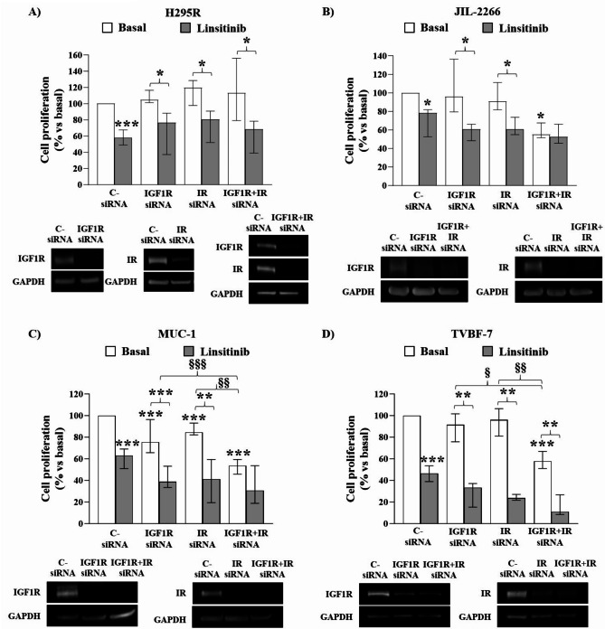
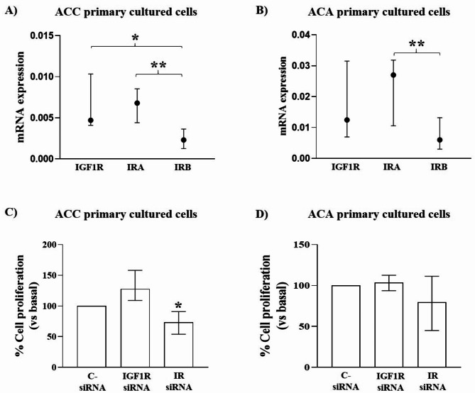
Methods: Immunohistochemistry staining of IGF1R and IR was performed on 118 ACC and 22 ACA to evaluate their expression and cellular localisation and statistical analyses were carried out to assess correlations with clinicopathological data. The expression of IRA and IRB in ACC and ACA tissues, ACC cell lines and ACC and ACA primary cultures was determined by RT-qPCR. To appraise the specific role of IGF1R and IR in mediating IGF2 mitogenic pathway, single and double silencing of receptors and their inhibition in 2 ACC cell lines derived from primary tumours (H295R and JIL-2266) and 2 derived from metastatic tumours (MUC-1 and TVBF-7) as well as in ACC and ACA primary cultures were performed.

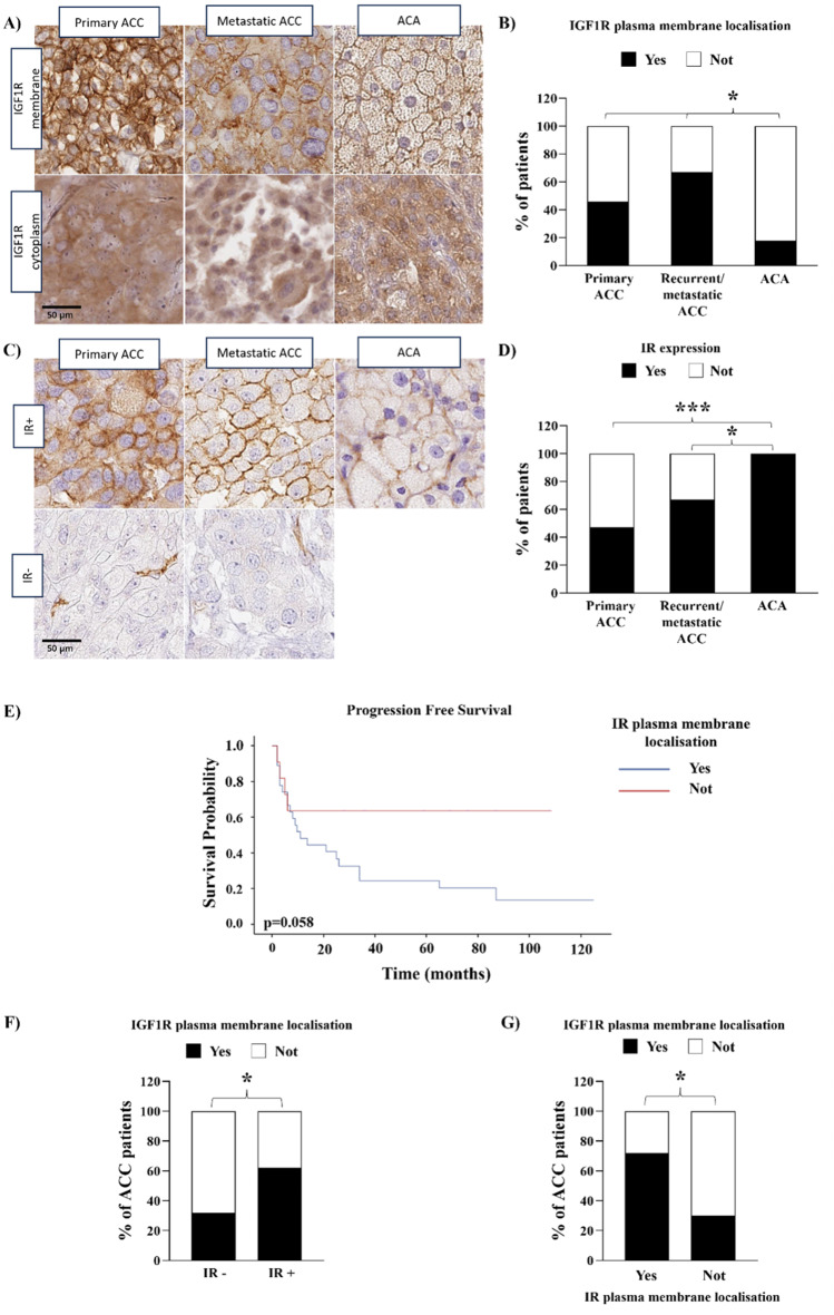
Results: We found a higher IGF1R plasma membrane localisation in ACC compared to ACA. In ACC this localisation was associated with higher Ki67 and Weiss score. IR was expressed in about half of ACC and in all ACA but, in ACC, it was associated with higher Ki67 and Weiss score. RT-qPCR revealed that the prevalent isoform of IR was IRA in ACC and ACA, but not in normal adrenals. In ACC cell lines, double IGF1R + IR silencing reduced cell proliferation in JIL-2266, MUC-1 and TVBF-7 but not in H295R. In ACC, but not ACA, primary cultures, cell proliferation was reduced after IR but not IGF1R knockdown.

Conclusions: Overall, these data suggest that IGF1R localisation and IR expression represent new biomarkers predicting tumour aggressiveness, as well as possible molecular markers useful to patients' stratification for more individualized IGF1R-IR targeted therapies or for novel pharmacological approaches specifically targeting IRA isoform.

Keywords: Adrenocortical carcinoma; Biomarker; Cellular localisation; IGF1R; Insulin receptor.


Conflict of interest statement

Declarations. Ethics approval and consent to participate: The study was conducted in accordance with the Declaration of Helsinki and approved by Fondazione IRCCS Ca’ Granda Ospedale Maggiore Policlinico Ethics Committee (Comitato Etico Milano Area 2, protocol code 20_2019bis, date of approval 18 January 2019 and protocol code 554_2022bis, date of approval 9 June 2022), by the local ethics committee of the University of Wuerzburg (#88/11 for the ENSAT Registry), by Careggi University Hospital Ethical Committee (Protocol code 2011/0020149—Rif CEAVC Em.2019 − 201, date of approval 26 November 2019), by the Ethical Committee of the Institutional Board of Laikon General Hospital (Protocol code 4/17.01.2022, date of approval 17 January 2022) and by the Ethical Committee of the Area Vasta Emilia Centro (CE-AVEC, Protocol code: 105/2017/U/Tess). Consent for publication: Not applicable. Competing interests: The authors declare no competing interests.

Figures

Fig. 1
Fig. 1
IGF1R and IR expression and localisation in ACC and ACA. A) Composite panel depicting representative cases of primary and metastatic ACC and ACA showing either presence or absence of IGF1R expression on the cell membrane. B) The stacked bar chart shows the percentage of patients with only or at least IGF1R plasma membrane localisation (black bars) and IGF1R other cellular localisations (cytoplasm and/or nucleus, white bars) in primary ACC (n = 90), recurrent/metastatic ACC (n = 9), and ACA (n = 17). *=p < 0.05, chi-squared test. C) Composite panel showing representative cases of primary and metastatic ACC and ACA, featuring either expression or negativity for insulin receptor (internal positive control represented by endothelial cells). D) The graph shows the percentage of patients with IR expression (black bars) in primary ACC (n = 92), recurrent/metastatic ACC (n = 9), and ACA (n = 19). *=p < 0.001, chi-squared test. E) Primary ACC with IR plasma membrane localisation shows a worse progression free survival. Kaplan-Meier analysis of the progression free survival in patients with IR plasma membrane localisation (n = 27) and in those without plasma membrane localisation (n = 11). F&G) IGF1R plasma membrane localisation is associated with IR expression and plasma membrane localisation. The bar charts show the association between the percentage of ACC patients with only or at least IGF1R plasma membrane localisation and the IR expression (F) (n = 76; “IR-” = no immunoreactivity to IR antibody, “IR+” = IR expression) or the IR plasma membrane localisation (G) (n = 39). *=p < 0.05, chi-squared test
Fig. 2
Fig. 2
Normalized IRA and IRB mRNA levels in ACC, ACA and NAG. IRA and IRB mRNA levels within the same sample in (A) adrenocortical carcinoma (ACC) (n = 36), (B) adrenocortical adenoma (ACA) (n = 18), and (C) normal adrenal gland (NAG) (n = 14). Difference in IRA (D), IRB (E) and IRA/IRB ratio (F) among the three groups
Fig. 3
Fig. 3
Insulin-like growth factor (IGF) system in H295R, JIL-2266, MUC-1, and TVBF-7. IGF1R mRNA (A) and protein (B) expression in ACC cell lines. A) The vertical scatter plot shows the normalized IGF1R mRNA expression for each cell line. GAPDH was used as reference gene. Horizontal bars represent median (IQR) values. ** = p < 0.01 vs. H295R; §= p < 0.05 vs. JIL-2266. B) On the left the result of the densitometrical analyses of at least three experiments (median (IQR)). IGF1R was normalized on GAPDH. On the right representative immunoblot. The membranes were incubated with IGF1R antibody and then reprobed with anti-GAPDH antibody. JIL-2266, MUC-1 and TVBF-7 were normalized on H295R. * = p < 0.05; ** = p < 0.01 vs. H295R. C&D) The charts illustrate the different IR isoforms mRNA expression between H295R, JIL-2266, MUC-1, and TVBF-7. Horizontal bars represent median values. * = p < 0.05; ** = p < 0.01 vs. H295R and § = p < 0.05 vs. JIL-2266. E) IGF2 secretion in culture media measured by ELISA assay in H295R, JIL-2266, MUC-1, and TVBF-7. Cells were incubated for 2 h with a serum-deprived cell culture media and, in case of H295R, also with insulin-transferrin-selenium (ITS) + Premix deprivation. Starved base medium of each cell line was used as negative control. Secretion was expressed as ng of IGF2 secreted by 1 * 106 cells
Fig. 4
Fig. 4
Effect of IGF1R and/or IR gene knockdown or Linsitinib inhibition on proliferation in human ACC cell lines. H295R (A), JIL-2266 (B), MUC-1 (C) and TVBF-7 (D) proliferation assays. Cells were seeded at the density of 1.4 × 104 for H295R, 4.5 × 103 for JIL-2266 and MUC-1 and 1.6 × 104 for TVBF-7 in a 96-well. The following day they were silenced for IGF1R, IR, or IGF1R + IR for 72 h and then, a serum starvation for 2 h followed by incubation with Linsitinib 1µM for 24 h was performed. BrdU was added for 2 h and its incorporation in newly synthesized DNA was measured. Western blot was performed in parallel with each experiment to evaluate the silencing efficiency of IGF1R and IR and only experiments achieving a silencing efficiency ≥ 70% were considered for analysis. Experiments were repeated at least 3 times and each determination was done in triplicate. Graphs show the median and IQR expressed as percentage of basal C- siRNA of cell proliferation in H295R (A), JIL-2266 (B), MUC-1 (C) and TVBF-7 (D). * = p < 0.05; ** = p < 0.01; *** = p < 0.001 vs. c- siRNA; § = p < 0.05; §§ = p < 0.01; §§§ = p < 0.001. Below each graph representative immunoblots are present to illustrate the efficiency of cell silencing after IGF1R, IR and IGF1R + IR knockdown in H295R (A), JIL-2266 (B), MUC-1 (C) and TVBF-7 (D)
Fig. 5
Fig. 5
IGF system in ACC and ACA primary cultures. A&B) IGF1R, IRA, and IRB expression in ACC (n = 4) (A) and ACA (n = 8) (B) primary cultures. The graphs show ΔCt of genes normalized on GAPDH with median and IQR. *=p < 0.05 C&D) 1.4 × 104 ACC (n = 4) and ACA (n = 8) primary cultured cells were seeded and silenced the following day for IGF1R or IR for 72 h. At the end an incubation with a serum-free media for 24 h followed by addition of BrdU for additional 24 h was performed. Each determination was done at least in triplicate. The charts values represent median and IQR expressed as percentage of cell proliferation respect to basal C- siRNA. *=p < 0.05 vs. C- siRNA

References

    1. Fassnacht M, Johanssen S, Quinkler M, Bucsky P, Willenberg HS, Beuschlein F et al. German Adrenocortical Carcinoma Registry Group; European Network for the Study of Adrenal Tumors. Limited prognostic value of the 2004 International Union Against Cancer staging classification for adrenocortical carcinoma: proposal for a Revised TNM Classification. Cancer 2009;115:243– 50. 10.1002/cncr.24030. PMID: 19025987. - PubMed
    1. Lippert J, Fassnacht M, Ronchi CL. The role of molecular profiling in adrenocortical carcinoma. Clin Endocrinol (Oxf) 2022;97:460– 72. 10.1111/cen.14629. PMID: 34750847. - PubMed
    1. Berruti A, Tiberio GAM, Sigala S. Adrenocortical Carcinoma. Cancers (Basel) 2021;13:1077. 10.3390/cancers13051077. PMID: 33802418. - PMC - PubMed
    1. Else T, Kim AC, Sabolch A, Raymond VM, Kandathil A, Caoili EM et al. Adrenocortical carcinoma. Endocr Rev 2014;35:282–326. 10.1210/er.2013-1029. PMID: 24423978. - PMC - PubMed
    1. Amodru V, Garcia ME, Libe R, Brue T, Reznik Y, Castinetti F. Medical management of adrenocortical carcinoma: Current recommendations, new therapeutic options and future perspectives. Ann Endocrinol (Paris). 2021;82:52– 8. 10.1016/j.ando.2020.12.003. PMID: 33279475. - PubMed

MeSH terms