. 2025 Mar 5;22(1):64.
doi: 10.1186/s12974-025-03387-6.

The protective PLCγ2-P522R variant mitigates Alzheimer's disease-associated pathologies by enhancing beneficial microglial functions

Affiliations

The protective PLCγ2-P522R variant mitigates Alzheimer's disease-associated pathologies by enhancing beneficial microglial functions

Mari Takalo et al. J Neuroinflammation. .

Abstract

Background: Phospholipase C gamma 2, proline 522 to arginine (PLCγ2-P522R) is a protective variant that reduces the risk of Alzheimer's disease (AD). Recently, it was shown to mitigate β-amyloid pathology in a 5XFAD mouse model of AD. Here, we investigated the protective functions of the PLCγ2-P522R variant in a less aggressive APP/PS1 mouse model of AD and assessed the underlying cellular mechanisms using mouse and human microglial models.

Methods: The effects of the protective PLCγ2-P522R variant on microglial activation, AD-associated β-amyloid and neuronal pathologies, and behavioral changes were investigated in PLCγ2-P522R knock-in variant mice crossbred with APP/PS1 mice. Transcriptomic, proteomic, and functional studies were carried out using microglia isolated from mice carrying the PLCγ2-P522R variant. Finally, microglia-like cell models generated from human blood and skin biopsy samples of PLCγ2-P522R variant carriers were employed.

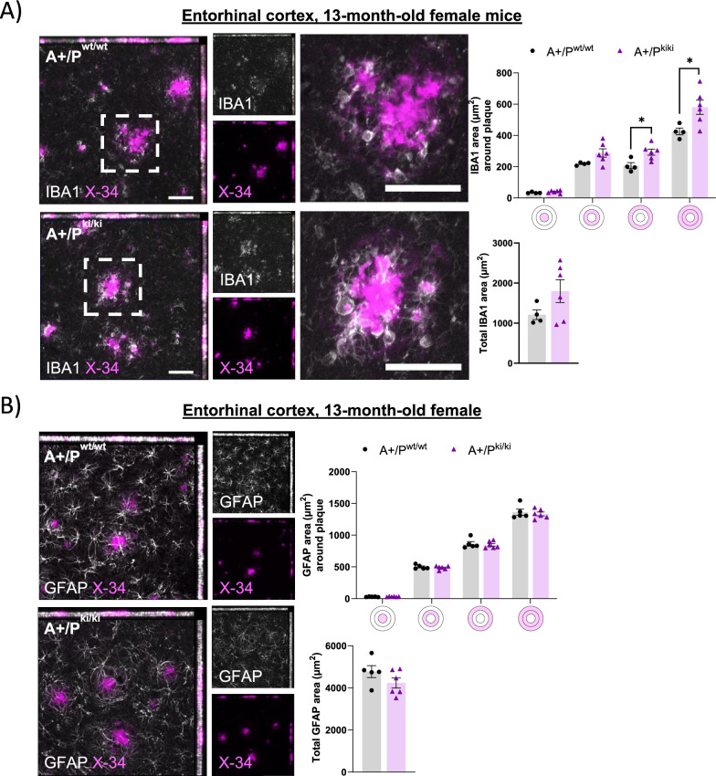
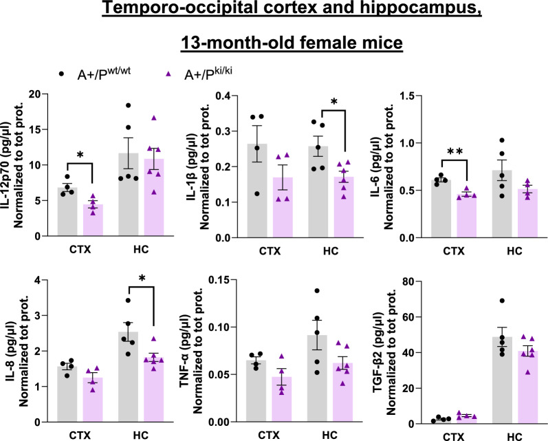
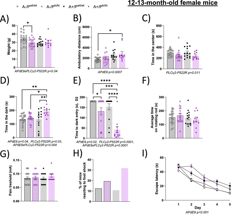
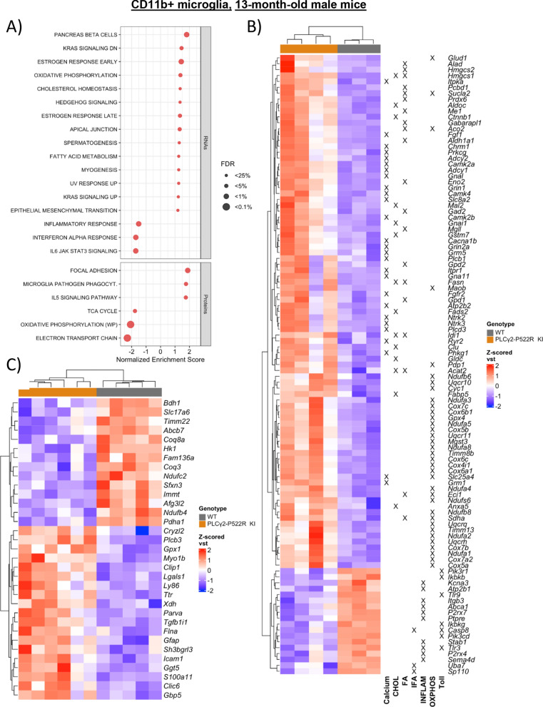
Results: The PLCγ2-P522R variant decreased β-amyloid plaque count and coverage in female APP/PS1 mice. Moreover, the PLCγ2-P522R variant promoted anxiety in these mice. The area of the microglia around β-amyloid plaques was also increased in mice carrying the PLCγ2-P522R variant, while β-amyloid plaque-associated neuronal dystrophy and the levels of certain cytokines, including IL-6 and IL-1β, were reduced. These alterations were revealed through [18F]FEPPA PET imaging and behavioral studies, as well as various cytokine immunoassays, transcriptomic and proteomic analyses, and immunohistochemical analyses using mouse brain tissues. In cultured mouse primary microglia, the PLCγ2-P522R variant reduced the size of lipid droplets. Furthermore, transcriptomic and proteomic analyses revealed that the PLCγ2-P522R variant regulated key targets and pathways involved in lipid metabolism, mitochondrial fatty acid oxidation, and inflammatory/interferon signaling in acutely isolated adult mouse microglia and human monocyte-derived microglia-like cells. Finally, the PLCγ2-P522R variant also increased mitochondrial respiration in human iPSC-derived microglia.

Conclusions: These findings suggest that the PLCγ2-P522R variant exerts protective effects against β-amyloid and neuronal pathologies by increasing microglial responsiveness to β-amyloid plaques in APP/PS1 mice. The changes observed in lipid/fatty acid and mitochondrial metabolism revealed by the omics and metabolic assessments of mouse and human microglial models suggest that the protective effects of the PLCγ2-P522R variant are potentially associated with increased metabolic capacity of microglia.

Keywords: Alzheimer’s disease; Lipid droplets; Microglia; PLCγ2-P522R variant; Phospholipase C gamma 2; β-amyloid pathology.


Conflict of interest statement

Declarations. Ethics approval and consent to participate: The animals were raised and handled at the Laboratory Animal Center of the University of Eastern Finland, Kuopio, Finland. All mouse studies were carried out in accordance with the guidelines of the European Community Council Directives 86/609/EEC and approved by the Regional State Administrative Agency and Project Authorization Board (ESAVI/7315/2024, EKS-004–2019). All study protocols concerning human samples were approved by the Medical Research Ethics Committee of Wellbeing Services County of North Savo (formerly the Medical Research Ethics Committee of North Savo Hospital District, 833/2023, 123/2016). Participant recontacting was approved by the Scientific Steering Committee of Auria Biobank (BB_2020-0062). Consent for publication: All the authors have approved the final version of the manuscript and provided their consent for publication. Competing interests: CH collaborates with Denali Therapeutics and is a member of the advisory boards of AviadoBio and Cure Ventures.

Figures

Fig. 1
Fig. 1
PLCγ2-P522R variant increases microglia activation in the brain of 13-month-old female and male APP/PS1 mice. A Illustration of the PLCγ2 protein domain and the location of the PLCγ2-P522R variant in the protein domain. Created in https://BioRender.com. B-C Example positron emission (PET) images of the uptake (percent of the injected dose per ml of tissue) of [18F]FEPPA for both APP/PS1 (A+/Pwt/wt) and APP/PS1xPLCγ2-P522R (A+/Pki/ki) mice at (B) 7 months and (C) 13 months of age with an anatomical CT reference image in the background. [18F]FEPPA uptake remains unaltered between the genotypes at the 7-month timepoint. A+/Pwt/wt n = 12 and A+/Pki/ki n = 17. Compared with that in A+/Pwt/wt mice, [18F]FEPPA uptake in the whole brain (*p = 0.032), pons (*p = 0.013), and hippocampus (*p = 0.012) was significantly greater in 13-month-old A+/Pki/ki mice. A+/Pwt/wt n = 10 and A+/Pki/ki n = 13. The colored circles indicate data obtained from female mice, and the hollow circles indicate data obtained from male mice. The data were normalized to those of A+/Pwt/wt mice of each sex
Fig. 2
Fig. 2
β-amyloid pathology is mitigated in female APP/PS1 mice carrying the PLCγ2-P522R variant. A X-34-positive β-amyloid plaque count (*p = 0.005) and coverage (**p = 0.006, total plaque area, % of whole analyzed area) were lower in the entorhinal cortex of APP/PS1xPLCγ2-P522R (A+/Pki/ki) mice than in that of APP/PS1 (A+/Pwt/wt) mice. The size (µm2) of the individual plaques did not differ between the genotypes. A+/Pwt/wt n = 4 and A+/Pki/ki n = 6. B Insoluble but not soluble Aβ40 and −42 levels are slightly but not significantly lower in the temporo-occipital cortex of A+/Pki/ki mice than in that of A+/Pwt/wt mice. Aβ40 and Aβ42 levels were normalized to the total protein concentration in the same sample. A+/Pwt/wt n = 4 and A+/Pki/ki = 4. C Representative Western blots and corresponding quantification showing no differences in the levels of full-length APP (APPtot, normalized to GAPDH), APP C-terminal fragments (C99 and C83, normalized to APPtot), or soluble APPα and APPβ (sAPPα, sAPPβ, normalized to APPtot) species in the temporo‒occipital cortex of A+/Pki/ki and A+/Pwt/wt mice. A+/Pwt/wt n = 4 and A+/Pki/ki n = 4. The scale bars in the representative immunofluorescence images are 657 µm for the whole area and 164 µm for the zoomed view. Unpaired samples t test. All the data are presented as the means ± SEMs. Each data point represents an individual mouse
Fig. 3
Fig. 3
PLCγ2-P522R variant increases the area of microglia around β-amyloid plaques in female APP/PS1 mice. A The area (µm2) of IBA1-positive microglia was greater at distances of 20–40 µm (*p = 0.017) and 0–40 µm (*p = 0.03) from the plaque outline in the entorhinal cortex of APP/PS1xPLCγ2‒P522R (A+/Pki/ki) mice than in that of APP/PS1 (A+/Pwt/wt) mice. Simultaneously, a trend toward an increase in the total IBA1 area is observed. A+/Pwt/wt n = 4 and A+/Pki/ki n = 6. B GFAP-positive astrocyte area (µm2) around plaques (within 0–20 µm and 20–40 µm from the plaque outline) and total GFAP area remain unaltered between the genotypes. A+/Pwt/wt n = 5, A+/Pki/ki n = 6. The colored disks on the x-axis of A and B indicate the analyzed area overlapping (middle circle) and surrounding β-amyloid plaques. The scale bar in the representative immunofluorescence images is 50 μm. Unpaired samples t test. All the data are presented as the means ± SEMs. Each data point represents an individual mouse
Fig. 4
Fig. 4
PLCγ2-P522R variant decreases β-amyloid plaque-associated dystrophic neurites in female APP/PS1 mice. A Total area (µm2) of diffuse β-amyloid (6E10) and composition β-amyloid plaques, as indicated by the percentage of compact (X-34) and diffuse (6E10) β-amyloid plaques, remaining unaltered in the entorhinal cortex of the APP/PS1xPLCγ2-P522R (A+/Pki/ki) mice compared with the APP/PS1 (A+/Pwt/wt) mice. A+/Pwt/wt n = 5 and A+/Pki/ki n = 6. B) A 3D reconstruction showing the 6E10 and IBA1 (microglia) signals and their colocalization. Quantification of the 6E10 signal within the IBA1-positive area as a percentage of all the 6E10 signal in the entorhinal cortices of the A+/Pki/ki mice compared with that in the A+/Pwt/wt mice. C The area (µm2) of 22C11-labeled dystrophic neurites around β-amyloid plaques strongly correlates with plaque size (r = 0.8113, ****p < 0.0001). The 22C11-positive area was decreased within the β-amyloid plaque area (p = 0.033), and a similar decreasing trend was observed within the 0–14 µm radius from the β-amyloid plaque outline in the entorhinal cortices of the A+/Pki/ki mice compared with the A+/Pwt/wt mice when the plaque size was normalized. A+/Pwt/wt n = 4 and A+/Pki/ki n = 6. Colored disks on the x-axis indicate the analyzed area overlapping (middle circle) and surrounding β-amyloid plaques. Scale bars in the representative immunofluorescence images are 50 μm. Pearson correlation and unpaired samples t tests were used. All the data are presented as the means ± SEMs. Each data point represents an individual mouse
Fig. 5
Fig. 5
PLCγ2-P522R variant alleviates neuroinflammation in the brain tissue of female APP/PS1 mice. A IL-12p70 (p = 0.049) and IL-6 (p = 0.003) levels are significantly lower in the temporo-occipital cortex (CTX), whereas IL-1β (p = 0.021) and IL-8 (p = 0.026) levels are significantly lower in the hippocampus (HC) of APP/PS1xPLCγ2-P522R (A+/Pki/ki) mice than in those of APP/PS1 (A+/Pwt/wt) mice. In addition, the IL-1β, IL-8, and TNF-α levels tended to decrease in the CTX group, whereas the IL-6, TNF-α, and TGF-β2 levels tended to decrease in the HCs of the A+/Pki/ki mice compared with those of the A+/Pwt/wt mice. CTX A+/Pwt/wt n = 4 and A+/Pki/ki n = 4; HC A+/Pwt/wt n = 5 and HC A+/Pki/ki n = 6. Unpaired samples t test. All the data are presented as the means ± SEMs. Each data point represents an individual mouse
Fig. 6
Fig. 6
PLCγ2-P522R variant does not increase the plaque-associated APOE or DAM signature in female APP/PS1 mice. A Analysis of total APOE and APOE areas (µm2) within and surrounding (within 0–10- and 10–20-µm distances from the amyloid plaque outline) β-amyloid plaques revealed no differences between the APP/PS1xPLCγ2-P522R (A+/Pki/ki) and APP/PS1 (A+/Pwt/wt) mice. A+/Pwt/wt n = 5, A+/Pki/ki n = 6. Colored disks on the x-axis indicate the analyzed area overlapping (middle circle) and surrounding β-amyloid plaques. The scale bar in the representative immunofluorescence images is 50 μm. Unpaired samples t test. All the data are presented as the means ± SEMs. Each data point represents an individual mouse. B Heatmap of z scored vst-normalized expression of homeostatic as well as stage 1 and stage 2 disease-associated microglia (DAM) signature genes in the bulk temporo-occipital cortex and C) CD11b+ microglia isolated from the whole brain tissue of 13-month-old wild-type (A-/Pwt/wt), A+/Pwt/wt and A+/Pki/ki female mice. Cortex A-/Pwt/wt n = 1, A+/Pwt/wt n = 4, and A+/Pki/ki n = 4; CD11b+ A-/Pwt/wt n = 7, A+/Pwt/wt n = 4, and A+/P.ki/ki n = 4
Fig. 7
Fig. 7
PLCγ2-P522R female mice exhibit avoidance responses to open places, bright environments, and sudden unpleasant stimuli. A Body weights of the PLCγ2-P522R (Pki/ki) mice are lower than those of their wild-type (Pwt/wt) littermates on the APP/PS1-negative background (A-, *p = 0.024) but not on the APP/PS1 hemizygous background (A+). B PLCγ2-P522R does not influence the ambulatory distance traveled in the spontaneous activity test. C Pki/ki mice spent significantly less time in the center of the novel test box than did control Pwt/wt mice, regardless of the APP/PS1 genotype. D In the passive avoidance test, the Pki/ki mice showed increased avoidance of the illuminated chamber, which was further accentuated by the APP/PS1 hemizygous genotype. E Even after receiving a foot shock, Pki/ki mice have a shorter latency to enter the dark chamber as compared to Pwt/wt mice. This tendency was further accentuated by the APP/PS1 hemizygous genotype. F No difference in motor skills was observed between the genotypes, as indicated by the time spent on the rotating rod. G Pki/ki and Pwt/wt mice have similar thresholds for pain (foot shock), but H) Pki/ki mice have a stronger behavioral response (crab-like walking backward) to electric foot shock. I) PLCγ2-P522R does not influence spatial learning or memory in the Morris water maze. The APP/PS1 hemizygous mice presented strongly impaired learning in terms of escape latency (F1,64 = 46.8, p < 0.001). A-/Pwt/wt n = 20, A-/Pki/ki n = 18, A+/Pwt/wt n = 14, and A+/Pki/ki n = 11. Two-way ANOVA with Šídák's multiple comparisons test. All the data are presented as the means ± SEMs. Each data point represents an individual mouse
Fig. 8
Fig. 8
Fatty acid metabolism and mitochondrial function-related targets are upregulated in male mouse PLCγ2-P522R microglia. A Dot plot of normalized enrichment scores for enriched and depleted gene sets in enrichment analyses by GSEA for gene (MSigDB Hallmark, FDR < 0.25) and protein (Wikipathways, FDR < 0.05) expression in CD11b+ microglia isolated from 13-month-old PLCγ2-P522R KI and wild-type (WT) mice. B Heatmap of z scored vst-normalized expression of up- and downregulated differentially expressed genes (DEGs) that were core enrichment genes in a GSEA of gene sets for calcium signaling (Calcium), cholesterol homeostasis (CHOL), fatty acid metabolism (FA), the IFN-α response (IFA), the inflammatory response (INFLAM), oxidative phosphorylation (OXPHOS), and Toll-like receptor signaling (Toll). C Heatmap of z scored vs. normalized expression of significantly up- and downregulated differentially expressed proteins (DEPs, expressed as their encoding gene symbols) derived from a proteomics study. RNA WT n = 3 and PLCγ2-P522R KI n = 4; protein WT n = 5 and PLCγ2-P522R KI n = 6
Fig. 9
Fig. 9
PLCγ2-P522R variant changes lipid accumulation in mouse primary microglia. A The percentage of lipid droplet (LD)-positive cells was greater in mouse primary microglia after 24 h of lipopolysaccharide (LPS) and 48 h of myelin treatment than in untreated cells (UNT). The percentage of LD-positive cells did not differ between wild-type (WT) and PLCγ2-P522R knock-in (KI) microglia. PLCγ2-P522R KI decreases the size (µm2) of individual LDs under myelin-treated conditions (****p < 0.0001). n (analyzed images) WT UNT n = 11, LPS n = 22, myelin n = 42; PLCγ2-P522R KI UNT n = 6, LPS n = 12, myelin n = 35. Two-way ANOVA with Tukey’s multiple comparisons test. All the data are presented as the means ± SEMs
Fig. 10
Fig. 10
PLCγ2-P522R variant improves mitochondrial function in human microglial models. Volcano plot of differentially expressed genes (DEGs) in blood monocyte-derived microglia-like cells (MDMi) from PLCγ2-P522R variant carriers (CG) and matched controls (CC) under A) untreated (UNT), B myelin-treated and C lipopolysaccharide (LPS)-treated conditions. FDR < 0.05. Horizontal dashed line: adjusted p value of 0.05; vertical dashed lines: |log2FC|= 0.1. D Dot plot of normalized enrichment scores for enriched and depleted gene sets in enrichment analyses by GSEA for gene (hallmarks, FDR < 0.25) expression in MDMi cells of CG and CC individuals upon UNT, LPS, and myelin treatments. E Heatmap of z scored vst-normalized expression of up- and downregulated DEGs in the LPS-treated condition that were core enrichment genes in a GSEA of gene sets for the IFN-γ response (IFG), inflammatory response (INFLAM), Kras signaling up (KRASup), MYC-target (MYC), oxidative phosphorylation (OXPHOS), and TNF-α signaling (TNFA). UNT CC n = 5 and CG n = 4; Myelin CC n = 5 and CG n = 4; LPS CC n = 5 and CG n = 4. F) The mitochondrial oxygen consumption rate (OCR, maximal respiration and spare respiratory capacity) is greater in homozygous PLCγ2-P522R (GG)-induced pluripotent stem cell-derived microglia (iMGL) than in isogenic controls (CCs) with the APOE3/3 genetic background but not the APOE4/4 genetic background. iMGL n = 1 line per group, 7–9 technical replicates per line. Two-way ANOVA with Tukey’s multiple comparisons test. The data are presented as the means ± SEMs

References

    1. Bill CA, Vines CM, Phospholipase C. Calcium Signaling. Cham: Springer International Publishing; 2020.
    1. Sims R, Van Der Lee SJ, Naj AC, Bellenguez C, et al. Rare coding variants in PLCG2, ABI3, and TREM2 implicate microglial-mediated innate immunity in Alzheimer’s disease. Nat Genet. 2017;49(9):1373–84. - PMC - PubMed
    1. Magno L, Lessard CB, Martins M, Lang V, Cruz P, Asi Y, et al. Alzheimer’s disease phospholipase C-gamma-2 (PLCG2) protective variant is a functional hypermorph. Alz Res Therapy. 2019;11(1):16. - PMC - PubMed
    1. Hansen DV, Hanson JE, Sheng M. Microglia in Alzheimer’s disease. J Cell Biol. 2018;217(2):459–72. - PMC - PubMed
    1. Keren-Shaul H, Spinrad A, Weiner A, Matcovitch-Natan O, Dvir-Szternfeld R, Ulland TK, et al. A unique microglia type associated with restricting development of alzheimer’s disease. Cell. 2017;169(7):1276-1290.e17. - PubMed

Substances

LinkOut - more resources